Pleckstrin homology domain

Last updated
1bwn opm.png
PH domain of tyrosine-protein kinase BTK
Identifiers
SymbolPH
Pfam PF00169
Pfam clan CL0266
InterPro IPR001849
SMART PH
PROSITE PDOC50003
SCOP2 1dyn / SCOPe / SUPFAM
OPM superfamily 49
OPM protein 1pls
CDD cd00821
Available protein structures:
Pfam   structures / ECOD  
PDB RCSB PDB; PDBe; PDBj
PDBsum structure summary

Pleckstrin homology domain (PH domain) or (PHIP) is a protein domain of approximately 120 amino acids that occurs in a wide range of proteins involved in intracellular signaling or as constituents of the cytoskeleton. [1] [2] [3] [4] [5] [6] [7]

Contents

This domain can bind phosphatidylinositol lipids within biological membranes (such as phosphatidylinositol (3,4,5)-trisphosphate and phosphatidylinositol (4,5)-bisphosphate), [8] and proteins such as the βγ-subunits of heterotrimeric G proteins, [9] and protein kinase C. [10] Through these interactions, PH domains play a role in recruiting proteins to different membranes, thus targeting them to appropriate cellular compartments or enabling them to interact with other components of the signal transduction pathways.

Lipid binding specificity

Individual PH domains possess specificities for phosphoinositides phosphorylated at different sites within the inositol ring, e.g., some bind phosphatidylinositol (4,5)-bisphosphate but not phosphatidylinositol (3,4,5)-trisphosphate or phosphatidylinositol (3,4)-bisphosphate, while others may possess the requisite affinity. This is important because it makes the recruitment of different PH domain containing proteins sensitive to the activities of enzymes that either phosphorylate or dephosphorylate these sites on the inositol ring, such as phosphoinositide 3-kinase or PTEN, respectively. Thus, such enzymes exert a part of their effect on cell function by modulating the localization of downstream signaling proteins that possess PH domains that are capable of binding their phospholipid products.

Structure

The 3D structure of several PH domains has been determined. [11] All known cases have a common structure consisting of two perpendicular anti-parallel beta sheets, followed by a C-terminal amphipathic helix. The loops connecting the beta-strands differ greatly in length, making the PH domain relatively difficult to detect while providing the source of the domain's specificity. The only conserved residue among PH domains is a single tryptophan located within the alpha helix that serves to nucleate the core of the domain.

Proteins containing PH domain

PH domains can be found in many different proteins, such as OSBP or ARF. Recruitment to the Golgi apparatus in this case is dependent on both PtdIns and ARF. A large number of PH domains have poor affinity for phosphoinositides and are hypothesized to function as protein binding domains. A Genome-wide look in Saccharomyces cerevisiae showed that most of the 33 yeast PH domains are indeed promiscuous in binding to phosphoinositides, while only one (Num1-PH) behaved highly specific . [12] Proteins reported to contain PH domains belong to the following families:

Subfamilies

Examples

Human genes encoding proteins containing this domain include:

See also

Related Research Articles

<span class="mw-page-title-main">Phosphoinositide phospholipase C</span>

Phosphoinositide phospholipase C is a family of eukaryotic intracellular enzymes that play an important role in signal transduction processes. These enzymes belong to a larger superfamily of Phospholipase C. Other families of phospholipase C enzymes have been identified in bacteria and trypanosomes. Phospholipases C are phosphodiesterases.

<span class="mw-page-title-main">Phosphatidylinositol (3,4,5)-trisphosphate</span> Chemical compound

Phosphatidylinositol (3,4,5)-trisphosphate (PtdIns(3,4,5)P3), abbreviated PIP3, is the product of the class I phosphoinositide 3-kinases' (PI 3-kinases) phosphorylation of phosphatidylinositol (4,5)-bisphosphate (PIP2). It is a phospholipid that resides on the plasma membrane.

<span class="mw-page-title-main">Phosphoinositide 3-kinase</span> Class of enzymes

Phosphoinositide 3-kinases (PI3Ks), also called phosphatidylinositol 3-kinases, are a family of enzymes involved in cellular functions such as cell growth, proliferation, differentiation, motility, survival and intracellular trafficking, which in turn are involved in cancer.

<span class="mw-page-title-main">Phosphatidylinositol 4,5-bisphosphate</span> Chemical compound

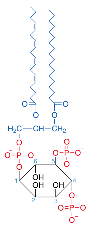
Phosphatidylinositol 4,5-bisphosphate or PtdIns(4,5)P2, also known simply as PIP2 or PI(4,5)P2, is a minor phospholipid component of cell membranes. PtdIns(4,5)P2 is enriched at the plasma membrane where it is a substrate for a number of important signaling proteins. PIP2 also forms lipid clusters that sort proteins.

<span class="mw-page-title-main">Phosphatidylinositol 3-phosphate</span> Chemical compound

Phosphatidylinositol 3-phosphate (PtdIns3P) is a phospholipid found in cell membranes that helps to recruit a range of proteins, many of which are involved in protein trafficking, to the membranes. It is the product of both the class II and III phosphoinositide 3-kinases activity on phosphatidylinositol.

<span class="mw-page-title-main">Phosphatidylinositol 3,4-bisphosphate</span>

Phosphatidylinositol (3,4)-bisphosphate is a minor phospholipid component of cell membranes, yet an important second messenger. The generation of PtdIns(3,4)P2 at the plasma membrane activates a number of important cell signaling pathways.

<span class="mw-page-title-main">Phosphatidylinositol 3,5-bisphosphate</span> Chemical compound

Phosphatidylinositol 3,5-bisphosphate is one of the seven phosphoinositides found in eukaryotic cell membranes. In quiescent cells, the PtdIns(3,5)P2 levels, typically quantified by HPLC, are the lowest amongst the constitutively present phosphoinositides. They are approximately 3 to 5-fold lower as compared to PtdIns3P and PtdIns5P levels, and more than 100-fold lower than the abundant PtdIns4P and PtdIns(4,5)P2. PtdIns(3,5)P2 was first reported to occur in mouse fibroblasts and budding yeast S. cerevisiae in 1997. In S. cerevisiae PtdIns(3,5)P2 levels increase dramatically during hyperosmotic shock. The response to hyperosmotic challenge is not conserved in most tested mammalian cells except for differentiated 3T3L1 adipocytes.

Phospholipase D (EC 3.1.4.4, lipophosphodiesterase II, lecithinase D, choline phosphatase, PLD; systematic name phosphatidylcholine phosphatidohydrolase) is an enzyme of the phospholipase superfamily that catalyses the following reaction

<span class="mw-page-title-main">PIK3R1</span> Protein-coding gene in the species Homo sapiens

Phosphatidylinositol 3-kinase regulatory subunit alpha is an enzyme that in humans is encoded by the PIK3R1 gene.

In enzymology, a ceramide kinase, also abbreviated as CERK, is an enzyme that catalyzes the chemical reaction:

<span class="mw-page-title-main">INPP5D</span> Protein-coding gene in the species Homo sapiens

Src homology 2 (SH2) domain containing inositol polyphosphate 5-phosphatase 1(SHIP1) is an enzyme with phosphatase activity. SHIP1 is structured by multiple domain and is encoded by the INPP5D gene in humans. SHIP1 is expressed predominantly by hematopoietic cells but also, for example, by osteoblasts and endothelial cells. This phosphatase is important for the regulation of cellular activation. Not only catalytic but also adaptor activities of this protein are involved in this process. Its movement from the cytosol to the cytoplasmic membrane, where predominantly performs its function, is mediated by tyrosine phosphorylation of the intracellular chains of cell surface receptors that SHIP1 binds. Insufficient regulation of SHIP1 leads to different pathologies.

<span class="mw-page-title-main">Phospholipase C</span> Class of enzymes

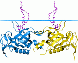
Phospholipase C (PLC) is a class of membrane-associated enzymes that cleave phospholipids just before the phosphate group (see figure). It is most commonly taken to be synonymous with the human forms of this enzyme, which play an important role in eukaryotic cell physiology, in particular signal transduction pathways. Phospholipase C's role in signal transduction is its cleavage of phosphatidylinositol 4,5-bisphosphate (PIP2) into diacyl glycerol (DAG) and inositol 1,4,5-trisphosphate (IP3), which serve as second messengers. Activators of each PLC vary, but typically include heterotrimeric G protein subunits, protein tyrosine kinases, small G proteins, Ca2+, and phospholipids.

<span class="mw-page-title-main">PI4KB</span> Protein-coding gene in the species Homo sapiens

Phosphatidylinositol 4-kinase beta is an enzyme that in humans is encoded by the PI4KB gene.

<span class="mw-page-title-main">COL4A3BP</span> Protein-coding gene in the species Homo sapiens

Collagen type IV alpha-3-binding protein, also known as ceramide transfer protein (CERT) or StAR-related lipid transfer protein 11 (STARD11) is a protein that in humans is encoded by the COL4A3BP gene. The protein contains a pleckstrin homology domain at its amino terminus and a START domain towards the end of the molecule. It is a member of the StarD2 subfamily of START domain proteins.

<span class="mw-page-title-main">DAPP1</span> Protein-coding gene in the species Homo sapiens

Dual adapter for phosphotyrosine and 3-phosphotyrosine and 3-phosphoinositide is a protein that in humans is encoded by the DAPP1 gene.

<span class="mw-page-title-main">RASA3</span> Protein-coding gene in the species Homo sapiens

Ras GTPase-activating protein 3 is an enzyme that in humans is encoded by the RASA3 gene.

<span class="mw-page-title-main">OSBP</span> Protein-coding gene in the species Homo sapiens

Oxysterol-binding protein 1 is a protein that in humans is encoded by the OSBP gene.

<span class="mw-page-title-main">PLEKHA1</span> Protein-coding gene in the species Homo sapiens

Pleckstrin homology domain-containing family A member 1 is a protein that in humans is encoded by the PLEKHA1 gene.

The Akt signaling pathway or PI3K-Akt signaling pathway is a signal transduction pathway that promotes survival and growth in response to extracellular signals. Key proteins involved are PI3K and Akt.

<span class="mw-page-title-main">Phosphoinositide-dependent kinase-1</span> Protein-coding gene in the species Homo sapiens

In the field of biochemistry, PDPK1 refers to the protein 3-phosphoinositide-dependent protein kinase-1, an enzyme which is encoded by the PDPK1 gene in humans. It is implicated in the development and progression of melanomas.

References

  1. Mayer BJ, Ren R, Clark KL, Baltimore D (May 1993). "A putative modular domain present in diverse signaling proteins". Cell. 73 (4): 629–30. doi:10.1016/0092-8674(93)90244-K. PMID   8500161. S2CID   44282241.
  2. Haslam RJ, Koide HB, Hemmings BA (May 1993). "Pleckstrin domain homology". Nature. 363 (6427): 309–10. Bibcode:1993Natur.363..309H. doi:10.1038/363309b0. PMID   8497315. S2CID   4334376.
  3. Musacchio A, Gibson T, Rice P, Thompson J, Saraste M (September 1993). "The PH domain: a common piece in the structural patchwork of signalling proteins". Trends in Biochemical Sciences. 18 (9): 343–8. doi:10.1016/0968-0004(93)90071-T. PMID   8236453.
  4. Gibson TJ, Hyvönen M, Musacchio A, Saraste M, Birney E (September 1994). "PH domain: the first anniversary". Trends in Biochemical Sciences. 19 (9): 349–53. doi: 10.1016/0968-0004(94)90108-2 . PMID   7985225.
  5. Pawson T (February 1995). "Protein modules and signalling networks". Nature. 373 (6515): 573–80. Bibcode:1995Natur.373..573P. doi:10.1038/373573a0. PMID   7531822. S2CID   4324726.
  6. Ingley E, Hemmings BA (December 1994). "Pleckstrin homology (PH) domains in signal transduction". Journal of Cellular Biochemistry. 56 (4): 436–43. doi:10.1002/jcb.240560403. PMID   7890802. S2CID   23154429.
  7. Saraste M, Hyvönen M (June 1995). "Pleckstrin homology domains: a fact file". Current Opinion in Structural Biology. 5 (3): 403–8. doi:10.1016/0959-440X(95)80104-9. PMID   7583640.
  8. Wang DS, Shaw G (December 1995). "The association of the C-terminal region of beta I sigma II spectrin to brain membranes is mediated by a PH domain, does not require membrane proteins, and coincides with a inositol-1,4,5 triphosphate binding site". Biochemical and Biophysical Research Communications. 217 (2): 608–15. doi:10.1006/bbrc.1995.2818. PMID   7503742.
  9. Wang DS, Shaw R, Winkelmann JC, Shaw G (August 1994). "Binding of PH domains of beta-adrenergic receptor kinase and beta-spectrin to WD40/beta-transducin repeat containing regions of the beta-subunit of trimeric G-proteins". Biochemical and Biophysical Research Communications. 203 (1): 29–35. doi:10.1006/bbrc.1994.2144. PMID   8074669.
  10. Yao L, Kawakami Y, Kawakami T (September 1994). "The pleckstrin homology domain of Bruton tyrosine kinase interacts with protein kinase C". Proceedings of the National Academy of Sciences of the United States of America. 91 (19): 9175–9. Bibcode:1994PNAS...91.9175Y. doi: 10.1073/pnas.91.19.9175 . PMC   44770 . PMID   7522330.
  11. Riddihough G (November 1994). "More meanders and sandwiches". Nature Structural Biology. 1 (11): 755–7. doi:10.1038/nsb1194-755. PMID   7634082. S2CID   5410578.
  12. Yu JW, Mendrola JM, Audhya A, Singh S, Keleti D, DeWald DB, Murray D, Emr SD, Lemmon MA (March 2004). "Genome-wide analysis of membrane targeting by S. cerevisiae pleckstrin homology domains". Molecular Cell. 13 (5): 677–88. doi: 10.1016/S1097-2765(04)00083-8 . PMID   15023338.
  13. Fort P, Blangy A (June 2017). "The Evolutionary Landscape of Dbl-Like RhoGEF Families: Adapting Eukaryotic Cells to Environmental Signals". Genome Biol Evol. 9 (6): 1471–1486. doi:10.1093/gbe/evx100. PMC   5499878 . PMID   28541439.
  14. Sugiura M, Kono K, Liu H, Shimizugawa T, Minekura H, Spiegel S, Kohama T (June 2002). "Ceramide kinase, a novel lipid kinase. Molecular cloning and functional characterization". The Journal of Biological Chemistry. 277 (26): 23294–300. doi: 10.1074/jbc.M201535200 . PMID   11956206.
  15. Komolov KE, Benovic JL (January 2018). "G protein-coupled receptor kinases: Past, present and future". Cellular Signalling. 41: 17–24. doi:10.1016/j.cellsig.2017.07.004. PMC   5722692 . PMID   28711719.