Dynamin

Last updated
Dynamin family
PDB 2aka EBI.jpg
Structure of the nucleotide-free myosin II motor domain from Dictyostelium discoideum fused to the GTPase domain of dynamin I from Rattus norvegicus
Identifiers
SymbolDynamin_N
Pfam PF00350
Pfam clan CL0023
InterPro IPR001401
PROSITE PDOC00362
Available protein structures:
Pfam   structures / ECOD  
PDB RCSB PDB; PDBe; PDBj
PDBsum structure summary
Dynamin central region
PDB 2aka EBI.jpg
Structure of the nucleotide-free myosin II motor domain from Dictyostelium discoideum fused to the GTPase domain of dynamin I from Rattus norvegicus
Identifiers
SymbolDynamin_M
Pfam PF01031
InterPro IPR000375
Available protein structures:
Pfam   structures / ECOD  
PDB RCSB PDB; PDBe; PDBj
PDBsum structure summary

Dynamin is a GTPase responsible for endocytosis in the eukaryotic cell. Dynamin is part of the "dynamin superfamily", which includes classical dynamins, dynamin-like proteins, Mx proteins, OPA1, mitofusins, and GBPs. Members of the dynamin family are principally involved in the scission of newly formed vesicles from the membrane of one cellular compartment and their targeting to, and fusion with, another compartment, both at the cell surface (particularly caveolae internalization) as well as at the Golgi apparatus. [1] [2] [3] Dynamin family members also play a role in many processes including division of organelles, [4] cytokinesis and microbial pathogen resistance.

Contents

Structure

Dynamin assembled into helical polymers as visualized by negative stain electron microscopy. Dynamin assembles into spirals.jpg
Dynamin assembled into helical polymers as visualized by negative stain electron microscopy.

Dynamin itself is a 96 kDa enzyme, and was first isolated when researchers were attempting to isolate new microtubule-based motors from the bovine brain. Dynamin has been extensively studied in the context of clathrin-coated vesicle budding from the cell membrane. [3] [6] Beginning from the N-terminus, Dynamin consists of a GTPase domain connected to a helical stalk domain via a flexible neck region containing a Bundle Signalling Element and GTPase Effector Domain. At the opposite end of the stalk domain is a loop that links to a membrane-binding Pleckstrin homology domain. The protein strand then loops back towards the GTPase domain and terminates with a Proline Rich Domain that binds to the Src Homology domains of many proteins.

Function

During clathrin-mediated endocytosis, the cell membrane invaginates to form a budding vesicle. Dynamin binds to and assembles around the neck of the endocytic vesicle, forming a helical polymer arranged such that the GTPase domains dimerize in an asymmetric manner across helical rungs. [7] [8] The polymer constricts the underlying membrane upon GTP binding and hydrolysis via conformational changes emanating from the flexible neck region that alters the overall helical symmetry. [8] Constriction around the vesicle neck leads to the formation of a hemi-fission membrane state that ultimately results in membrane scission. [2] [6] [9] Constriction may be in part the result of the twisting activity of dynamin, which makes dynamin the only molecular motor known to have a twisting activity. [10]

Types

In mammals, three different dynamin genes have been identified with key sequence differences in their Pleckstrin homology domains leading to differences in the recognition of lipid membranes:

Pharmacology

Small molecule inhibitors of dynamin activity have been developed, including Dynasore [11] [12] and photoswitchable derivatives (Dynazo) [13] for spatiotemporal control of endocytosis with light (photopharmacology).

Disease implications

Mutations in Dynamin II have been found to cause dominant intermediate Charcot-Marie-Tooth disease. [14] Epileptic encephalopathy–causing de novo mutations in dynamin have been suggested to cause dysfunction of vesicle scission during synaptic vesicle endocytosis. [15]

Related Research Articles

<span class="mw-page-title-main">Endocytosis</span> Cellular process

Endocytosis is a cellular process in which substances are brought into the cell. The material to be internalized is surrounded by an area of cell membrane, which then buds off inside the cell to form a vesicle containing the ingested material. Endocytosis includes pinocytosis and phagocytosis. It is a form of active transport.

GTPases are a large family of hydrolase enzymes that bind to the nucleotide guanosine triphosphate (GTP) and hydrolyze it to guanosine diphosphate (GDP). The GTP binding and hydrolysis takes place in the highly conserved P-loop "G domain", a protein domain common to many GTPases.
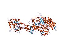
<span class="mw-page-title-main">Clathrin</span> Protein playing a major role in the formation of coated vesicles

Clathrin is a protein that plays a major role in the formation of coated vesicles. Clathrin was first isolated and named by Barbara Pearse in 1976. It forms a triskelion shape composed of three clathrin heavy chains and three light chains. When the triskelia interact they form a polyhedral lattice that surrounds the vesicle, hence the protein's name, which is derived from the Latin clathrum meaning lattice. Coat-proteins, like clathrin, are used to build small vesicles in order to transport molecules within cells. The endocytosis and exocytosis of vesicles allows cells to communicate, to transfer nutrients, to import signaling receptors, to mediate an immune response after sampling the extracellular world, and to clean up the cell debris left by tissue inflammation. The endocytic pathway can be hijacked by viruses and other pathogens in order to gain entry to the cell during infection.

<span class="mw-page-title-main">Receptor-mediated endocytosis</span>

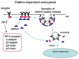
Receptor-mediated endocytosis (RME), also called clathrin-mediated endocytosis, is a process by which cells absorb metabolites, hormones, proteins – and in some cases viruses – by the inward budding of the plasma membrane (invagination). This process forms vesicles containing the absorbed substances and is strictly mediated by receptors on the surface of the cell. Only the receptor-specific substances can enter the cell through this process.

<span class="mw-page-title-main">LDL receptor</span> Mammalian protein found in Homo sapiens

The low-density lipoprotein receptor (LDL-R) is a mosaic protein of 839 amino acids that mediates the endocytosis of cholesterol-rich low-density lipoprotein (LDL). It is a cell-surface receptor that recognizes apolipoprotein B100 (ApoB100), which is embedded in the outer phospholipid layer of very low-density lipoprotein (VLDL), their remnants—i.e. intermediate-density lipoprotein (IDL), and LDL particles. The receptor also recognizes apolipoprotein E (ApoE) which is found in chylomicron remnants and IDL. In humans, the LDL receptor protein is encoded by the LDLR gene on chromosome 19. It belongs to the low density lipoprotein receptor gene family. It is most significantly expressed in bronchial epithelial cells and adrenal gland and cortex tissue.

<span class="mw-page-title-main">Pleckstrin homology domain</span> Protein domain

Pleckstrin homology domain or (PHIP) is a protein domain of approximately 120 amino acids that occurs in a wide range of proteins involved in intracellular signaling or as constituents of the cytoskeleton.

Phospholipase D (EC 3.1.4.4, lipophosphodiesterase II, lecithinase D, choline phosphatase, PLD; systematic name phosphatidylcholine phosphatidohydrolase) is an enzyme of the phospholipase superfamily that catalyses the following reaction

<span class="mw-page-title-main">BAR domain</span> Group of highly conserved protein dimerisation domains

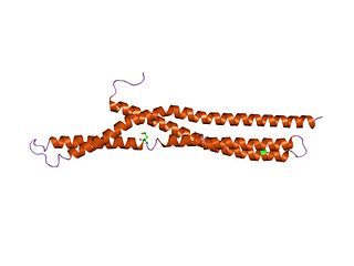
In molecular biology, BAR domains are highly conserved protein dimerisation domains that occur in many proteins involved in membrane dynamics in a cell. The BAR domain is banana-shaped and binds to membrane via its concave face. It is capable of sensing membrane curvature by binding preferentially to curved membranes. BAR domains are named after three proteins that they are found in: Bin, Amphiphysin and Rvs.

Synaptojanin is a protein involved in vesicle uncoating in neurons. This is an important regulatory lipid phosphatase. It dephosphorylates the D-5 position phosphate from phosphatidylinositol (3,4,5)-trisphosphate (PIP3) and Phosphatidylinositol (4,5)-bisphosphate(PIP2). It belongs to family of 5-phosphatases, which are structurally unrelated to D-3 inositol phosphatases like PTEN. Other members of the family of 5'phosphoinositide phosphatases include OCRL, SHIP1, SHIP2, INPP5J, INPP5E, INPP5B, INPP5A and SKIP.

<span class="mw-page-title-main">DNM2</span> Protein-coding gene in the species Homo sapiens

Dynamin-2 is a protein that in humans is encoded by the DNM2 gene.

<span class="mw-page-title-main">Intersectin 1</span> Protein-coding gene in the species Homo sapiens

Intersectin-1 is a protein that, in humans, is encoded by the ITSN1 gene.

<span class="mw-page-title-main">DNM1</span> Protein-coding gene in the species Homo sapiens

Dynamin-1 is a protein that in humans is encoded by the DNM1 gene.

<span class="mw-page-title-main">CYTH2</span> Protein-coding gene in the species Homo sapiens

Cytohesin-2 is a protein that in humans is encoded by the CYTH2 gene.

<span class="mw-page-title-main">DNM1L</span> Protein-coding gene in humans

Dynamin-1-like protein is a GTPase that regulates mitochondrial fission. In humans, dynamin-1-like protein, which is typically referred to as dynamin-related protein 1 (Drp1), is encoded by the DNM1L gene and is part of the dynamin superfamily (DSP) family of proteins.

<span class="mw-page-title-main">ELMO1</span> Protein-coding gene in the species Homo sapiens

Engulfment and cell motility protein 1 is a protein that in humans is encoded by the ELMO1 gene. ELMO1 is located on chromosome number seven in humans and is located on chromosome number thirteen in mice.

<span class="mw-page-title-main">RAPH1</span> Protein-coding gene in the species Homo sapiens

Ras-associated and pleckstrin homology domains-containing protein 1 is a protein that in humans is encoded by the RAPH1 gene.

The EHD protein family is a relatively small group of proteins which have been shown to play a role in several physiological functions, the most notable being the regulation of endocytotic vesicles. This family is recognized by its highly conserved EH domain, a structural motif that has been shown to facilitate specificity and interaction between protein and ligand. The four mammalian EHD proteins that have been classified are: EHD1, EHD2, EHD3, and EHD4.

Bulk endocytosis refers to a form of endocytosis of synaptic vesicles at nerve terminals. In bulk endocytosis, compared to clathrin-mediated endocytosis, a larger area of presynaptic plasma membrane is internalised as cisternae or endosomes from which multiple synaptic vesicles can subsequently bud off. Bulk endocytosis is activated specifically during intense stimulation, such as during high-frequency trains of action potentials or in response to membrane depolarization by high extracellular concentrations of potassium.
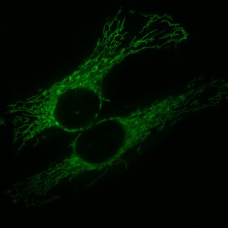
<span class="mw-page-title-main">Mitochondrial fission</span>

Mitochondrial fission is the process where mitochondria divide or segregate into two separate mitochondrial organelles. Mitochondrial fission is counteracted by the process of mitochondrial fusion, whereby two separate mitochondria can fuse together to form a large one. Mitochondrial fusion in turn can result in elongated mitochondrial networks. Both mitochondrial fission and fusion are balanced in the cell, and mutations interfering with either processes are associated with a variety of diseases. Mitochondria can divide by prokaryotic binary fission and since they require mitochondrial DNA for their function, fission is coordinated with DNA replication. Some of the proteins that are involved in mitochondrial fission have been identified and some of them are associated with mitochondrial diseases. Mitochondrial fission has significant implications in stress response and apoptosis.

Dynamin Superfamily Protein (DSP) is a protein superfamily includes classical dynamins, GBPs, Mx proteins, OPA1, mitofusins in Eukaryote, and bacterial dynamin-like proteins (BDLPs) in Prokaryote. DSPs mediate eukaryotic membrane fusion and fission necessary for endocytosis, organelle biogenesis and maintenance, Mitochondrial fusion and fission, as well as for prokaryotic cytokinesis.

References

  1. 1 2 Henley, John R.; Cao, Hong; McNiven, Mark A. (December 16, 1999). "Participation of dynamin in the biogenesis of cytoplasmic vesicles". The FASEB Journal. 13 (9002): S243-7. doi:10.1096/fasebj.13.9002.S243. PMID   10619136. S2CID   24401725.
  2. 1 2 Hinshaw, J. "Research statement, Jenny E. Hinshaw, Ph.D." National Institute of Diabetes & Digestive & Kidney Diseases, Laboratory of Cell Biochemistry and Biology. Accessed 19 March 2013.
  3. 1 2 Urrutia R, Henley JR, Cook T, McNiven MA (January 1997). "The dynamins: redundant or distinct functions for an expanding family of related GTPases?". Proceedings of the National Academy of Sciences of the United States of America. 94 (2): 377–384. Bibcode:1997PNAS...94..377U. doi: 10.1073/pnas.94.2.377 . PMC   34135 . PMID   9012790.
  4. Thoms S, Erdmann R (October 2005). "Dynamin-related proteins and Pex11 proteins in peroxisome division and proliferation". The FEBS Journal. 272 (20): 5169–5181. doi:10.1111/j.1742-4658.2005.04939.x. PMID   16218949.
  5. Hinshaw JE, Schmid SL (March 1995). "Dynamin self-assembles into rings suggesting a mechanism for coated vesicle budding". Nature. 374 (6518): 190–192. Bibcode:1995Natur.374..190H. doi:10.1038/374190a0. PMID   7877694. S2CID   4365628.
  6. 1 2 3 Praefcke GJ, McMahon HT (February 2004). "The dynamin superfamily: universal membrane tubulation and fission molecules?". Nature Reviews. Molecular Cell Biology. 5 (2): 133–147. doi:10.1038/nrm1313. PMID   15040446. S2CID   6305282.
  7. Sundborger AC, Fang S, Heymann JA, Ray P, Chappie JS, Hinshaw JE (August 2014). "A dynamin mutant defines a superconstricted prefission state". Cell Reports. 8 (3): 734–742. doi:10.1016/j.celrep.2014.06.054. PMC   4142656 . PMID   25088425.
  8. 1 2 Kong L, Sochacki KA, Wang H, Fang S, Canagarajah B, Kehr AD, et al. (August 2018). "Cryo-EM of the dynamin polymer assembled on lipid membrane". Nature. 560 (7717): 258–262. Bibcode:2018Natur.560..258K. doi:10.1038/s41586-018-0378-6. PMC   6121775 . PMID   30069048.
  9. Mattila JP, Shnyrova AV, Sundborger AC, Hortelano ER, Fuhrmans M, Neumann S, et al. (August 2015). "A hemi-fission intermediate links two mechanistically distinct stages of membrane fission". Nature. 524 (7563): 109–113. Bibcode:2015Natur.524..109M. doi:10.1038/nature14509. PMC   4529379 . PMID   26123023.
  10. Roux A, Uyhazi K, Frost A, De Camilli P (May 2006). "GTP-dependent twisting of dynamin implicates constriction and tension in membrane fission". Nature. 441 (7092): 528–531. Bibcode:2006Natur.441..528R. doi:10.1038/nature04718. PMID   16648839. S2CID   4413887.
  11. Macia E, Ehrlich M, Massol R, Boucrot E, Brunner C, Kirchhausen T (June 2006). "Dynasore, a cell-permeable inhibitor of dynamin". Developmental Cell. 10 (6): 839–850. doi: 10.1016/j.devcel.2006.04.002 . PMID   16740485.
  12. Eschenburg S, Reubold TF (November 2018). "Modulation of dynamin function by small molecules". Biological Chemistry. 399 (12): 1421–1432. doi: 10.1515/hsz-2018-0257 . PMID   30067507. S2CID   51895475.
  13. Camarero N, Trapero A, Pérez-Jiménez A, Macia E, Gomila-Juaneda A, Martín-Quirós A, et al. (September 2020). "Correction: Photoswitchable dynasore analogs to control endocytosis with light". Chemical Science. 11 (35): 9712. doi:10.1039/D0SC90189J. PMC   7495901 . PMID   33016959.
  14. Züchner S, Noureddine M, Kennerson M, Verhoeven K, Claeys K, De Jonghe P, et al. (March 2005). "Mutations in the pleckstrin homology domain of dynamin 2 cause dominant intermediate Charcot-Marie-Tooth disease". Nature Genetics. 37 (3): 289–294. doi:10.1038/ng1514. PMID   15731758. S2CID   19191581.
  15. Dhindsa RS, Bradrick SS, Yao X, Heinzen EL, Petrovski S, Krueger BJ, et al. (June 2015). "Epileptic encephalopathy-causing mutations in DNM1 impair synaptic vesicle endocytosis". Neurology. Genetics. 1 (1): e4. doi:10.1212/01.NXG.0000464295.65736.da. PMC   4821085 . PMID   27066543.