Cytokeratin

Last updated
Keratin intermediate filaments in epithelial cells (red stain). Epithelial-cells.jpg
Keratin intermediate filaments in epithelial cells (red stain).

Cytokeratins are keratin proteins found in the intracytoplasmic cytoskeleton of epithelial tissue. They are an important component of intermediate filaments, which help cells resist mechanical stress. [1] Expression of these cytokeratins within epithelial cells is largely specific to particular organs or tissues. Thus they are used clinically to identify the cell of origin of various human tumors.

Contents

Naming

The term cytokeratin began to be used in the late 1970s, when the protein subunits of keratin intermediate filaments inside cells were first being identified and characterized. [2] In 2006 a new systematic nomenclature for mammalian keratins was created, and the proteins previously called cytokeratins are simply called keratins (human epithelial category). For example, cytokeratin-4 (CK-4) has been renamed keratin-4 (K4). [3] However, they are still commonly referred to as cytokeratins in clinical practice.

Types

Micrograph showing low molecular weight cytokeratin (LMWCK) staining of intermediate trophoblast (placental tissue) and endometrial glands. Intermediate trophoblast - lmwk - high mag.jpg
Micrograph showing low molecular weight cytokeratin (LMWCK) staining of intermediate trophoblast (placental tissue) and endometrial glands.

There are two categories of cytokeratins: the acidic type I cytokeratins and the basic or neutral type II cytokeratins. Within each category, cytokeratins are numbered in order of decreasing size, from high molecular weight (HMWCKs) to low molecular weight (LMWCKs). Cytokeratins are usually found in heterodimeric pairs of acidic and basic subunits of similar size. [4]

Basic CK
(Type B / Class II)
Acidic CK
(Type A / Class I)
HMWCK
"squamous keratins"
CK-1
CK-2
CK-3
CK-4
CK-5
CK-6
CK-9
CK-10
CK-11
CK-12
CK-13
CK-14
CK-15
CK-16
CK-17
LMWCK
"simple keratins"
CK-7
CK-8
CK-18
CK-19
CK-20

Expression of these cytokeratins is largely organ or tissue specific. The subsets of cytokeratins which an epithelial cell expresses depends mainly on the type of epithelium, the moment in the course of terminal differentiation and the stage of development. Thus a specific cytokeratin expression profile allows the identification of epithelial cells. Furthermore, this applies also to the malignant counterparts of the epithelia, (carcinomas), as the cytokeratin profile is generally retained. Thus the study of cytokeratin expression by immunohistochemistry techniques is a tool of immense value widely used for tumor diagnosis and characterization in surgical pathology. [5]

CytokeratinSites
Cytokeratin 4
Cytokeratin 7
Cytokeratin 8
  • Glandular epithelia of the digestive, respiratory and urogenital tracts, both endocrine and exocrine cells, as well as mesothelial cells
  • Adenocarcinomas originating from those above [6]
Cytokeratin 10
Cytokeratin 13
  • Non-keratinized squamous epithelia, except cornea [6]
Cytokeratin 14
Cytokeratin 18
  • Glandular epithelia of the digestive, respiratory, and urogenital tracts, both endocrine and exocrine cells, as well as mesothelial cells
  • Adenocarcinomas originating from those above [6]
Cytokeratin 19
  • Glandular-type epithelia [6]
  • Carcinomas [6]

Does not react with hepatocytes and hepatocellular carcinoma [6]

Cytokeratin 20
  • Glandular-type epithelia. Signet ring/round clear cells [6]
  • GI stromal tumor (Krukenberg) [6]

Molecular biology

The cytokeratins are encoded by a family encompassing 30 genes. Among them, 20 are epithelial genes and the remaining 10 are specific for trichocytes.

All cytokeratin chains are composed of a central α-helix-rich domain (with a 50 to 90% sequence identity among cytokeratins of the same type and around 30% between cytokeratins of different type) with non-α-helical N- and C-terminal domains. The α-helical domain has 310-150 amino acids and comprises four segments in which a seven-residue pattern repeats. Into this repeated pattern, the first and fourth residues are hydrophobic and the charged residues show alternate positive and negative polarity, resulting in the polar residues being located on one side of the helix. This central domain of the chain provides the molecular alignment in the keratin structure and makes the chains form coiled dimers in solution.

The end-domain sequences of type I and II cytokeratin chains contain in both sides of the rod domain the subdomains V1 and V2, which have variable size and sequence. The type II also presents the conserved subdomains H1 and H2, encompassing 36 and 20 residues respectively. The subdomains V1 and V2 contain residues enriched by glycines and/or serines, the former providing the cytokeratin chain a strong insoluble character and facilitating the interaction with other molecules. These terminal domains are also important in defining the function of the cytokeratin chain characteristic of a particular epithelial cell type.

Two dimers of cytokeratin group into a keratin tetramer by anti-parallel binding. This cytokeratin tetramer is considered to be the main building block of the cytokeratin chain. By head-to-tail linking of the cytokeratin tetramers, the protofilaments are originated, which in turn intertwine in pairs to form protofibrils. Four protofibrils give place to one cytokeratin filament.

Cytokeratin filaments in the human epithelial cell Cytokeratin filaments.jpg
Cytokeratin filaments in the human epithelial cell

Cell biology

In the cytoplasm, keratin filaments associate laterally with each other to create bundles of ~50 nm radius. The radius of these bundles is set by the interplay between long range electrostatic repulsion and short range hydrophobic attraction. [7] These keratin bundles span a complex network which extends from the surface of the nucleus to the cell membrane. Numerous accessory proteins are involved in the genesis and maintenance of such structure.

This association between the plasma membrane and the nuclear surface provides important implications for the organization of the cytoplasm and cellular communication mechanisms. Apart from the relatively static functions provided in terms of supporting the nucleus and providing tensile strength to the cell, the cytokeratin networks undergo rapid phosphate exchanges mediated depolymerization, with important implications in the more dynamic cellular processes such as mitosis and post-mitotic period, cell movement and differentiation.

Cytokeratins interact with desmosomes and hemidesmosomes, thus collaborating to cell–cell adhesion and basal cell–underlying connective tissue connection.

The intermediate filaments of the eukaryotic cytoskeleton, which the cytokeratins are one of its three components, have been probed to associate also with the ankyrin and spectrin complex protein network that underlies the cell membrane.[ citation needed ]

Related Research Articles

<span class="mw-page-title-main">Keratin</span> One of a family of fibrous structural proteins

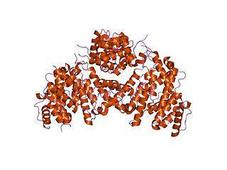
Keratin is one of a family of structural fibrous proteins also known as scleroproteins. Alpha-keratin (α-keratin) is a type of keratin found in vertebrates. It is the key structural material making up scales, hair, nails, feathers, horns, claws, hooves, and the outer layer of skin among vertebrates. Keratin also protects epithelial cells from damage or stress. Keratin is extremely insoluble in water and organic solvents. Keratin monomers assemble into bundles to form intermediate filaments, which are tough and form strong unmineralized epidermal appendages found in reptiles, birds, amphibians, and mammals. Excessive keratinization participate in fortification of certain tissues such as in horns of cattle and rhinos, and armadillos' osteoderm. The only other biological matter known to approximate the toughness of keratinized tissue is chitin. Keratin comes in two types, the primitive, softer forms found in all vertebrates and harder, derived forms found only among sauropsids.

<span class="mw-page-title-main">Cytoskeleton</span> Network of filamentous proteins that forms the internal framework of cells

The cytoskeleton is a complex, dynamic network of interlinking protein filaments present in the cytoplasm of all cells, including those of bacteria and archaea. In eukaryotes, it extends from the cell nucleus to the cell membrane and is composed of similar proteins in the various organisms. It is composed of three main components: microfilaments, intermediate filaments, and microtubules, and these are all capable of rapid growth or disassembly depending on the cell's requirements.

<span class="mw-page-title-main">Intermediate filament</span> Cytoskeletal structure

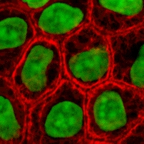
Intermediate filaments (IFs) are cytoskeletal structural components found in the cells of vertebrates, and many invertebrates. Homologues of the IF protein have been noted in an invertebrate, the cephalochordate Branchiostoma.

<span class="mw-page-title-main">Keratin 7</span>

Keratin, type II cytoskeletal 7 also known as cytokeratin-7 (CK-7) or keratin-7 (K7) or sarcolectin (SCL) is a protein that in humans is encoded by the KRT7 gene. Keratin 7 is a type II keratin. It is specifically expressed in the simple epithelia lining the cavities of the internal organs and in the gland ducts and blood vessels.

<span class="mw-page-title-main">Keratin 6A</span>

Keratin 6A is one of the 27 different type II keratins expressed in humans. Keratin 6A was the first type II keratin sequence determined. Analysis of the sequence of this keratin together with that of the first type I keratin led to the discovery of the four helical domains in the central rod of keratins. In humans Keratin 6A is encoded by the KRT6A gene.

Type II keratins constitutes the Type II intermediate filaments (IFs) of the intracytoplasmatic cytoskeleton, which is present in all mammalian epithelial cells. The type 2 cytokeratins consist of basic or neutral, high molecular weight proteins which in vivo are arranged in pairs of heterotypic Type I and Type II keratin chains, coexpressed during differentiation of simple and stratified epithelial tissues. It has been seen that Type II Keratins are developed before Type 1 keratins during human embryonic development.

Type I keratins are cytokeratins that constitute the Type I intermediate filaments (IFs) of the intracytoplasmatic cytoskeleton, which is present in all mammalian epithelial cells. Most of the type I keratins consist of acidic, low molecular weight proteins which in vivo are arranged in pairs of heterotypic Type I and Type II keratin chains, coexpressed during differentiation of simple and stratified epithelial tissues.

<span class="mw-page-title-main">Keratin 10</span> Protein-coding gene in the species Homo sapiens

Keratin, type I cytoskeletal 10 also known as cytokeratin-10 (CK-10) or keratin-10 (K10) is a protein that in humans is encoded by the KRT10 gene. Keratin 10 is a type I keratin.

<span class="mw-page-title-main">Keratin 19</span> Protein-coding gene in the species Homo sapiens

Keratin, type I cytoskeletal 19 also known as cytokeratin-19 (CK-19) or keratin-19 (K19) is a 40 kDa protein that in humans is encoded by the KRT19 gene. Keratin 19 is a type I keratin.

<span class="mw-page-title-main">Keratin 18</span>

Keratin 18 is a type I cytokeratin. It is, together with its filament partner keratin 8, perhaps the most commonly found products of the intermediate filament gene family. They are expressed in single layer epithelial tissues of the body. Mutations in this gene have been linked to cryptogenic cirrhosis. Two transcript variants encoding the same protein have been found for this gene.

Hair keratin is a type of keratin found in hair and the nails.

Internexin, alpha-internexin, is a Class IV intermediate filament approximately 66 KDa. The protein was originally purified from rat optic nerve and spinal cord. The protein copurifies with other neurofilament subunits, as it was originally discovered, however in some mature neurons it can be the only neurofilament expressed. The protein is present in developing neuroblasts and in the central nervous system of adults. The protein is a major component of the intermediate filament network in small interneurons and cerebellar granule cells, where it is present in the parallel fibers.

<span class="mw-page-title-main">Vimentin</span> Type III intermediate filament protein

Vimentin is a structural protein that in humans is encoded by the VIM gene. Its name comes from the Latin vimentum which refers to an array of flexible rods.

Neurofilaments (NF) are classed as type IV intermediate filaments found in the cytoplasm of neurons. They are protein polymers measuring 10 nm in diameter and many micrometers in length. Together with microtubules (~25 nm) and microfilaments (7 nm), they form the neuronal cytoskeleton. They are believed to function primarily to provide structural support for axons and to regulate axon diameter, which influences nerve conduction velocity. The proteins that form neurofilaments are members of the intermediate filament protein family, which is divided into six types based on their gene organization and protein structure. Types I and II are the keratins which are expressed in epithelia. Type III contains the proteins vimentin, desmin, peripherin and glial fibrillary acidic protein (GFAP). Type IV consists of the neurofilament proteins NF-L, NF-M, NF-H and α-internexin. Type V consists of the nuclear lamins, and type VI consists of the protein nestin. The type IV intermediate filament genes all share two unique introns not found in other intermediate filament gene sequences, suggesting a common evolutionary origin from one primitive type IV gene.

<span class="mw-page-title-main">Fimbrin</span> Protein-coding gene in the species Homo sapiens

Fimbrin also known as is plastin 1 is a protein that in humans is encoded by the PLS1 gene. Fimbrin is an actin cross-linking protein important in the formation of filopodia.

Crescentin is a protein which is a bacterial relative of the intermediate filaments found in eukaryotic cells. Just as tubulins and actins, the other major cytoskeletal proteins, have prokaryotic homologs in, respectively, the FtsZ and MreB proteins, intermediate filaments are linked to the crescentin protein. Some of its homologs are erroneously labelled Chromosome segregation protein ParA. This protein family is found in Caulobacter and Methylobacterium.

<span class="mw-page-title-main">Villin-1</span> Actin-binding protein

Villin-1 is a 92.5 kDa tissue-specific actin-binding protein associated with the actin core bundle of the brush border. Villin-1 is encoded by the VIL1 gene. Villin-1 contains multiple gelsolin-like domains capped by a small "headpiece" at the C-terminus consisting of a fast and independently folding three-helix bundle that is stabilized by hydrophobic interactions. The headpiece domain is a commonly studied protein in molecular dynamics due to its small size and fast folding kinetics and short primary sequence.

<span class="mw-page-title-main">Keratin 8</span>

Keratin, type II cytoskeletal 8 also known as cytokeratin-8 (CK-8) or keratin-8 (K8) is a keratin protein that is encoded in humans by the KRT8 gene. It is often paired with keratin 18.

<span class="mw-page-title-main">Keratin 5</span>

Keratin 5, also known as KRT5, K5, or CK5, is a protein that is encoded in humans by the KRT5 gene. It dimerizes with keratin 14 and forms the intermediate filaments (IF) that make up the cytoskeleton of basal epithelial cells. This protein is involved in several diseases including epidermolysis bullosa simplex and breast and lung cancers.

Alpha-keratin, or α-keratin, is a type of keratin found in mammalian vertebrates. This protein is the primary component in hairs, horns, claws, nails and the epidermis layer of the skin. α-keratin is a fibrous structural protein, meaning it is made up of amino acids that form a repeating secondary structure. The secondary structure of α-keratin is very similar to that of a traditional protein α-helix and forms a coiled coil. Due to its tightly wound structure, it can function as one of the strongest biological materials and has various functions in mammals, from predatory claws to hair for warmth. α-keratin is synthesized through protein biosynthesis, utilizing transcription and translation, but as the cell matures and is full of α-keratin, it dies, creating a strong non-vascular unit of keratinized tissue.

References

  1. Herrmann H, Bär H, Kreplak L, Strelkov SV, Aebi U (July 2007). "Intermediate filaments: from cell architecture to nanomechanics". Nat. Rev. Mol. Cell Biol. 8 (7): 562–73. doi:10.1038/nrm2197. PMID   17551517. S2CID   27115011.
  2. Franke WW, Schmid E, Osborn M, Weber K (June 1979). "Intermediate-sized filaments of human endothelial cells". The Journal of Cell Biology. 81 (3): 570–80. doi:10.1083/jcb.81.3.570. PMC   2110384 . PMID   379021.
  3. Schweizer J, Bowden PE, Coulombe PA, et al. (July 2006). "New consensus nomenclature for mammalian keratins". The Journal of Cell Biology. 174 (2): 169–74. doi:10.1083/jcb.200603161. PMC   2064177 . PMID   16831889.
  4. Rekhtman, Natasha; Bishop, Justin A. (2011). Quick Reference Handbook for Surgical Pathologists. Heidelberg: Springer. pp. 4–8. ISBN   978-3-642-20085-4.
  5. Dabbs, DJ (2010). Diagnostic Immunohistochemistry: Theranostic and Genomic Applications (3rd ed.). New York: Saunders.
  6. 1 2 3 4 5 6 7 8 9 10 11 12 13 MUbio > MONOCLONAL ANTIBODIES TO CYTOKERATINS [ permanent dead link ] Retrieved October 2010
  7. Haimov, Ehud (July 2020). "Model for Bundling of Keratin Intermediate Filaments". Biophysical Journal. 119 (1): 65–74. doi: 10.1016/j.bpj.2020.05.024 . PMC   7335914 . PMID   32533940.