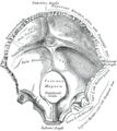
Squamous part of occipital bone

Last updated
Squamous part of occipital bone
Squamous part of occipital bone05.png
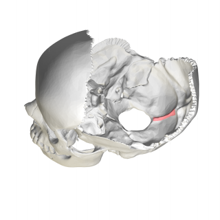
Human skull seen from above (parietal bones removed). Squamous part is shown in red.
Gray131 Squama occipitalis.png
Occipital bone at birth, seen from below. (Squamous part is top half, portion above foramen magnum, shown in yellow.)
Details
Identifiers
Latin squama occipitalis
TA98 A02.1.04.010
TA2 565
FMA 52860
Anatomical terms of bone

The squamous part of occipital bone is situated above and behind the foramen magnum, and is curved from above downward and from side to side.

Contents

External surface

The external surface is convex and presents midway between the summit of the bone and the foramen magnum a prominence, the external occipital protuberance and inion.

Extending lateralward from this on either side are two curved lines, one a little above the other. The upper, often faintly marked, is named the highest nuchal line, and to it the epicranial aponeurosis is attached.

The lower is termed the superior nuchal line. That area of the squamous part, which lies above the highest nuchal lines is named the occipital plane(planum occipitale) and is covered by the occipitalis muscle. That below, termed the nuchal plane, is rough and irregular for the attachment of several muscles.

From the external occipital protuberance, an often faintly marked ridge or crest, the median nuchal line, descends to the foramen magnum and affords attachment to the nuchal ligament. Running from the middle of this line across either half of the nuchal plane is the inferior nuchal line.

Several muscles are attached to the outer surface of the squamous part, thus the superior nuchal line gives origin to the occipitalis and trapezius muscles, and insertion to the sternocleidomastoid and splenius capitis muscles. Into the surface between the superior and inferior nuchal lines the semispinalis capitis and the obliquus capitis superior are inserted, while the inferior nuchal line and the area below it receive the insertions of the rectus capitis posterior major and minor.

The posterior atlantooccipital membrane is attached around the postero-lateral part of the foramen magnum, just outside the margin of the foramen.

Internal surface

The internal surface is deeply concave and divided into four fossae by the cruciform eminence.

The upper two fossae are triangular and lodge the occipital lobes of the cerebrum; the lower two are quadrilateral and accommodate the hemispheres of the cerebellum.

At the point of intersection of the four divisions of the cruciform eminence is the internal occipital protuberance.

From this protuberance the upper division of the cruciform eminence runs to the superior angle of the bone, and on one side of it (generally the right) is a deep groove, the sagittal sulcus, which lodges the hinder part of the superior sagittal sinus. To the margins of this sulcus the falx cerebri is attached.

The lower division of the cruciform eminence is prominent and is named the internal occipital crest; it bifurcates near the foramen magnum and gives attachment to the falx cerebelli. In the attached margin of this falx is the occipital sinus, which is sometimes duplicated.

In the upper part of the internal occipital crest, a small depression is sometimes distinguishable; it is termed the vermian fossa since it is occupied by part of the vermis of the cerebellum. Transverse grooves, one on either side, extend from the internal occipital protuberance to the lateral angles of the bone; those grooves accommodate the transverse sinuses, and their prominent margins give attachment to the tentorium cerebelli.

The groove on the right side is usually larger than that on the left and is continuous with that for the superior sagittal sinus.

Exceptions to this condition are, however, not infrequent: the left may be larger than the right or the two may be almost equal in size.

The angle of union of the superior sagittal and transverse sinuses is named the confluence of the sinuses, and its position is indicated by a depression situated on one or other side of the protuberance.

Additional images

Related Research Articles

<span class="mw-page-title-main">Atlas (anatomy)</span> First cervical vertebra of the spine which supports the skull

In anatomy, the atlas (C1) is the most superior (first) cervical vertebra of the spine and is located in the neck.

<span class="mw-page-title-main">Occipital bone</span> Saucer-shaped membrane bone situated at the back and lower part of the cranium

The occipital bone is a cranial dermal bone and the main bone of the occiput. It is trapezoidal in shape and curved on itself like a shallow dish. The occipital bone overlies the occipital lobes of the cerebrum. At the base of the skull in the occipital bone, there is a large oval opening called the foramen magnum, which allows the passage of the spinal cord.

<span class="mw-page-title-main">Parietal bone</span> Bone in the human skull which, when joined together, forms the sides and roof of the cranium

The parietal bones are two bones in the skull which, when joined at a fibrous joint, form the sides and roof of the cranium. In humans, each bone is roughly quadrilateral in form, and has two surfaces, four borders, and four angles. It is named from the Latin paries (-ietis), wall.
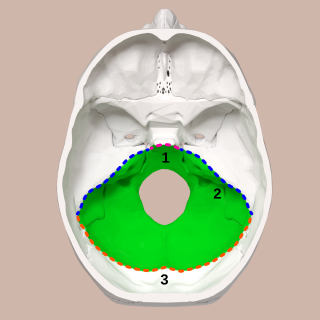
<span class="mw-page-title-main">Posterior cranial fossa</span> Area of the cranium containing the brainstem and cerebellum

The posterior cranial fossa is the part of the cranial cavity located between the foramen magnum, and tentorium cerebelli. It is formed by the sphenoid bones, temporal bones, and occipital bone. It lodges the cerebellum, and parts of the brainstem.

<span class="mw-page-title-main">Falx cerebri</span> Anatomical structure of the brain

The falx cerebri is a large, crescent-shaped fold of dura mater that descends vertically into the longitudinal fissure between the cerebral hemispheres of the human brain, separating the two hemispheres and supporting dural sinuses that provide venous and CSF drainage to the brain. It is attached to the crista galli anteriorly, and blends with the tentorium cerebelli posteriorly.

<span class="mw-page-title-main">Cerebellar tentorium</span> Vertebrate brain structure separating the cerebellum from the occipital lobes

The cerebellar tentorium or tentorium cerebelli is an extension of the dura mater between the inferior aspect of the occipital lobes and the superior aspect of the cerebellum. The free border of the tentorium gives passage to the midbrain.

<span class="mw-page-title-main">Occipital sinus</span>

The occipital sinus is the smallest of the dural venous sinuses. It is usually unpaired, and is sometimes altogether absent. It is situated in the attached margin of the falx cerebelli. It commences near the foramen magnum, and ends by draining into the confluence of sinuses.

<span class="mw-page-title-main">Superior sagittal sinus</span> Anatomical structure of the brain

The superior sagittal sinus, within the human head, is an unpaired area along the attached margin of the falx cerebri. It allows blood to drain from the lateral aspects of anterior cerebral hemispheres to the confluence of sinuses. Cerebrospinal fluid drains through arachnoid granulations into the superior sagittal sinus and is returned to venous circulation.

<span class="mw-page-title-main">Falx cerebelli</span> Skull anatomy

The falx cerebelli is a small sickle-shaped fold of dura mater projecting forwards into the posterior cerebellar notch as well as projecting into the vallecula of the cerebellum between the two cerebellar hemispheres.

<span class="mw-page-title-main">Nuchal lines</span>

The nuchal lines are four curved lines on the external surface of the occipital bone:

<span class="mw-page-title-main">Transverse sinuses</span>

The transverse sinuses, within the human head, are two areas beneath the brain which allow blood to drain from the back of the head. They run laterally in a groove along the interior surface of the occipital bone. They drain from the confluence of sinuses to the sigmoid sinuses, which ultimately connect to the internal jugular vein. See diagram : labeled under the brain as "SIN. TRANS.".

<span class="mw-page-title-main">Petrous part of the temporal bone</span> Feature at the base of the human skull

The petrous part of the temporal bone is pyramid-shaped and is wedged in at the base of the skull between the sphenoid and occipital bones. Directed medially, forward, and a little upward, it presents a base, an apex, three surfaces, and three angles, and houses in its interior, the components of the inner ear. The petrous portion is among the most basal elements of the skull and forms part of the endocranium. Petrous comes from the Latin word petrosus, meaning "stone-like, hard". It is one of the densest bones in the body. In other mammals, it is a separate bone, the petrosal bone.

<span class="mw-page-title-main">Basilar part of occipital bone</span> Section of the main skull bone that extends forward and upward

The basilar part of the occipital bone extends forward and upward from the foramen magnum, and presents in front an area more or less quadrilateral in outline.

<span class="mw-page-title-main">Lateral parts of occipital bone</span>

The lateral parts of the occipital bone are situated at the sides of the foramen magnum; on their under surfaces are the condyles for articulation with the superior facets of the atlas.
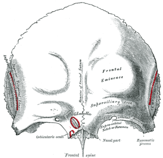
<span class="mw-page-title-main">Squamous part of the frontal bone</span> Upper two-thirds of the forward skull

The squamous part of the frontal bone is the superior portion when viewed in standard anatomical orientation. There are two surfaces of the squamous part of the frontal bone: the external surface, and the internal surface.
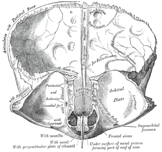
<span class="mw-page-title-main">Sagittal sulcus</span> Groove inside the upper two-thirds of the front of the skull, holding a key vein

The sagittal sulcus is a midline groove that runs across the internal surfaces of part of the squamous part of the frontal bone, the parietal bones, and part of the occipital bones. The sagittal sulcus accommodates the superior sagittal sinus. The falx cerebri attaches to the edge of the sagittal sulcus on either side.
<span class="mw-page-title-main">Internal occipital crest</span>

In the occipital bone, the lower division of the cruciate eminence is prominent, and is named the internal occipital crest; it bifurcates near the foramen magnum and gives attachment to the falx cerebelli; in the attached margin of this falx is the occipital sinus, which is sometimes duplicated.

<span class="mw-page-title-main">Body of sphenoid bone</span>

The body of the sphenoid bone, more or less cubical in shape, is hollowed out in its interior to form two large cavities, the sphenoidal sinuses, which are separated from each other by a septum.

<span class="mw-page-title-main">Base of skull</span> Inferior area of the skull, composed of the endocranium and lower parts of the skull roof

The base of skull, also known as the cranial base or the cranial floor, is the most inferior area of the skull. It is composed of the endocranium and the lower parts of the calvaria.

<span class="mw-page-title-main">Outline of human anatomy</span> Overview of and topical guide to human anatomy

The following outline is provided as an overview of and topical guide to human anatomy:

References

PD-icon.svgThis article incorporates text in the public domain from page 129 of the 20th edition of Gray's Anatomy (1918)