
A finger is a prominent digit on the forelimbs of most tetrapod vertebrate animals, especially those with prehensile extremities such as humans and other primates. Most tetrapods have five digits (pentadactyly), and short digits are typically referred to as toes, while those that are notably elongated are called fingers. In humans, the fingers are flexibly articulated and opposable, serving as an important organ of tactile sensation and fine movements, which are crucial to the dexterity of the hands and the ability to grasp and manipulate objects.
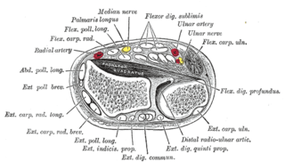
Flexor digitorum superficialis or flexor digitorum communis sublimis is an extrinsic flexor muscle of the fingers at the proximal interphalangeal joints.

Psoriatic arthritis (PsA) is a long-term inflammatory arthritis that occurs in people affected by the autoimmune disease psoriasis. The classic features of psoriatic arthritis include dactylitis, skin lesions, and nail lesions. Damage to the nails can include small depressions in the nail (pitting), thickening of the nails, and detachment of the nail from the nailbed. Skin damage consistent with psoriasis frequently occur before the onset of psoriatic arthritis but psoriatic arthritis can precede the rash in 15% of affected individuals. It is classified as a type of seronegative spondyloarthropathy.
Osteophytes are exostoses that form along joint margins. They should not be confused with enthesophytes, which are bony projections that form at the attachment of a tendon or ligament. Osteophytes are not always distinguished from exostoses in any definite way, although in many cases there are a number of differences. Osteophytes are typically intra-articular.

A hammer toe, hammertoe or contracted toe is a deformity of the muscles and ligaments of the proximal interphalangeal joint of the second, third, fourth, or fifth toe, bending it into a shape resembling a hammer. In the early stage, a flexible hammertoe is movable at the joints; a rigid hammertoe joint cannot be moved and usually requires surgery.

Heberden's nodes are hard or bony swellings that can develop in the distal interphalangeal joints (DIP). They are a sign of osteoarthritis and are caused by formation of osteophytes of the articular (joint) cartilage in response to repeated trauma at the joint.

William Heberden FRS was an English physician.

Froment sign is a special test of the wrist for palsy of the ulnar nerve, specifically, the action of adductor pollicis.
Rat-bite fever (RBF) is an acute, febrile human illness caused by bacteria transmitted by rodents, in most cases, which is passed from rodent to human by the rodent's urine or mucous secretions. Alternative names for rat-bite fever include streptobacillary fever, streptobacillosis, spirillary fever, bogger, and epidemic arthritic erythema. It is a rare disease spread by infected rodents and caused by two specific types of bacteria:
- Streptobacillus moniliformis, the only reported bacteria that causes RBF in North America
- Spirillum minus, common in Asia. Most cases occur in Japan, but specific strains of the disease are present in the United States, Europe, Australia, and Africa.

The knuckles are the joints of the fingers. The word is cognate to similar words in other Germanic languages, such as the Dutch "knokkel" (knuckle) or German "Knöchel" (ankle), i.e., Knöchlein, the diminutive of the German word for bone (Knochen). Anatomically, it is said that the knuckles consist of the metacarpophalangeal (MCP) and interphalangeal (IP) joints of the finger. The knuckles at the base of the fingers may be referred to as the 1st or major knuckles while the knuckles at the midfinger are known as the 2nd and 3rd, or minor, knuckles. However, the ordinal terms are used inconsistently and may refer to any of the knuckles.
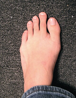
Morton's toe is the condition of having a first metatarsal bone that is shorter than the second metatarsal. It is a type of brachymetatarsia. This condition is the result of a premature closing of the first metatarsal's growth plate, resulting in a short big toe, giving the second toe the appearance of being long compared to the first toe.

Boutonniere deformity is a deformed position of the fingers or toes, in which the joint nearest the knuckle is permanently bent toward the palm while the farthest joint is bent back away. Causes include injury, inflammatory conditions like rheumatoid arthritis, and genetic conditions like Ehlers-Danlos syndrome.

Charles Jacques Bouchard was a French pathologist and an esperantist born in Montier-en-Der, a commune the department of Haute-Marne.

Jammed finger is a common term used to describe various types of finger joint injuries. It happens from a forceful impact originating at the tip of the finger directed towards the base. This type of directional force is called axial loading. It occurs most often when the finger is fully extended. This kind of impact can stretch or strain the ligaments in the joint beyond their normal limits. The severity of damage to the finger increases with the amount of force on the fingertip. In severe cases, injury to bone may occur. When experiencing a jammed finger, the extent of injury is not always obvious and one should be evaluated by a medical professional. Toes may become jammed as well, with similar results.

An ulnar claw, also known as claw hand or Spinster’s Claw, is a deformity or an abnormal attitude of the hand that develops due to ulnar nerve damage causing paralysis of the lumbricals. A claw hand presents with a hyperextension at the metacarpophalangeal joints and flexion at the proximal and distal interphalangeal joints of the 4th and 5th fingers. The patients with this condition can make a full fist but when they extend their fingers, the hand posture is referred to as claw hand. The ring- and little finger can usually not fully extend at the proximal interphalangeal joint (PIP).

Hypertrophic osteoarthropathy is a medical condition combining clubbing and periostitis of the small hand joints, especially the distal interphalangeal joints and the metacarpophalangeal joints. Distal expansion of the long bones as well as painful, swollen joints and synovial villous proliferation are often seen. The condition may occur alone (primary), or it may be secondary to diseases like lung cancer. Among patients with lung cancer, it is most associated with adenocarcinoma and least associated with small cell lung cancer. These patients often get clubbing and increased bone deposition on long bones. Their presenting signs and symptoms are sometimes only clubbing and painful ankles.
Knuckle pads, also known as heloderma, meaning similar to the skin of the Gila monster lizard for which it is named, are circumscribed, keratotic, fibrous growths over the dorsa of the interphalangeal joints. They are described as well-defined, round, plaque-like, fibrous thickening that may develop at any age, and grow to be 10 to 15mm in diameter in the course of a few weeks or months, then go away over time.
Garrod's pads, also known as violinist's pads, are a cutaneous condition characterized by calluses on the dorsal aspect of the interphalangeal joints, i.e. the back side of the finger joints. They are often seen in violin, viola, and cello players, along with fiddler's neck and other dermatologic conditions peculiar to string musicians. Although Garrod's pads are conventionally described as appearing on the proximal interphalangeal joint, distal interphalangeal joint involvement has also been described.

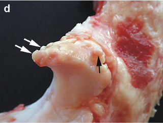
The limbs of the horse are structures made of dozens of bones, joints, muscles, tendons, and ligaments that support the weight of the equine body. They include two apparatuses: the suspensory apparatus, which carries much of the weight, prevents overextension of the joint and absorbs shock, and the stay apparatus, which locks major joints in the limbs, allowing horses to remain standing while relaxed or asleep. The limbs play a major part in the movement of the horse, with the legs performing the functions of absorbing impact, bearing weight, and providing thrust. In general, the majority of the weight is borne by the front legs, while the rear legs provide propulsion. The hooves are also important structures, providing support, traction and shock absorption, and containing structures that provide blood flow through the lower leg. As the horse developed as a cursorial animal, with a primary defense mechanism of running over hard ground, its legs evolved to the long, sturdy, light-weight, one-toed form seen today.
Acquired hand deformity refers to the structural or functional abnormalities that develop in the hand. There are multiple varying causes of acquired hand deformity, triggering significant consequences and complications. Trauma, including blunt force, penetrating injuries, burns, and sports-related incidents, is a primary cause of acquired hand deformities. Inflammatory conditions such as rheumatoid arthritis, gouty arthritis, and systemic lupus erythematosus can also contribute to hand deformities by affecting the joints. Degenerative arthritis, specifically osteoarthritis, functions to evoke impaired hand function due to the gradual deterioration of cartilage. Neurological disorders like cerebral palsy can result in hand contractures due to increased muscle tone and stiffness. There are different types of acquired hand deformities, each with distinct characteristics and underlying causes, such as boutonnière deformity, Dupuytren's contracture, gamekeeper's thumb, hand osteoarthritis deformity, mallet finger, swan-neck deformity, ulnar claw hand, among many others.
