Chronic wound

Last updated

A chronic wound is a wound that does not heal in an orderly set of stages and in a predictable amount of time the way most wounds do; wounds that do not heal within three months are often considered chronic. [1] Chronic wounds seem to be detained in one or more of the phases of wound healing. For example, chronic wounds often remain in the inflammatory stage for too long. [2] [3] To overcome that stage and jump-start the healing process, a number of factors need to be addressed such as bacterial burden, necrotic tissue, and moisture balance of the whole wound. [4] In acute wounds, there is a precise balance between production and degradation of molecules such as collagen; in chronic wounds this balance is lost and degradation plays too large a role. [5] [6]

Contents

Chronic wounds may never heal or may take years to do so. These wounds can cause patients severe emotional and physical stress and create a significant financial burden on patients and the whole healthcare system. [7]

Acute and chronic wounds are at opposite ends of a spectrum of wound-healing types that progress toward being healed at different rates. [8]

Signs and symptoms

Chronic wound patients often report pain as dominant in their lives. [9] It is recommended that healthcare providers handle the pain related to chronic wounds as one of the main priorities in chronic wound management (together with addressing the cause). Six out of ten venous leg ulcer patients experience pain with their ulcer, [10] and similar trends are observed for other chronic wounds.

Persistent pain (at night, at rest, and with activity) is the main problem for patients with chronic ulcers. [11] Frustrations regarding ineffective analgesics and plans of care that they were unable to adhere to were also identified.

Cause

In addition to poor circulation, neuropathy, and difficulty moving, factors that contribute to chronic wounds include systemic illnesses, age, and repeated trauma. The genetic skin disorders collectively known as epidermolysis bullosa display skin fragility and a tendency to develop chronic, non-healing wounds. [12] Comorbid ailments that may contribute to the formation of chronic wounds include vasculitis (an inflammation of blood vessels), immune suppression, pyoderma gangrenosum, and diseases that cause ischemia. [2] Immune suppression can be caused by illnesses or medical drugs used over a long period, like steroids. [2] Emotional stress can also negatively affect the healing of a wound, possibly by raising blood pressure and levels of cortisol, which lowers immunity. [7]

What appears to be a chronic wound may also be a malignancy; for example, cancerous tissue can grow until blood cannot reach the cells and the tissue becomes an ulcer. [13] Cancer, especially squamous cell carcinoma, may also form as the result of chronic wounds, probably due to repetitive tissue damage that stimulates rapid cell proliferation. [13]

Another factor that may contribute to chronic wounds is old age. [14] The skin of older people is more easily damaged, and older cells do not proliferate as fast and may not have an adequate response to stress in terms of gene upregulation of stress-related proteins. [14] In older cells, stress response genes are overexpressed when the cell is not stressed, but when it is, the expression of these proteins is not upregulated by as much as in younger cells. [14]

Comorbid factors that can lead to ischemia are especially likely to contribute to chronic wounds. Such factors include chronic fibrosis, edema, sickle cell disease, and peripheral artery disease such as by atherosclerosis. [2]

Repeated physical trauma plays a role in chronic wound formation by continually initiating the inflammatory cascade. The trauma may occur by accident, for example when a leg is repeatedly bumped against a wheelchair rest, or it may be due to intentional acts. Heroin users who lose venous access may resort to 'skin popping', or injecting the drug subcutaneously, which is highly damaging to tissue and frequently leads to chronic ulcers. [15] Children who are repeatedly seen for a wound that does not heal are sometimes found to be victims of a parent with Munchausen syndrome by proxy, a disease in which the abuser may repeatedly inflict harm on the child in order to receive attention. [16]

Periwound skin damage caused by excessive amounts of exudate and other bodily fluids can perpetuate the non-healing status of chronic wounds. Maceration, excoriation, dry (fragile) skin, hyperkeratosis, callus and eczema are frequent problems [17] that interfere with the integrity of periwound skin. They can create a gateway for infection as well as cause wound edge deterioration preventing wound closure.

Pathophysiology

Chronic wounds may affect only the epidermis and dermis, or they may affect tissues all the way to the fascia. [18] They may be formed originally by the same things that cause acute ones, such as surgery or accidental trauma, or they may form as the result of systemic infection, vascular, immune, or nerve insufficiency, or comorbidities such as neoplasias or metabolic disorders. [18] The reason a wound becomes chronic is that the body's ability to deal with the damage is overwhelmed by factors such as repeated trauma, continued pressure, ischemia, or illness. [8] [18]

Though much progress has been accomplished in the study of chronic wounds lately, advances in the study of their healing have lagged behind expectations. This is partly because animal studies are difficult because animals do not get chronic wounds, since they usually have loose skin that quickly contracts, and they normally do not get old enough or have contributing diseases such as neuropathy or chronic debilitating illnesses. [14] Nonetheless, current researchers now understand some of the major factors that lead to chronic wounds, among which are ischemia, reperfusion injury, and bacterial colonization. [14]

Ischemia

Ischemia is an important factor in the formation and persistence of wounds, especially when it occurs repetitively (as it usually does) or when combined with a patient's old age. [14] Ischemia causes tissue to become inflamed and cells to release factors that attract neutrophils such as interleukins, chemokines, leukotrienes, and complement factors. [14]

While they fight pathogens, neutrophils also release inflammatory cytokines and enzymes that damage cells. [2] [14] One of their important jobs is to produce Reactive Oxygen Species (ROS) to kill bacteria, for which they use an enzyme called myeloperoxidase. [14] The enzymes and ROS produced by neutrophils and other leukocytes damage cells and prevent cell proliferation and wound closure by damaging DNA, lipids, proteins, [19] the extracellular matrix (ECM), and cytokines that speed healing. [14] Neutrophils remain in chronic wounds for longer than they do in acute wounds, and contribute to the fact that chronic wounds have higher levels of inflammatory cytokines and ROS. [3] [6] Since wound fluid from chronic wounds has an excess of proteases and ROS, the fluid itself can inhibit healing by inhibiting cell growth and breaking down growth factors and proteins in the ECM. This impaired healing response is considered uncoordinated. [20] However, soluble mediators of the immune system (growth factors), cell-based therapies and therapeutic chemicals can propagate coordinated healing. [21]

It has been suggested that the three fundamental factors underlying chronic wound pathogenesis are cellular and systemic changes of aging, repeated bouts of ischemia-reperfusion injury, and bacterial colonization with resulting inflammatory host response. [22]

Bacterial colonization

Since more oxygen in the wound environment allows white blood cells to produce ROS to kill bacteria, patients with inadequate tissue oxygenation, for example those who developed hypothermia during surgery, are at higher risk for infection. [14] The host's immune response to the presence of bacteria prolongs inflammation, delays healing, and damages tissue. [14] Infection can lead not only to chronic wounds but also to gangrene, loss of the infected limb, and death of the patient. More recently, an interplay between bacterial colonization and increases in reactive oxygen species leading to formation and production of biofilms has been shown to generate chronic wounds. [23]

Like ischemia, bacterial colonization and infection damage tissue by causing a greater number of neutrophils to enter the wound site. [2] In patients with chronic wounds, bacteria with resistances to antibiotics may have time to develop. [24] In addition, patients that carry drug resistant bacterial strains such as methicillin-resistant Staphylococcus aureus (MRSA) have more chronic wounds. [24]

Growth factors and proteolytic enzymes

Chronic wounds also differ in makeup from acute wounds in that their levels of proteolytic enzymes such as elastase. [5] and matrix metalloproteinases (MMPs) are higher, while their concentrations of growth factors such as Platelet-derived growth factor and Keratinocyte Growth Factor are lower. [6] [18]

Since growth factors (GFs) are imperative in timely wound healing, inadequate GF levels may be an important factor in chronic wound formation. [18] In chronic wounds, the formation and release of growth factors may be prevented, the factors may be sequestered and unable to perform their metabolic roles, or degraded in excess by cellular or bacterial proteases. [18]

Chronic wounds such as diabetic and venous ulcers are also caused by a failure of fibroblasts to produce adequate ECM proteins and by keratinocytes to epithelialize the wound. [25] Fibroblast gene expression is different in chronic wounds than in acute wounds. [25]

Though all wounds require a certain level of elastase and proteases for proper healing, too high a concentration is damaging. [5] Leukocytes in the wound area release elastase, which increases inflammation, destroys tissue, proteoglycans, and collagen, [26] and damages growth factors, fibronectin, and factors that inhibit proteases. [5] The activity of elastase is increased by human serum albumin, which is the most abundant protein found in chronic wounds. [5] However, chronic wounds with inadequate albumin are especially unlikely to heal, so regulating the wound's levels of that protein may in the future prove helpful in healing chronic wounds. [5]

Excess matrix metalloproteinases, which are released by leukocytes, may also cause wounds to become chronic. MMPs break down ECM molecules, growth factors, and protease inhibitors, and thus increase degradation while reducing construction, throwing the delicate compromise between production and degradation out of balance. [6] [27]

Diagnosis

Infection

If a chronic wound becomes more painful this is a good indication that it is infected. [28] A lack of pain however does not mean that it is not infected. [28] Other methods of determination are less effective. [28]

Classification

The vast majority of chronic wounds can be classified into three categories: venous ulcers, diabetic, and pressure ulcers. [8] [14] A small number of wounds that do not fall into these categories may be due to causes such as radiation poisoning or ischemia. [14]

Venous and arterial ulcers

Venous ulcers, which usually occur in the legs, account for about 70% to 90% of chronic wounds [2] and mostly affect the elderly. They are thought to be due to venous hypertension caused by improper function of valves that exist in the veins to prevent blood from flowing backward. Ischemia results from the dysfunction and, combined with reperfusion injury, causes the tissue damage that leads to the wounds.

Diabetic ulcers

Another major cause of chronic wounds, diabetes, is increasing in prevalence. [29] Diabetics have a 15% higher risk for amputation than the general population [2] due to chronic ulcers. Diabetes causes neuropathy, which inhibits nociception and the perception of pain. [2] Thus patients may not initially notice small wounds to legs and feet, and may therefore fail to prevent infection or repeated injury. [8] Further, diabetes causes immune compromise and damage to small blood vessels, preventing adequate oxygenation of tissue, which can cause chronic wounds. [8] Pressure also plays a role in the formation of diabetic ulcers. [14]

Pressure ulcers

Another leading type of chronic wounds is pressure ulcers, [30] which usually occur in people with conditions such as paralysis that inhibit movement of body parts that are commonly subjected to pressure such as the heels, shoulder blades, and sacrum. [31] [32] Pressure ulcers are caused by ischemia that occurs when pressure on the tissue is greater than the pressure in capillaries, and thus restricts blood flow into the area. [30] Muscle tissue, which needs more oxygen and nutrients than skin does, shows the worst effects from prolonged pressure. [32] As in other chronic ulcers, reperfusion injury damages tissue.

Treatment

Though treatment of the different chronic wound types varies slightly, appropriate treatment seeks to address the problems at the root of chronic wounds, including ischemia, bacterial load, and imbalance of proteases. [14] Periwound skin issues should be assessed and their abatement included in a proposed treatment plan. [17] Various methods exist to ameliorate these problems, including antibiotic and antibacterial use, debridement, irrigation, vacuum-assisted closure, warming, oxygenation, moist wound healing (the term pioneered by George D. Winter), removing mechanical stress, and adding cells or other materials to secrete or enhance levels of healing factors. [29]

It is uncertain whether intravenous metronidazole is useful in reducing foul smelling from malignant wounds. [33] There is insufficient evidence to use silver-containing dressings or topical agents for the treatment of infected or contaminated chronic wounds. [34]

The challenge of any treatment is to address as many adverse factors as possible simultaneously, so each of them receives equal attention and does not continue to impede healing as the treatment progresses. [35] [36]

Preventing and treating infection

To lower the bacterial count in wounds, therapists may use topical antibiotics, which kill bacteria and can also help by keeping the wound environment moist, [37] [38] which is important for speeding the healing of chronic wounds. [3] [31] Some researchers have experimented with the use of tea tree oil, an antibacterial agent which also has anti-inflammatory effects. [24] Disinfectants are contraindicated because they damage tissues and delay wound contraction. [38] Further, they are rendered ineffective by organic matter in wounds like blood and exudate and are thus not useful in open wounds. [38]

A greater amount of exudate and necrotic tissue in a wound increases likelihood of infection by serving as a medium for bacterial growth away from the host's defenses. [14] Since bacteria thrive on dead tissue, wounds are often surgically debrided to remove the devitalized tissue. [37] Debridement and drainage of wound fluid are an especially important part of the treatment for diabetic ulcers, which may create the need for amputation if infection gets out of control. Mechanical removal of bacteria and devitalized tissue is also the idea behind wound irrigation, which is accomplished using pulsed lavage. [14]

Removing necrotic or devitalized tissue is also the aim of maggot therapy, the intentional introduction by a health care practitioner of live, disinfected maggots into non-healing wounds. Maggots dissolve only necrotic, infected tissue; disinfect the wound by killing bacteria; and stimulate wound healing. Maggot therapy has been shown to accelerate debridement of necrotic wounds and reduce the bacterial load of the wound, leading to earlier healing, reduced wound odor and less pain. The combination and interactions of these actions make maggots an extremely potent tool in chronic wound care.

Negative pressure wound therapy (NPWT) is a treatment that improves ischemic tissues and removes wound fluid used by bacteria. [8] [14] This therapy, also known as vacuum-assisted closure, reduces swelling in tissues, which brings more blood and nutrients to the area, as does the negative pressure itself. [8] The treatment also decompresses tissues and alters the shape of cells, causes them to express different mRNAs and to proliferate and produce ECM molecules. [2] [8]

Recent technological advancements produced novel approaches such as self-adaptive wound dressings [39] that rely on properties of smart polymers sensitive to changes in humidity levels. The dressing delivers absorption or hydration as needed over each independent wound area and aids in the natural process of autolytic debridement. It effectively removes liquefied slough and necrotic tissue, disintegrated bacterial biofilm as well as harmful exudate components, known to slow the healing process. [40] The treatment also reduces bacterial load by effective evacuation and immobilization of microorganisms from the wound bed, and subsequent chemical binding of available water that is necessary for their replication. [41] Self-adaptive dressings protect periwound skin from extrinsic factors and infection while regulating moisture balance over vulnerable skin around the wound.

Treating trauma and painful wounds

Persistent chronic pain associated with non-healing wounds is caused by tissue (nociceptive) or nerve (neuropathic) damage and is influenced by dressing changes and chronic inflammation. Chronic wounds take a long time to heal and patients can experience chronic wounds for many years. [42] Chronic wound healing may be compromised by coexisting underlying conditions, such as venous valve backflow, peripheral vascular disease, uncontrolled edema and diabetes mellitus.

If wound pain is not assessed and documented it may be ignored and/or not addressed properly. It is important to remember that increased wound pain may be an indicator of wound complications that need treatment, and therefore practitioners must constantly reassess the wound as well as the associated pain.

Optimal management of wounds requires holistic assessment. Documentation of the patient's pain experience is critical and may range from the use of a patient diary, (which should be patient driven), to recording pain entirely by the healthcare professional or caregiver. [43] Effective communication between the patient and the healthcare team is fundamental to this holistic approach. The more frequently healthcare professionals measure pain, the greater the likelihood of introducing or changing pain management practices.

At present there are few local options for the treatment of persistent pain, whilst managing the exudate levels present in many chronic wounds. Important properties of such local options are that they provide an optimal wound healing environment, while providing a constant local low dose release of ibuprofen while worn.

If local treatment does not provide adequate pain reduction, it may be necessary for patients with chronic painful wounds to be prescribed additional systemic treatment for the physical component of their pain. Clinicians should consult with their prescribing colleagues referring to the WHO pain relief ladder of systemic treatment options for guidance. For every pharmacological intervention there are possible benefits and adverse events that the prescribing clinician will need to consider in conjunction with the wound care treatment team.

Ischemia and hypoxia

Blood vessels constrict in tissue that becomes cold and dilate in warm tissue, altering blood flow to the area. Thus keeping the tissues warm is probably necessary to fight both infection and ischemia. [31] Some healthcare professionals use 'radiant bandages' to keep the area warm, and care must be taken during surgery to prevent hypothermia, which increases rates of post-surgical infection. [14]

Underlying ischemia may also be treated surgically by arterial revascularization, for example in diabetic ulcers, and patients with venous ulcers may undergo surgery to correct vein dysfunction.

Diabetics that are not candidates for surgery (and others) may also have their tissue oxygenation increased by Hyperbaric Oxygen Therapy, or HBOT, which may provide a short-term improvement in healing by improving the oxygenated blood supply to the wound. [19] [44] In addition to killing bacteria, higher oxygen content in tissues speeds growth factor production, fibroblast growth, and angiogenesis. [2] [19] However, increased oxygen levels also means increased production of ROS. [19] Antioxidants, molecules that can lose an electron to free radicals without themselves becoming radicals, can lower levels of oxidants in the body and have been used with some success in wound healing. [6]

Low level laser therapy has been repeatedly shown to significantly reduce the size and severity of diabetic ulcers as well as other pressure ulcers.

Pressure wounds are often the result of local ischemia from the increased pressure. Increased pressure also plays a roles in many diabetic foot ulcerations as changes due to the disease causes the foot to have limited joint mobility and creates pressure points on the bottom of the foot. Effective measures to treat this includes a surgical procedure called the gastrocnemius recession in which the calf muscle is lengthened to decrease the fulcrum created by this muscle and resulting in a decrease in plantar forefoot pressure. [45]

Growth factors and hormones

Since chronic wounds underexpress growth factors necessary for healing tissue, chronic wound healing may be speeded by replacing or stimulating those factors and by preventing the excessive formation of proteases like elastase that break them down. [5] [6]

One way to increase growth factor concentrations in wounds is to apply the growth factors directly. This generally takes many repetitions and requires large amounts of the factors, although biomaterials are being developed that control the delivery of growth factors over time. [46] Another way is to spread onto the wound a gel of the patient's own blood platelets, which then secrete growth factors such as vascular endothelial growth factor (VEGF), insulin-like growth factor 1–2 (IGF), PDGF, transforming growth factor-β (TGF-β), and epidermal growth factor (EGF). [18] Other treatments include implanting cultured keratinocytes into the wound to reepithelialize it and culturing and implanting fibroblasts into wounds. [29] [37] Some patients are treated with artificial skin substitutes that have fibroblasts and keratinocytes in a matrix of collagen to replicate skin and release growth factors.

In other cases, skin from cadavers is grafted onto wounds, providing a cover to keep out bacteria and preventing the buildup of too much granulation tissue, which can lead to excessive scarring. Though the allograft (skin transplanted from a member of the same species) is replaced by granulation tissue and is not actually incorporated into the healing wound, it encourages cellular proliferation and provides a structure for epithelial cells to crawl across. [2] On the most difficult chronic wounds, allografts may not work, requiring skin grafts from elsewhere on the patient, which can cause pain and further stress on the patient's system. [3]

Collagen dressings are another way to provide the matrix for cellular proliferation and migration, while also keeping the wound moist and absorbing exudate. [6] Additionally Collagen has been shown to be chemotactic to human blood monocytes, which can enter the wound site and transform into beneficial wound-healing cells. [47]

Since levels of protease inhibitors are lowered in chronic wounds, some researchers are seeking ways to heal tissues by replacing these inhibitors in them. [27] Secretory leukocyte protease inhibitor (SLPI), which inhibits not only proteases but also inflammation and microorganisms like viruses, bacteria, and fungi, may prove to be an effective treatment. [27]

Research into hormones and wound healing has shown estrogen to speed wound healing in elderly humans and in animals that have had their ovaries removed, possibly by preventing excess neutrophils from entering the wound and releasing elastase. [26] Thus the use of estrogen is a future possibility for treating chronic wounds.

Epidemiology

Chronic wounds mostly affect people over the age of 60. [14] The incidence is 0.78% of the population and the prevalence ranges from 0.18 to 0.32%. [18] As the population ages, the number of chronic wounds is expected to rise. [30] Ulcers that heal within 12 weeks are usually classified as acute, and longer-lasting ones as chronic.

Related Research Articles

<span class="mw-page-title-main">Gangrene</span> Type of tissue death by a lack of blood supply

Gangrene is a type of tissue death caused by a lack of blood supply. Symptoms may include a change in skin color to red or black, numbness, swelling, pain, skin breakdown, and coolness. The feet and hands are most commonly affected. If the gangrene is caused by an infectious agent, it may present with a fever or sepsis.

<span class="mw-page-title-main">Ulcer (dermatology)</span> Type of cutaneous condition

An ulcer is a sore on the skin or a mucous membrane, accompanied by the disintegration of tissue. Ulcers can result in complete loss of the epidermis and often portions of the dermis and even subcutaneous fat. Ulcers are most common on the skin of the lower extremities and in the gastrointestinal tract. An ulcer that appears on the skin is often visible as an inflamed tissue with an area of reddened skin. A skin ulcer is often visible in the event of exposure to heat or cold, irritation, or a problem with blood circulation.

<span class="mw-page-title-main">Wound</span> Acute injury from laceration, puncture, blunt force, or compression

A wound is a rapid onset of injury that involves lacerated or punctured skin, or a contusion from blunt force trauma or compression. In pathology, a wound is an acute injury that damages the epidermis of the skin. To heal a wound, the body undertakes a series of actions collectively known as the wound healing process.

<span class="mw-page-title-main">Peripheral artery disease</span> Medical condition

Peripheral artery disease (PAD) is a vascular disorder abnormal narrowing of arteries other than those that supply the heart or brain. PAD can happen in any blood vessel, but it is more common in the legs than the arms.

<span class="mw-page-title-main">Wound healing</span> Series of events that restore integrity to damaged tissue after an injury

Wound healing refers to a living organism's replacement of destroyed or damaged tissue by newly produced tissue.

<span class="mw-page-title-main">Elastase</span> Enzyme

In molecular biology, elastase is an enzyme from the class of proteases (peptidases) that break down proteins. In particular, it is a serine protease.

<span class="mw-page-title-main">Pressure ulcer</span> Skin ulcer (bed sore)

Pressure ulcers, also known as pressure sores, bed sores or pressure injuries, are localised damage to the skin and/or underlying tissue that usually occur over a bony prominence as a result of usually long-term pressure, or pressure in combination with shear or friction. The most common sites are the skin overlying the sacrum, coccyx, heels, and hips, though other sites can be affected, such as the elbows, knees, ankles, back of shoulders, or the back of the cranium.
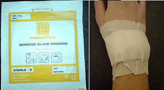
<span class="mw-page-title-main">Dressing (medicine)</span> Sterile pad or compress applied to wounds

A dressing or compress is a sterile pad applied to a wound to promote healing and protect the wound from further harm. A dressing is designed to be in direct contact with the wound, as distinguished from a bandage, which is most often used to hold a dressing in place. Many modern dressings are self-adhesive.

<span class="mw-page-title-main">Maggot therapy</span> Wound care by maggot therapy

Maggot therapy is a type of biotherapy involving the introduction of live, disinfected maggots into non-healing skin and soft-tissue wounds of a human or other animal for the purpose of cleaning out the necrotic (dead) tissue within a wound (debridement), and disinfection.

<span class="mw-page-title-main">Venous ulcer</span> Medical condition

Venous ulcer is defined by the American Venous Forum as "a full-thickness defect of skin, most frequently in the ankle region, that fails to heal spontaneously and is sustained by chronic venous disease, based on venous duplex ultrasound testing." Venous ulcers are wounds that are thought to occur due to improper functioning of venous valves, usually of the legs. They are an important cause of chronic wounds, affecting 1% of the population. Venous ulcers develop mostly along the medial distal leg, and can be painful with negative effects on quality of life.

The history of wound care spans from prehistory to modern medicine. Wounds naturally heal by themselves, but hunter-gatherers would have noticed several factors and certain herbal remedies would speed up or assist the process, especially if it was grievous. In ancient history, this was followed by the realisation of the necessity of hygiene and the halting of bleeding, where wound dressing techniques and surgery developed. Eventually the germ theory of disease also assisted in improving wound care.

<span class="mw-page-title-main">Negative-pressure wound therapy</span> Therapeutic technique

Negative-pressure wound therapy (NPWT), also known as a vacuum assisted closure (VAC), is a therapeutic technique using a suction pump, tubing, and a dressing to remove excess exudate and promote healing in acute or chronic wounds and second- and third-degree burns. The therapy involves the controlled application of sub-atmospheric pressure to the local wound environment using a sealed wound dressing connected to a vacuum pump. The use of this technique in wound management started in the 1990s and this technique is often recommended for treatment of a range of wounds including dehisced surgical wounds, closed surgical wounds, open abdominal wounds, open fractures, pressure injuries or pressure ulcers, diabetic foot ulcers, venous insufficiency ulcers, some types of skin grafts, burns, sternal wounds. It may also be considered after a clean surgery in a person who is obese.

<span class="mw-page-title-main">Calciphylaxis</span> Medical condition

Calciphylaxis, also known as calcific uremic arteriolopathy (CUA) or “Grey Scale”, is a rare syndrome characterized by painful skin lesions. The pathogenesis of calciphylaxis is unclear but believed to involve calcification of the small blood vessels located within the fatty tissue and deeper layers of the skin, blood clots, and eventual death of skin cells due to lack of blood flow. It is seen mostly in people with end-stage kidney disease but can occur in the earlier stages of chronic kidney disease and rarely in people with normally functioning kidneys. Calciphylaxis is a rare but serious disease, believed to affect 1-4% of all dialysis patients. It results in chronic non-healing wounds and indicates poor prognosis, with typical life expectancy of less than one year.

A hydrocolloid dressing is an opaque or transparent dressing for wounds. A hydrocolloid dressing is biodegradable, breathable, and depending on the dressing selected, may adhere to the skin, so no separate taping is needed.

The dermal equivalent, also known as dermal replacement or neodermis, is an in vitro model of the dermal layer of skin. There is no specific way of forming a dermal equivalent, however the first dermal equivalent was constructed by seeding dermal fibroblasts into a collagen gel. This gel may then be allowed to contract as a model of wound contraction. This collagen gel contraction assay may be used to screen for treatments which promote or inhibit contraction and thus affect the development of a scar. Other cell types may be incorporated into the dermal equivalent to increase the complexity of the model. For example, keratinocytes may be seeded on the surface to create a skin equivalent, or macrophages may be incorporated to model the inflammatory phase of wound healing.

Wound bed preparation (WBP) is a systematic approach to wound management by identifying and removing barriers to healing. The concept was originally developed in plastic surgery. During the year 2000, the concept was applied to systematizing the treatment of chronic wounds. The 2000 proposals recommended that wound management address the identifiable impediments to healing in order to achieve more successful outcomes. Three publications appeared that year that focused on the concept of managing the healing processes of a wound exudate, bioburden and devitalized tissue. Initially, emphasis was placed on debridement, moisture balance and bacterial balance as the three guiding principles of good wound care, while at the same time recognizing that the provision of care includes a vast array of patient, clinical and environmental variables.

Chronic wound pain is a condition described as unremitting, disabling, and recalcitrant pain experienced by individuals with various types of chronic wounds. Chronic wounds such as venous leg ulcers, arterial ulcers, diabetic foot ulcers, pressure ulcers, and malignant wounds can have an enormous impact on an individual’s quality of life with pain being one of the most distressing symptoms.

<span class="mw-page-title-main">Diabetic foot ulcer</span> Medical condition

Diabetic foot ulcer is a breakdown of the skin and sometimes deeper tissues of the foot that leads to sore formation. It may occur due to a variety of mechanisms. It is thought to occur due to abnormal pressure or mechanical stress chronically applied to the foot, usually with concomitant predisposing conditions such as peripheral sensory neuropathy, peripheral motor neuropathy, autonomic neuropathy or peripheral arterial disease. It is a major complication of diabetes mellitus, and it is a type of diabetic foot disease. Secondary complications to the ulcer, such as infection of the skin or subcutaneous tissue, bone infection, gangrene or sepsis are possible, often leading to amputation.

Chronic limb threatening ischemia (CLTI), also known as critical limb ischemia (CLI), is an advanced stage of peripheral artery disease (PAD). It is defined as ischemic rest pain, arterial insufficiency ulcers, and gangrene. The latter two conditions are jointly referred to as tissue loss, reflecting the development of surface damage to the limb tissue due to the most severe stage of ischemia. Compared to the other manifestation of PAD, intermittent claudication, CLI has a negative prognosis within a year after the initial diagnosis, with 1-year amputation rates of approximately 12% and mortality of 50% at 5 years and 70% at 10 years.

Wound assessment is a component of wound management. As far as may be practical, the assessment is to be accomplished before prescribing any treatment plan. The objective is to collect information about the patient and about the wound, that may be relevant to planning and implementing the treatment.

References

  1. Mustoe T (March 17–18, 2005). "Dermal ulcer healing: Advances in understanding" (PDF). Tissue repair and ulcer/wound healing: molecular mechanisms, therapeutic targets and future directions. Paris, France: EUROCONFERENCES. Archived from the original (PDF) on October 27, 2005.
  2. 1 2 3 4 5 6 7 8 9 10 11 12 Snyder RJ (2005). "Treatment of nonhealing ulcers with allografts". Clinics in Dermatology. 23 (4): 388–95. doi:10.1016/j.clindermatol.2004.07.020. PMID   16023934.
  3. 1 2 3 4 Taylor JE, Laity PR, Hicks J, Wong SS, Norris K, Khunkamchoo P, et al. (October 2005). "Extent of iron pick-up in deforoxamine-coupled polyurethane materials for therapy of chronic wounds". Biomaterials. 26 (30): 6024–33. doi:10.1016/j.biomaterials.2005.03.015. PMID   15885771.
  4. Gist S, Tio-Matos I, Falzgraf S, Cameron S, Beebe M (June 2009). "Wound care in the geriatric client". Clinical Interventions in Aging. 4: 269–87. doi: 10.2147/CIA.S4726 . PMC   2697592 . PMID   19554098.
  5. 1 2 3 4 5 6 7 Edwards JV, Howley P, Cohen IK (October 2004). "In vitro inhibition of human neutrophil elastase by oleic acid albumin formulations from derivatized cotton wound dressings". International Journal of Pharmaceutics. 284 (1–2): 1–12. doi:10.1016/j.ijpharm.2004.06.003. PMID   15454291.[ permanent dead link ]
  6. 1 2 3 4 5 6 7 Schönfelder U, Abel M, Wiegand C, Klemm D, Elsner P, Hipler UC (November 2005). "Influence of selected wound dressings on PMN elastase in chronic wound fluid and their antioxidative potential in vitro". Biomaterials. 26 (33): 6664–73. doi:10.1016/j.biomaterials.2005.04.030. PMID   15978664.
  7. 1 2 Augustin M, Maier K (2003). "Psychosomatic aspects of chronic wounds". Dermatology and Psychosomatics/Dermatologie und Psychosomatik. 4 (1): 5–13. doi:10.1159/000070529. S2CID   72066898.
  8. 1 2 3 4 5 6 7 8 Moreo K (2005). "Understanding and overcoming the challenges of effective case management for patients with chronic wounds". The Case Manager. 16 (2): 62–3, 67. doi:10.1016/j.casemgr.2005.01.014. PMID   15818347.
  9. Krasner D (May 1998). "Painful venous ulcers: themes and stories about living with the pain and suffering". Journal of Wound, Ostomy, and Continence Nursing. 25 (3): 158–68. doi:10.1097/00152192-199805000-00008. PMID   9678007.
  10. Hofman D, Ryan TJ, Arnold F, Cherry GW, Lindholm C, Bjellerup M, Glynn C (May 1997). "Pain in venous leg ulcers". Journal of Wound Care. 6 (5): 222–4. doi:10.12968/jowc.1997.6.5.222. PMID   9256727.
  11. Walshe C (December 1995). "Living with a venous leg ulcer: a descriptive study of patients' experiences". Journal of Advanced Nursing. 22 (6): 1092–100. doi:10.1111/j.1365-2648.1995.tb03110.x. PMID   8675863.
  12. Bardhan, Ajoy; Bruckner-Tuderman, Leena; Chapple, Iain L. C.; Fine, Jo-David; Harper, Natasha; Has, Cristina; Magin, Thomas M.; Marinkovich, M. Peter; Marshall, John F.; McGrath, John A.; Mellerio, Jemima E. (2020-09-24). "Epidermolysis bullosa". Nature Reviews Disease Primers. 6 (1): 78. doi:10.1038/s41572-020-0210-0. ISSN   2056-676X. PMID   32973163. S2CID   221861310.
  13. 1 2 Trent, JT. 2003. Wounds and malignancy. Archived 2016-01-13 at the Wayback Machine Advances in Skin & Wound Care. Accessed January 1, 2007.
  14. 1 2 3 4 5 6 7 8 9 10 11 12 13 14 15 16 17 18 19 20 21 Mustoe T (May 2004). "Understanding chronic wounds: a unifying hypothesis on their pathogenesis and implications for therapy". American Journal of Surgery. 187 (5A): 65S–70S. doi:10.1016/S0002-9610(03)00306-4. PMID   15147994.
  15. Williams AM, Southern SJ (October 2005). "Conflicts in the treatment of chronic ulcers in drug addicts--case series and discussion". British Journal of Plastic Surgery. 58 (7): 997–9. doi: 10.1016/j.bjps.2005.04.024 . PMID   16040018.
  16. Vennemann B, Perdekamp MG, Weinmann W, Faller-Marquardt M, Pollak S, Brandis M (May 2006). "A case of Munchausen syndrome by proxy with subsequent suicide of the mother". Forensic Science International. 158 (2–3): 195–9. doi:10.1016/j.forsciint.2005.07.014. PMID   16169176.
  17. 1 2 Dowsett C, Gronemann MN, Harding K (2015). "Taking wound assessment beyond the edge". Wounds International. 6 (1). Archived from the original on 2018-05-04. Retrieved 2017-03-31.
  18. 1 2 3 4 5 6 7 8 Crovetti G, Martinelli G, Issi M, Barone M, Guizzardi M, Campanati B, et al. (April 2004). "Platelet gel for healing cutaneous chronic wounds". Transfusion and Apheresis Science. 30 (2): 145–51. doi:10.1016/j.transci.2004.01.004. PMID   15062754.
  19. 1 2 3 4 Alleva R, Nasole E, Di Donato F, Borghi B, Neuzil J, Tomasetti M (July 2005). "alpha-Lipoic acid supplementation inhibits oxidative damage, accelerating chronic wound healing in patients undergoing hyperbaric oxygen therapy". Biochemical and Biophysical Research Communications. 333 (2): 404–10. doi:10.1016/j.bbrc.2005.05.119. PMC   2136431 . PMID   15950945.
  20. Krishnaswamy VR, Manikandan M, Munirajan AK, Vijayaraghavan D, Korrapati PS (December 2014). "Expression and integrity of dermatopontin in chronic cutaneous wounds: a crucial factor in impaired wound healing". Cell and Tissue Research. 358 (3): 833–41. doi:10.1007/s00441-014-2000-z. PMID   25260909. S2CID   16355532.
  21. Lasagni L, Sagrinati C, Ronconi E, Angelotti ML, Parente E, Ballerini L, et al. (2010). "Novel strategies of regenerative medicine using chemical compounds". Current Medicinal Chemistry. 17 (34): 4134–49. doi:10.2174/092986710793348590. PMID   20939819.
  22. Mustoe T (May 2004). "Understanding chronic wounds: a unifying hypothesis on their pathogenesis and implications for therapy". American Journal of Surgery (review). 187 (5A): 65S–70S. doi:10.1016/S0002-9610(03)00306-4. PMID   15147994.
  23. Dhall S, Do D, Garcia M, Wijesinghe DS, Brandon A, Kim J, et al. (2014). "A novel model of chronic wounds: importance of redox imbalance and biofilm-forming bacteria for establishment of chronicity". PLOS ONE. 9 (10): e109848. Bibcode:2014PLoSO...9j9848D. doi: 10.1371/journal.pone.0109848 . PMC   4196950 . PMID   25313558.
  24. 1 2 3 Halcón L, Milkus K (November 2004). "Staphylococcus aureus and wounds: a review of tea tree oil as a promising antimicrobial". American Journal of Infection Control. 32 (7): 402–8. doi:10.1016/j.ajic.2003.12.008. PMID   15525915.
  25. 1 2 Foy Y, Li J, Kirsner R, Eaglstein W (2004). "Analysis of fibroblast defects in extracellular matrix production in chronic wounds". Journal of the American Academy of Dermatology. 50 (3): P168. doi: 10.1016/j.jaad.2003.10.595 .
  26. 1 2 Kanda N, Watanabe S (April 2005). "Regulatory roles of sex hormones in cutaneous biology and immunology". Journal of Dermatological Science. 38 (1): 1–7. doi:10.1016/j.jdermsci.2004.10.011. PMID   15795118.
  27. 1 2 3 Lai JY, Borson ND, Strausbauch MA, Pittelkow MR (April 2004). "Mitosis increases levels of secretory leukocyte protease inhibitor in keratinocytes". Biochemical and Biophysical Research Communications. 316 (2): 407–10. doi:10.1016/j.bbrc.2004.02.065. PMID   15020232.
  28. 1 2 3 Reddy M, Gill SS, Wu W, Kalkar SR, Rochon PA (February 2012). "Does this patient have an infection of a chronic wound?". JAMA. 307 (6): 605–11. doi:10.1001/jama.2012.98. PMID   22318282.
  29. 1 2 3 Velander PE, Theopold C, Gheerardyn R, Bleiziffer O, Yao F, Eriksson E (2004). "Autologous cultured keratinocytes suspensions accelerate re-epithelialization in the diabetic pig". Journal of the American College of Surgeons. 199 (3): 58. doi:10.1016/j.jamcollsurg.2004.05.119.
  30. 1 2 3 Supp DM, Boyce ST (2005). "Engineered skin substitutes: practices and potentials". Clinics in Dermatology. 23 (4): 403–12. doi:10.1016/j.clindermatol.2004.07.023. PMID   16023936.
  31. 1 2 3 Thomas DR, Diebold MR, Eggemeyer LM (2005). "A controlled, randomized, comparative study of a radiant heat bandage on the healing of stage 3-4 pressure ulcers: a pilot study". Journal of the American Medical Directors Association. 6 (1): 46–9. doi:10.1016/j.jamda.2004.12.007. PMID   15871870.
  32. 1 2 Pressure ulcers: Surgical treatment and principles at eMedicine
  33. Ramasubbu DA, Smith V, Hayden F, Cronin P, et al. (Cochrane Wounds Group) (August 2017). "Systemic antibiotics for treating malignant wounds". The Cochrane Database of Systematic Reviews. 2017 (8): CD011609. doi:10.1002/14651858.CD011609.pub2. PMC   6483739 . PMID   28837757.
  34. Vermeulen, Hester; van Hattem, Jarne M; Storm-Versloot, Marja N; Ubbink, Dirk T; Westerbos, Stijn Joël (2007-01-24). Cochrane Wounds Group (ed.). "Topical silver for treating infected wounds". Cochrane Database of Systematic Reviews (1): CD005486. doi:10.1002/14651858.CD005486.pub2. PMID   17253557.
  35. Jones V, Grey JE, Harding KG (April 2006). "Wound dressings". BMJ. 332 (7544): 777–80. doi:10.1136/bmj.332.7544.777. PMC   1420733 . PMID   16575081.
  36. Cutting K (May 2010). "Wound dressings: 21st century performance requirements". Journal of Wound Care. 19(Sup 1): 4–9. doi:10.12968/jowc.2010.19.Sup1.48258.
  37. 1 2 3 Brem H, Kirsner RS, Falanga V (July 2004). "Protocol for the successful treatment of venous ulcers". American Journal of Surgery. 188 (1A Suppl): 1–8. doi: 10.1016/S0002-9610(03)00284-8 . PMID   15223495.
  38. 1 2 3 Patel CV, Powell L, Wilson SE (2000). "Surgical wound infections". Current Treatment Options in Infectious Diseases. 2: 147–53. ISSN   1523-3820.
  39. Wolcott R, Fischenich RN (April 2014). "Ultimate Standardization of First-Line Wound Dressings to a Single Type". Today's Wound Clinic. 8 (3).
  40. Reyzelman AM, Vartivarian M (August 2015). "Evidence of Intensive Autolytic Debridement With a Self-Adaptive Wound Dressing". Wounds. 27 (8): 229–35. PMID   26284377.
  41. Newman GR, Walker M, Hobot JA, Bowler PG (March 2006). "Visualisation of bacterial sequestration and bactericidal activity within hydrating Hydrofiber wound dressings". Biomaterials. 27 (7): 1129–39. doi:10.1016/j.biomaterials.2005.07.046. PMID   16120458.
  42. Flanagan M, Vogensen H, and Haase L. 2006. Case series investigating the experience of pain in patients with chronic venous leg ulcers treated with a foam dressing releasing ibuprofen. World Wide Wounds. 2006
  43. Osterbrink J (2003). "Der Deutsche Schmerzstandard und seine Auswirkungen auf die Pflege". Die Schwester, der Pfleger. 42: 758–64.
  44. Kranke P, Bennett MH, Martyn-St James M, Schnabel A, Debus SE, Weibel S (June 2015). "Hyperbaric oxygen therapy for chronic wounds" (PDF). The Cochrane Database of Systematic Reviews. 2015 (6): CD004123. doi:10.1002/14651858.CD004123.pub4. PMC   7055586 . PMID   26106870.
  45. Greenhagen RM, Johnson AR, Peterson MC, Rogers LC, Bevilacqua NJ (2010). "Gastrocnemius recession as an alternative to tendoAchillis lengthening for relief of forefoot pressure in a patient with peripheral neuropathy: a case report and description of a technical modification". The Journal of Foot and Ankle Surgery. 49 (2): 159.e9–13. doi:10.1053/j.jfas.2009.07.002. PMID   20137982.
  46. Pop MA, Almquist BD (September 2017). "Biomaterials: A potential pathway to healing chronic wounds?". Experimental Dermatology. 26 (9): 760–763. doi:10.1111/exd.13290. PMC   5500184 . PMID   28094868.
  47. Postlethwaite AE, Kang AH (June 1976). "Collagen-and collagen peptide-induced chemotaxis of human blood monocytes". The Journal of Experimental Medicine. 143 (6): 1299–307. doi:10.1084/jem.143.6.1299. PMC   2190221 . PMID   1271012.

Further reading