Commissure of inferior colliculus

Last updated
Commissure of inferior colliculus
Details
Part of Inferior colliculus
Identifiers
Latin Commissura colliculi inferioris
NeuroNames 481
NeuroLex ID birnlex_935
TA A14.1.06.613
TE E5.14.3.3.1.4.7
FMA 71115
Anatomical terms of neuroanatomy

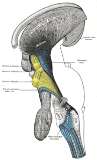
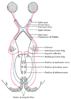
The commissure of inferior colliculus, also called the commissure of inferior colliculi is a thin white matter structure consisting of myelinated axons of neurons and joining together the paired inferior colliculi.

White matter part of the brain

White matter refers to areas of the central nervous system (CNS) that are mainly made up of myelinated axons, also called tracts. Long thought to be passive tissue, white matter affects learning and brain functions, modulating the distribution of action potentials, acting as a relay and coordinating communication between different brain regions.

Myelin fatty white substance, multi-layered membrane, unique to the nervous system, that functions as an insulator to greatly increase the velocity of axonal impulse conduction; it is an outgrowth of a type of glial cell

Myelin is a lipid-rich (fatty) substance formed in the central nervous system (CNS) by glial cells called oligodendrocytes, and in the peripheral nervous system (PNS) by Schwann cells. Myelin insulates nerve cell axons to increase the speed at which information travels from one nerve cell body to another or, for example, from a nerve cell body to a muscle. The myelinated axon can be likened to an electrical wire with insulating material (myelin) around it. However, unlike the plastic covering on an electrical wire, myelin does not form a single long sheath over the entire length of the axon. Rather, each myelin sheath insulates the axon over a single section and, in general, each axon comprises multiple long myelinated sections separated from each other by short gaps called Nodes of Ranvier. Each myelin sheath is formed by the concentric wrapping of an oligodendrocyte or Schwann cell process around the axon.

Axon The long process of a neuron that conducts nerve impulses, usually away from the cell body to the terminals and varicosities, which are sites of storage and release of neurotransmitter.

An axon, or nerve fiber, is a long, slender projection of a nerve cell, or neuron, in vertebrates, that typically conducts electrical impulses known as action potentials away from the nerve cell body. The function of the axon is to transmit information to different neurons, muscles, and glands. In certain sensory neurons, such as those for touch and warmth, the axons are called afferent nerve fibers and the electrical impulse travels along these from the periphery to the cell body, and from the cell body to the spinal cord along another branch of the same axon. Axon dysfunction has caused many inherited and acquired neurological disorders which can affect both the peripheral and central neurons. Nerve fibers are classed into three types – group A nerve fibers, group B nerve fibers, and group C nerve fibers. Groups A and B are myelinated, and group C are unmyelinated. These groups include both sensory fibers and motor fibers. Another classification groups only the sensory fibers as Type I, Type II, Type III, and Type IV.

It is evolutionarily one of the most ancient interhemispheric connections.

Evolution Change in the heritable characteristics of biological populations over successive generations

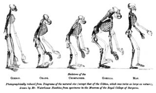
Evolution is change in the heritable characteristics of biological populations over successive generations. These characteristics are the expressions of genes that are passed on from parent to offspring during reproduction. Different characteristics tend to exist within any given population as a result of mutation, genetic recombination and other sources of genetic variation. Evolution occurs when evolutionary processes such as natural selection and genetic drift act on this variation, resulting in certain characteristics becoming more common or rare within a population. It is this process of evolution that has given rise to biodiversity at every level of biological organisation, including the levels of species, individual organisms and molecules.

Related Research Articles

Third ventricle

The third ventricle is one of four connected fluid-filled cavities comprising the ventricular system within the mammalian brain. It is a median cleft in the diencephalon between the two thalami, and is filled with cerebrospinal fluid (CSF).

Midbrain

The midbrain or mesencephalon is a portion of the central nervous system associated with vision, hearing, motor control, sleep/wake, arousal (alertness), and temperature regulation.

Cerebral aqueduct

The cerebral aqueduct, also known as the aqueductus mesencephali, mesencephalic duct, sylvian aqueduct, or aqueduct of Sylvius, is within the mesencephalon, contains cerebrospinal fluid (CSF), and connects the third ventricle in the diencephalon to the fourth ventricle within the region of the mesencephalon and metencephalon, located dorsal to the pons and ventral to the cerebellum.

Inferior colliculus Inferior colliculus

The inferior colliculus (IC) is the principal midbrain nucleus of the auditory pathway and receives input from several peripheral brainstem nuclei in the auditory pathway, as well as inputs from the auditory cortex. The inferior colliculus has three subdivisions: the central nucleus, a dorsal cortex by which it is surrounded, and an external cortex which is located laterally. Its bimodal neurons are implicated in auditory-somatosensory interaction, receiving projections from somatosensory nuclei. This multisensory integration may underlie a filtering of self-effected sounds from vocalization, chewing, or respiration activities.

Tectum A structure in the midbrain of chordate animals

The tectum is a region of the brain, specifically the dorsal (top) part of the midbrain (mesencephalon). The position of the tectum is contrasted with the tegmentum, which refers to the region ventral (lower) to the ventricular system. The tectum is responsible for auditory and visual reflexes.

Red nucleus

The red nucleus or nucleus ruber is a structure in the rostral midbrain involved in motor coordination. The red nucleus is pale pink, which is believed to be due to the presence of iron in at least two different forms: hemoglobin and ferritin. The structure is located in the tegmentum of the midbrain next to the substantia nigra and comprises caudal magnocellular and rostral parvocellular components. The red nucleus and substantia nigra are subcortical centers of the extrapyramidal motor system.

Spinocerebellar tract set of axonal fibers originating in the spinal cord and terminating in the ipsilateral cerebellum

The spinocerebellar tract is a nerve tract originating in the spinal cord and terminating in the same side (ipsilateral) of the cerebellum.

Corpora quadrigemina

In the brain, the corpora quadrigemina are the four colliculi—two inferior, two superior—located on the tectum of the dorsal aspect of the midbrain. They are respectively named the inferior and superior colliculus.

Cerebellar peduncles connect the cerebellum to the brain stem. There are six cerebellar peduncles in total, three on each side:

Spinotectal tract

The spinotectal tract arises in the anterolateral column and terminates in the inferior and superior colliculi.

Superior cerebellar peduncle

In the human brain, the superior cerebellar peduncle is a paired structure of white matter that connects the cerebellum to the midbrain. It consists mainly of efferent fibers, the cerebellothalamic tract that runs from a cerebellar hemisphere to the contralateral thalamus, and the cerebellorubral tract that runs from a cerebellar hemisphere to the red nucleus. It also contains afferent tracts, most prominent of which is the ventral spinocerebellar tract. Other afferent tracts are the trigeminothalamic fibers, tectocerebellar fibers, and noradrenergic fibers from the locus coeruleus. The superior peduncle emerges from the upper and medial parts of the white matter of each hemisphere and is placed under cover of the upper part of the cerebellum.

Ventral supraoptic decussation

The ventral supraoptic decussation is the crossover (decussation) point for signals from the left and right eye, en route respectively to the right and left sides of the visual cortex.

Superior medullary velum

The superior medullary velum is a thin, transparent lamina of white matter, which stretches between the superior cerebellar peduncles; on the dorsal surface of its lower half the folia and lingula are prolonged.

A commissure is the location at which two objects abut or are joined. The term is used especially in the fields of anatomy and biology.

Anterior white commissure

The anterior white commissure is a bundle of nerve fibers which cross the midline of the spinal cord just anterior to the gray commissure. A delta fibers and C fibers carrying pain sensation in the spinothalamic tract contribute to this commissure, as do fibers of the anterior corticospinal tract, which carry motor signals from the primary motor cortex.

The commissure is the corner of the mouth, where the vermillion border of the superior labium meets that of the inferior labium.

Anatomical terms of neuroanatomy

This article describes anatomical terminology that is used to describe the central and peripheral nervous systems - including the brain, brainstem, spinal cord, and nerves.

Brachium colliculi may refer to:

The commissure of superior colliculus, also called the commissure of superior colliculi is a thin white matter structure consisting of myelinated axons of neurons and joining together the paired superior colliculi.

References