Congenital disorder of glycosylation type IIc

Last updated
Congenital disorder of glycosylation type IIc
Other namesRambam-Hasharon syndrome, CDG-IIc, CDG2C
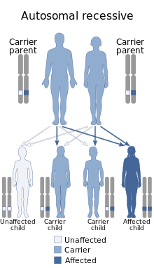
Autosomal recessive - en.svg
This condition ia inherited via autosomal recessive manner

Congenital disorder of glycosylation type IIc or Leukocyte adhesion deficiency-2 (LAD2) is a type of leukocyte adhesion deficiency attributable to the absence of neutrophil sialyl-LewisX, a ligand of P- and E-selectin on vascular endothelium. [1] It is associated with SLC35C1 . [2]

Contents

This disorder was discovered in two unrelated Israeli boys 3 and 5 years of age, each the offspring of consanguineous parents. Both had severe mental retardation, short stature, a distinctive facial appearance, and the Bombay (hh) blood phenotype, and both were secretor- and Lewis-negative. They both had had recurrent severe bacterial infections similar to those seen in patients with LAD1, including pneumonia, periodontitis, otitis media, and localized cellulitis. Similar to that in patients with LAD1, their infections were accompanied by pronounced leukocytosis (30,000 to 150,000/mm3) but an absence of pus formation at sites of recurrent cellulitis. In vitro studies revealed a pronounced defect in neutrophil motility. Because the genes for the red blood cell H antigen and for the secretor status encode for distinct α1,2-fucosyltransferases and the synthesis of Sialyl-LewisX requires an α1,3-fucosyltransferase, it was postulated that a general defect in fucose metabolism is the basis for this disorder. It was subsequently found that GDP-L-fucose transport into Golgi vesicles was specifically impaired, [3] and then missense mutations in the GDP-fucose transporter cDNA of three patients with LAD2 were discovered. Thus, GDP-fucose transporter deficiency is a cause of LAD2. [4]

See also

Related Research Articles

A congenital disorder of glycosylation is one of several rare inborn errors of metabolism in which glycosylation of a variety of tissue proteins and/or lipids is deficient or defective. Congenital disorders of glycosylation are sometimes known as CDG syndromes. They often cause serious, sometimes fatal, malfunction of several different organ systems in affected infants. The most common sub-type is PMM2-CDG where the genetic defect leads to the loss of phosphomannomutase 2 (PMM2), the enzyme responsible for the conversion of mannose-6-phosphate into mannose-1-phosphate.

Glycosylation is the reaction in which a carbohydrate, i.e. a glycosyl donor, is attached to a hydroxyl or other functional group of another molecule in order to form a glycoconjugate. In biology, glycosylation usually refers to an enzyme-catalysed reaction, whereas glycation may refer to a non-enzymatic reaction. Glycosylation is a form of co-translational and post-translational modification. Glycans serve a variety of structural and functional roles in membrane and secreted proteins. The majority of proteins synthesized in the rough endoplasmic reticulum undergo glycosylation. Glycosylation is also present in the cytoplasm and nucleus as the O-GlcNAc modification. Aglycosylation is a feature of engineered antibodies to bypass glycosylation. Five classes of glycans are produced:

Selectin Family of cell adhesion molecules

The selectins are a family of cell adhesion molecules. All selectins are single-chain transmembrane glycoproteins that share similar properties to C-type lectins due to a related amino terminus and calcium-dependent binding. Selectins bind to sugar moieties and so are considered to be a type of lectin, cell adhesion proteins that bind sugar polymers.

Leukocyte adhesion deficiency (LAD) is a rare autosomal recessive disorder characterized by immunodeficiency resulting in recurrent infections. LAD is currently divided into three subtypes: LAD1, LAD2, and the recently described LAD3, also known as LAD-1/variant. In LAD3, the immune defects are supplemented by a Glanzmann thrombasthenia-like bleeding tendency.

Glucose 6-phosphatase Enzyme

The enzyme glucose 6-phosphatase (EC 3.1.3.9, G6Pase; systematic name D-glucose-6-phosphate phosphohydrolase) catalyzes the hydrolysis of glucose 6-phosphate, resulting in the creation of a phosphate group and free glucose:

A fucosyltransferase is an enzyme that transfers an L-fucose sugar from a GDP-fucose donor substrate to an acceptor substrate. The acceptor substrate can be another sugar such as the transfer of a fucose to a core GlcNAc (N-acetylglucosamine) sugar as in the case of N-linked glycosylation, or to a protein, as in the case of O-linked glycosylation produced by O-fucosyltransferase. There are various fucosyltransferases in mammals, the vast majority of which, are located in the Golgi apparatus. The O-fucosyltransferases have recently been shown to localize to the endoplasmic reticulum (ER).

Sialyl-Lewis <sup>X</sup> Chemical compound

Sialyl LewisX (sLeX), also known as cluster of differentiation 15s (CD15s) or stage-specific embryonic antigen 1 (SSEA-1), is a tetrasaccharide carbohydrate which is usually attached to O-glycans on the surface of cells. It is known to play a vital role in cell-to-cell recognition processes. It is also the means by which an egg attracts sperm; first, to stick to it, then bond with it and eventually form a zygote. The discovery of the essential role that this tetrasaccharide plays in the fertilization process was reported in August 2011.

Lymphocyte function-associated antigen 1 (LFA-1) is an integrin found on lymphocytes and other leukocytes. LFA-1 plays a key role in emigration, which is the process by which leukocytes leave the bloodstream to enter the tissues. LFA-1 also mediates firm arrest of leukocytes. Additionally, LFA-1 is involved in the process of cytotoxic T cell mediated killing as well as antibody mediated killing by granulocytes and monocytes. As of 2007, LFA-1 has 6 known ligands: ICAM-1, ICAM-2, ICAM-3, ICAM-4, ICAM-5, and JAM-A. LFA-1/ICAM-1 interactions have recently been shown to stimulate signaling pathways that influence T cell differentiation. LFA-1 belongs to the integrin superfamily of adhesion molecules.

Leukocyte extravasation

Leukocyte extravasation is the movement of leukocytes out of the circulatory system and towards the site of tissue damage or infection. This process forms part of the innate immune response, involving the recruitment of non-specific leukocytes. Monocytes also use this process in the absence of infection or tissue damage during their development into macrophages.

An immune disorder is a dysfunction of the immune system. These disorders can be characterized in several different ways:

Fucosyltransferase 3 Protein-coding gene in the species Homo sapiens

Galactoside 3(4)-L-fucosyltransferase is an enzyme that in humans is encoded by the FUT3 gene.

FUT7 Protein-coding gene in the species Homo sapiens

Alpha-(1,3)-fucosyltransferase is an enzyme that in humans is encoded by the FUT7 gene.

GDP-fucose transporter 1

GDP-fucose transporter 1 is a protein that in humans is encoded by the SLC35C1 gene.

TSTA3 Protein-coding gene in the species Homo sapiens

GDP-L-fucose synthetase is an enzyme that in humans is encoded by the TSTA3 gene.

Leukocyte adhesion deficiency-1 Medical condition

Leukocyte adhesion deficiency-1 (LAD1) is a rare and often fatal genetic disorder in humans.

O-linked glycosylation is the attachment of a sugar molecule to the oxygen atom of serine (Ser) or threonine (Thr) residues in a protein. O-glycosylation is a post-translational modification that occurs after the protein has been synthesised. In eukaryotes, it occurs in the endoplasmic reticulum, Golgi apparatus and occasionally in the cytoplasm; in prokaryotes, it occurs in the cytoplasm. Several different sugars can be added to the serine or threonine, and they affect the protein in different ways by changing protein stability and regulating protein activity. O-glycans, which are the sugars added to the serine or threonine, have numerous functions throughout the body, including trafficking of cells in the immune system, allowing recognition of foreign material, controlling cell metabolism and providing cartilage and tendon flexibility. Because of the many functions they have, changes in O-glycosylation are important in many diseases including cancer, diabetes and Alzheimer's. O-glycosylation occurs in all domains of life, including eukaryotes, archaea and a number of pathogenic bacteria including Burkholderia cenocepacia, Neisseria gonorrhoeae and Acinetobacter baumannii.

FERMT3

Fermitin family homolog 3) (FERMT3), also known as kindlin-3 (KIND3), MIG2-like protein (MIG2B), or unc-112-related protein 2 (URP2) is a protein that in humans is encoded by the FERMT3 gene. The kindlin family of proteins, member of the B4.1 superfamily, comprises three conserved protein homologues, kindlin 1, 2, and 3. They each contain a bipartite FERM domain comprising four subdomains F0, F1, F2, and F3 that show homology with the FERM head (H) domain of the cytoskeletal Talin protein. Kindlins have been linked to Kindler syndrome, leukocyte adhesion deficiency, cancer and other acquired human diseases. They are essential in the organisation of focal adhesions that mediate cell-extracellular matrix junctions and are involved in other cellular compartments that control cell-cell contacts and nucleus functioning. Therefore, they are responsible for cell to cell crosstalk via cell-cell contacts and integrin mediated cell adhesion through focal adhesion proteins and as specialised adhesion structures of hematopoietic cells they are also present in podosome's F actin surrounding ring structure. Isoform 2 may act as a repressor of NF-kappa-B and apoptosis

The Ca2+:H+ antiporter-2 (CaCA2) family (TC# 2.A.106) is a member of the lysine exporter (LysE) superfamily. Note that this family differs from the calcium:cation antiporter (CaCA) family which belongs to the cation diffusion facilitator (CDF) superfamily. CaCA2 family proteins are found in bacteria, archaea, yeast, plants and animals. This family, previously called the uncharacterized Protein Family 0016 (UPF0016), is well conserved throughout prokaryotes and eukaryotes. They are usually 200-350 amino acyl residues long and exhibit 5-7 transmembrane segments (TMSs).

SLC35A1-CDG Medical condition

SLC35A1-CDG is a rare inherited disorder that mainly affects the vascular systems of the body. It forms part of a large group of disorders called congenital disorders of glycosylation. It is caused by mutations in the SLC35A1 gene, located in the sixth chromosome.

References

  1. Etzioni A, Harlan JM (2007). "Cell adhesion and leukocyte adhesion defects". In Ochs HD, Smith CI, Puck JM (eds.). Primary immunodeficiency diseases: a molecular and genetic approach. Oxford University Press. pp. 550–564.
  2. Yakubenia S, Frommhold D, Schölch D, et al. (August 2008). "Leukocyte trafficking in a mouse model for leukocyte adhesion deficiency II/congenital disorder of glycosylation IIc". Blood. 112 (4): 1472–81. doi: 10.1182/blood-2008-01-132035 . PMID   18541720.
  3. Sturla L, Puglielli L, Tonetti M, et al. (April 2001). "Impairment of the Golgi GDP-L-fucose transport and unresponsiveness to fucose replacement therapy in LAD II patients". Pediatr. Res. 49 (4): 537–42. doi: 10.1203/00006450-200104000-00016 . PMID   11264438.
  4. Lübke T, Marquardt T, Etzioni A, Hartmann E, von Figura K, Körner C (May 2001). "Complementation cloning identifies CDG-IIc, a new type of congenital disorders of glycosylation, as a GDP-fucose transporter deficiency". Nat. Genet. 28 (1): 73–6. doi:10.1038/88299. PMID   11326280.