GLUT2

Last updated
glucose transporter, type 2
Identifiers
Aliases Glc_transpt_2IPR002440GLUT2Glucosamine/glucose uniporterGlut-2Glucose Transporter Type 2
External IDs GeneCards:
Orthologs
SpeciesHumanMouse
Entrez
Ensembl
UniProt
RefSeq (mRNA)

n/a

n/a

RefSeq (protein)

n/a

n/a

Location (UCSC)n/an/a
PubMed searchn/an/a
Wikidata
View/Edit Human
Glut2basal.png Glut2basal.png
Glut2basal.png

Glucose transporter 2 (GLUT2) also known as solute carrier family 2 (facilitated glucose transporter), member 2 (SLC2A2) is a transmembrane carrier protein that enables protein facilitated glucose movement across cell membranes. It is the principal transporter for transfer of glucose between liver and blood [1] Unlike GLUT4, it does not rely on insulin for facilitated diffusion.

Contents

In humans, this protein is encoded by the SLC2A2 gene. [2] [3]

Tissue distribution

GLUT2 is found in cellular membranes of:

Function

GLUT2 has high capacity for glucose but low affinity (high KM, ca. 1520 mM) and thus functions as part of the "glucose sensor" in the pancreatic β-cells of rodents, though in human β-cells the role of GLUT2 seems to be a minor one. [8] It is a very efficient carrier for glucose. [9] [10] Similarly, a recent study showed that lack of GLUT2 in β-cells doesn't impair glucose homeostasis or glucose-stimulated insulin secretion in mice. [11]

GLUT2 also carries glucosamine. [12]

When the glucose concentration in the lumen of the small intestine goes above 30 mM, such as occurs in the fed-state, GLUT2 is up-regulated at the brush border membrane, enhancing the capacity of glucose transport. Basolateral GLUT2 in enterocytes also aids in the transport of fructose into the bloodstream through glucose-dependent cotransport. Recent studies show that renal GLUT2 contributes to systemic glucose homeostasis by regulating glucose reabsorption. [7] Lack of renal Glut2 reversed features of diabetes and obesity in mice. In addition, renal Glut2 deficiency caused knockdown of renal Sglt2 through the transcription factor Hnf1α. [7]

Clinical significance

Defects in the SLC2A2 gene are associated with a particular type of glycogen storage disease called Fanconi-Bickel syndrome. [13]

In drug-treated diabetic pregnancies in which glucose levels in the woman are uncontrolled, neural tube and cardiac defects in the early-developing brain, spine, and heart depend upon functional GLUT2 carriers, and defects in the GLUT2 gene have been shown to be protective against such defects in rats. [14] However, whilst a lack of GLUT2 adaptability [15] is negative, it is important to remember the fact that the main result of untreated gestational diabetes appears to cause babies to be of above-average size, which may well be an advantage that is managed very well with a healthy GLUT2 status.

Maintaining a regulated osmotic balance of sugar concentration between the blood circulation and the interstitial spaces is critical in some cases of edema including cerebral edema.

GLUT2 appears to be particularly important to osmoregulation, and preventing edema-induced stroke, transient ischemic attack or coma, especially when blood glucose concentration is above average. [16] GLUT2 could reasonably be referred to as the "diabetic glucose transporter" or a "stress hyperglycemia glucose transporter."

SLC2A2 was associated with clinical stages and independently associated with overall survival in patients with Hepatocellular carcinoma, and could be considered a new prognostic factor for HCC. [17]

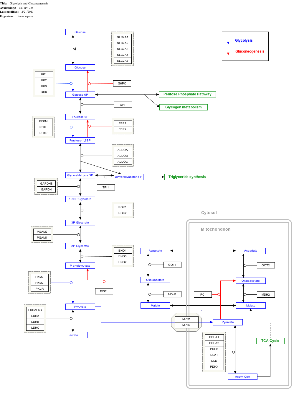
Interactive pathway map

Click on genes, proteins and metabolites below to link to respective articles. [§ 1]

[[File:
WP534.png go to articlego to articlego to articlego to articlego to articlego to articlego to articlego to articlego to articlego to articlego to articlego to articlego to articlego to articlego to articlego to articlego to articlego to articlego to articlego to articlego to articlego to articlego to articlego to articlego to articlego to articlego to articlego to articlego to articlego to articlego to articlego to articlego to articlego to articlego to articlego to articlego to articlego to articlego to articlego to articlego to articlego to articlego to articlego to articlego to articlego to articlego to articlego to articlego to articlego to articlego to articlego to articlego to articlego to articlego to articlego to articlego to articlego to articlego to articlego to articlego to articlego to articlego to articlego to articlego to articlego to articlego to articlego to articlego to articlego to articlego to articlego to article
[[
]]
[[
]]
[[
]]
[[
]]
[[
]]
[[
]]
[[
]]
[[
]]
[[
]]
[[
]]
[[
]]
[[
]]
[[
]]
[[
]]
[[
]]
[[
]]
[[
]]
[[
]]
[[
]]
[[
]]
[[
]]
[[
]]
[[
]]
[[
]]
[[
]]
[[
]]
[[
]]
[[
]]
[[
]]
[[
]]
[[
]]
[[
]]
[[
]]
[[
]]
[[
]]
[[
]]
[[
]]
[[
]]
[[
]]
[[
]]
[[
]]
[[
]]
[[
]]
[[
]]
[[
]]
[[
]]
[[
]]
[[
]]
[[
]]
[[
]]
[[
]]
[[
]]
[[
]]
[[
]]
[[
]]
[[
]]
[[
]]
[[
]]
[[
]]
[[
]]
[[
]]
[[
]]
[[
]]
[[
]]
[[
]]
[[
]]
[[
]]
[[
]]
[[
]]
[[
]]
[[
]]
[[
]]
[[
]]
[[
]]
[[
]]
WP534.png go to articlego to articlego to articlego to articlego to articlego to articlego to articlego to articlego to articlego to articlego to articlego to articlego to articlego to articlego to articlego to articlego to articlego to articlego to articlego to articlego to articlego to articlego to articlego to articlego to articlego to articlego to articlego to articlego to articlego to articlego to articlego to articlego to articlego to articlego to articlego to articlego to articlego to articlego to articlego to articlego to articlego to articlego to articlego to articlego to articlego to articlego to articlego to articlego to articlego to articlego to articlego to articlego to articlego to articlego to articlego to articlego to articlego to articlego to articlego to articlego to articlego to articlego to articlego to articlego to articlego to articlego to articlego to articlego to articlego to articlego to articlego to article
|alt=Glycolysis and Gluconeogenesis edit]]
Glycolysis and Gluconeogenesis edit
  1. The interactive pathway map can be edited at WikiPathways: "GlycolysisGluconeogenesis_WP534".

See also

Related Research Articles

<span class="mw-page-title-main">Insulin</span> Peptide hormone

Insulin is a peptide hormone produced by beta cells of the pancreatic islets encoded in humans by the insulin (INS) gene. It is considered to be the main anabolic hormone of the body. It regulates the metabolism of carbohydrates, fats and protein by promoting the absorption of glucose from the blood into liver, fat and skeletal muscle cells. In these tissues the absorbed glucose is converted into either glycogen via glycogenesis or fats (triglycerides) via lipogenesis, or, in the case of the liver, into both. Glucose production and secretion by the liver is strongly inhibited by high concentrations of insulin in the blood. Circulating insulin also affects the synthesis of proteins in a wide variety of tissues. It is therefore an anabolic hormone, promoting the conversion of small molecules in the blood into large molecules inside the cells. Low insulin levels in the blood have the opposite effect by promoting widespread catabolism, especially of reserve body fat.

<span class="mw-page-title-main">Beta cell</span> Type of cell found in pancreatic islets

Beta cells (β-cells), are specialized endocrine cells located within the pancreatic islets of Langerhans responsible for the production and release of insulin and amylin. Constituting ~50–70% of cells in human islets, beta cells play a vital role in maintaining blood glucose levels. Problems with beta cells can lead to disorders such as diabetes.

<span class="mw-page-title-main">Alpha cell</span> Glucagon secreting cell

Alpha cells(α cells) are endocrine cells that are found in the Islets of Langerhans in the pancreas. Alpha cells secrete the peptide hormone glucagon in order to increase glucose levels in the blood stream.

<span class="mw-page-title-main">Uniporter</span>

Uniporters, also known as solute carriers or facilitated transporters, are a type of membrane transport protein that passively transports solutes across a cell membrane. It uses facilitated diffusion for the movement of solutes down their concentration gradient from an area of high concentration to an area of low concentration. Unlike active transport, it does not require energy in the form of ATP to function. Uniporters are specialized to carry one specific ion or molecule and can be categorized as either channels or carriers. Facilitated diffusion may occur through three mechanisms: uniport, symport, or antiport. The difference between each mechanism depends on the direction of transport, in which uniport is the only transport not coupled to the transport of another solute.

<span class="mw-page-title-main">Glucose-dependent insulinotropic polypeptide</span> Mammalian protein found in Homo sapiens

Glucose-dependent insulinotropic polypeptide, abbreviated as GIP, is an inhibiting hormone of the secretin family of hormones. While it is a weak inhibitor of gastric acid secretion, its main role, being an incretin, is to stimulate insulin secretion.

<span class="mw-page-title-main">Amylin</span> Peptide hormone that plays a role in glycemic regulation

Amylin, or islet amyloid polypeptide (IAPP), is a 37-residue peptide hormone. It is co-secreted with insulin from the pancreatic β-cells in the ratio of approximately 100:1 (insulin:amylin). Amylin plays a role in glycemic regulation by slowing gastric emptying and promoting satiety, thereby preventing post-prandial spikes in blood glucose levels.

<span class="mw-page-title-main">Type 1 diabetes</span> Form of diabetes mellitus

Type 1 diabetes (T1D), formerly known as juvenile diabetes, is an autoimmune disease that originates when cells that make insulin are destroyed by the immune system. Insulin is a hormone required for the cells to use blood sugar for energy and it helps regulate glucose levels in the bloodstream. Before treatment this results in high blood sugar levels in the body. The common symptoms of this elevated blood sugar are frequent urination, increased thirst, increased hunger, weight loss, and other serious complications. Additional symptoms may include blurry vision, tiredness, and slow wound healing. Symptoms typically develop over a short period of time, often a matter of weeks if not months.

<span class="mw-page-title-main">Glucose 6-phosphatase</span> Enzyme

The enzyme glucose 6-phosphatase (EC 3.1.3.9, G6Pase; systematic name D-glucose-6-phosphate phosphohydrolase) catalyzes the hydrolysis of glucose 6-phosphate, resulting in the creation of a phosphate group and free glucose:

<span class="mw-page-title-main">Streptozotocin</span> Chemical compound

Streptozotocin or streptozocin (STZ) is a naturally occurring alkylating antineoplastic agent that is particularly toxic to the insulin-producing beta cells of the pancreas in mammals. It is used in medicine for treating certain cancers of the islets of Langerhans and used in medical research to produce an animal model for hyperglycemia and Alzheimer's in a large dose, as well as type 2 diabetes or type 1 diabetes with multiple low doses.

Glucose transporter type 4 (GLUT4), also known as solute carrier family 2, facilitated glucose transporter member 4, is a protein encoded, in humans, by the SLC2A4 gene. GLUT4 is the insulin-regulated glucose transporter found primarily in adipose tissues and striated muscle. The first evidence for this distinct glucose transport protein was provided by David James in 1988. The gene that encodes GLUT4 was cloned and mapped in 1989.

<span class="mw-page-title-main">Blood sugar regulation</span> Hormones regulating blood sugar levels

Blood sugar regulation is the process by which the levels of blood sugar, the common name for glucose dissolved in blood plasma, are maintained by the body within a narrow range.

<span class="mw-page-title-main">Zinc transporter 8</span> Protein found in humans

Zinc transporter 8 (ZNT8) is a protein that in humans is encoded by the SLC30A8 gene. ZNT8 is a zinc transporter related to insulin secretion in humans. In particular, ZNT8 is critical for the accumulation of zinc into beta cell secretory granules and the maintenance of stored insulin as tightly packaged hexamers. Certain alleles of the SLC30A8 gene may increase the risk for developing type 2 diabetes, but a loss-of-function mutation appears to greatly reduce the risk of diabetes.

GLUT8 also known as SLC2A8 is the eighth member of glucose transporter superfamily.

<span class="mw-page-title-main">PDX1</span> A protein involved in the pancreas and duodenum differentiation

PDX1, also known as insulin promoter factor 1, is a transcription factor in the ParaHox gene cluster. In vertebrates, Pdx1 is necessary for pancreatic development, including β-cell maturation, and duodenal differentiation. In humans this protein is encoded by the PDX1 gene, which was formerly known as IPF1. The gene was originally identified in the clawed frog Xenopus laevis and is present widely across the evolutionary diversity of bilaterian animals, although it has been lost in evolution in arthropods and nematodes. Despite the gene name being Pdx1, there is no Pdx2 gene in most animals; single-copy Pdx1 orthologs have been identified in all mammals. Coelacanth and cartilaginous fish are, so far, the only vertebrates shown to have two Pdx genes, Pdx1 and Pdx2.

<span class="mw-page-title-main">Major facilitator superfamily</span>

The major facilitator superfamily (MFS) is a superfamily of membrane transport proteins that facilitate movement of small solutes across cell membranes in response to chemiosmotic gradients.

<span class="mw-page-title-main">Forkhead box protein O1</span> Protein

Forkhead box protein O1 (FOXO1), also known as forkhead in rhabdomyosarcoma (FKHR), is a protein that in humans is encoded by the FOXO1 gene. FOXO1 is a transcription factor that plays important roles in regulation of gluconeogenesis and glycogenolysis by insulin signaling, and is also central to the decision for a preadipocyte to commit to adipogenesis. It is primarily regulated through phosphorylation on multiple residues; its transcriptional activity is dependent on its phosphorylation state.

The insulin transduction pathway is a biochemical pathway by which insulin increases the uptake of glucose into fat and muscle cells and reduces the synthesis of glucose in the liver and hence is involved in maintaining glucose homeostasis. This pathway is also influenced by fed versus fasting states, stress levels, and a variety of other hormones.

In recent years it has become apparent that the environment and underlying mechanisms affect gene expression and the genome outside of the central dogma of biology. It has been found that many epigenetic mechanisms are involved in the regulation and expression of genes such as DNA methylation and chromatin remodeling. These epigenetic mechanisms are believed to be a contributing factor to pathological diseases such as type 2 diabetes. An understanding of the epigenome of Diabetes patients may help to elucidate otherwise hidden causes of this disease.

Mladen Vranic, MD, DSc, O.C., O.Ont, FRSC, FRCP(C), FCAHS, Canadian Medical Hall of Fame[CMHF] April 3, 1930 – June 18, 2019, was a Croatian-born diabetes researcher, best known for his work in tracer methodology, exercise and stress in diabetes, the metabolic effects of hormonal interactions, glucagon physiology, extrapancreatic glucagon, the role of the direct and indirect metabolic effects of insulin and the prevention of hypoglycemia. Vranic was recognized by a number of national and international awards for his research contributions, mentoring and administration including the Orders of Canada (Officer) and Ontario.

<span class="mw-page-title-main">Alpha-1,3-mannosyl-glycoprotein 4-beta-N-acetylglucosaminyltransferase A</span> Protein-coding gene in the species Homo sapiens

Alpha-1,3-mannosyl-glycoprotein 4-beta-N-acetylglucosaminyltransferase A is a type II membrane protein and an enzyme – particularly a glycosyltransferase – that, in addition to the related isoenzyme B (MGAT4B), takes part in the transfer of N-acetylglucosamine (GlcNAc) to the core mannose residues of N-linked glycans in Golgi apparatus. Therefore, it is essential for the formation of tri- and tetra-antennary sugar chains. Furthermore, it is involved in glucose transport by mediating SLC2A2/GLUT2 glycosylation with controlling cell-surface expression of SLC2A2 in pancreatic beta cells and, as it is suggested, in regulating the availability of serum glycoproteins, oncogenesis, and differentiation.

References

  1. Gwyn W. Gould; Helen M. Thomas; Thomas J. Jess; Graeme I. Bell (May 1991). "Expression of human glucose transporters in Xenopus oocytes: kinetic characterization and substrate specificities of the erythrocyte, liver, and brain isoforms". Biochemistry. 30 (21): 5139–5145. doi:10.1021/bi00235a004. PMID   2036379.
  2. Froguel P, Zouali H, Sun F, Velho G, Fukumoto H, Passa P, Cohen D (July 1991). "CA repeat polymorphism in the glucose transporter GLUT 2 gene". Nucleic Acids Research. 19 (13): 3754. doi:10.1093/nar/19.13.3754-a. PMC   328421 . PMID   1852621.
  3. Uldry M, Thorens B (February 2004). "The SLC2 family of facilitated hexose and polyol transporters" (PDF). Pflügers Archiv. 447 (5): 480–9. doi:10.1007/s00424-003-1085-0. PMID   12750891. S2CID   25539725.
  4. McCulloch LJ, van de Bunt M, Braun M, Frayn KN, CLark A, Gloyn AL (December 2011). "GLUT2 (SLC2A2) is not the principal glucose transporter in human pancreatic beta cells: implications for understanding genetic association signals at this locus". Molecular Genetics and Metabolism. 104 (4): 648–53. doi:10.1016/j.ymgme.2011.08.026. PMID   21920790.
  5. Kellett GL, Brot-Laroche E (October 2005). "Apical GLUT2: a major pathway of intestinal sugar absorption". Diabetes. 54 (10): 3056–62. doi: 10.2337/diabetes.54.10.3056 . PMID   16186415.
  6. Freitas HS, Schaan BD, Seraphim PM, Nunes MT, Machado UF (June 2005). "Acute and short-term insulin-induced molecular adaptations of GLUT2 gene expression in the renal cortex of diabetic rats". Molecular and Cellular Endocrinology. 237 (1–2): 49–57. doi:10.1016/j.mce.2005.03.005. PMID   15869838. S2CID   44856595.
  7. 1 2 3 de Souza Cordeiro, Leticia Maria; Bainbridge, Lauren; Devisetty, Nagavardhini; McDougal, David H.; Peters, Dorien J. M.; Chhabra, Kavaljit H. (2022). "Loss of function of renal Glut2 reverses hyperglycaemia and normalises body weight in mouse models of diabetes and obesity". Diabetologia. 65 (6): 1032–1047. doi:10.1007/s00125-022-05676-8. PMC   9081162 . PMID   35290476.
  8. McCulloch LJ, van de Bunt M, Braun M, Frayn KN, Clark A, Gloyn AL (December 2011). "GLUT2 (SLC2A2) is not the principal glucose transporter in human pancreatic beta cells: implications for understanding genetic association signals at this locus". Molecular Genetics and Metabolism. 104 (4): 648–53. doi:10.1016/j.ymgme.2011.08.026. PMID   21920790.
  9. Guillam MT, Hümmler E, Schaerer E, Yeh JI, Birnbaum MJ, Beermann F, Schmidt A, Dériaz N, Thorens B, Wu JY (November 1997). "Early diabetes and abnormal postnatal pancreatic islet development in mice lacking Glut-2". Nature Genetics. 17 (3): 327–30. doi:10.1038/ng1197-327. PMID   9354799. S2CID   37328600.
  10. Efrat S (November 1997). "Making sense of glucose sensing". Nature Genetics. 17 (3): 249–50. doi:10.1038/ng1197-249. PMID   9354775. S2CID   13219161.
  11. Bathina, Siresha; Faniyan, Tumininu S.; Bainbridge, Lauren; Davis, Autumn; Chhabra, Kavaljit H. (2023). "Normal β-Cell Glut2 Expression Is not Required for Regulating Glucose-Stimulated Insulin Secretion and Systemic Glucose Homeostasis in Mice". Biomolecules. 13 (3): 540. doi: 10.3390/biom13030540 . PMC   10046365 . PMID   36979475.
  12. Uldry M, Ibberson M, Hosokawa M, Thorens B (July 2002). "GLUT2 is a high affinity glucosamine transporter". FEBS Letters. 524 (1–3): 199–203. doi: 10.1016/S0014-5793(02)03058-2 . PMID   12135767. S2CID   40913482.
  13. Santer R, Groth S, Kinner M, Dombrowski A, Berry GT, Brodehl J, Leonard JV, Moses S, Norgren S, Skovby F, Schneppenheim R, Steinmann B, Schaub J (January 2002). "The mutation spectrum of the facilitative glucose transporter gene SLC2A2 (GLUT2) in patients with Fanconi-Bickel syndrome". Human Genetics. 110 (1): 21–9. doi:10.1007/s00439-001-0638-6. PMID   11810292. S2CID   1767168.
  14. Li R, Thorens B, Loeken MR (March 2007). "Expression of the gene encoding the high-Km glucose transporter 2 by the early postimplantation mouse embryo is essential for neural tube defects associated with diabetic embryopathy". Diabetologia. 50 (3): 682–9. doi: 10.1007/s00125-006-0579-7 . PMID   17235524.
  15. Thomson AB, Wild G (March 1997). "Adaptation of intestinal nutrient transport in health and disease. Part I". Digestive Diseases and Sciences. 42 (3): 453–69. doi:10.1023/A:1018807120691. PMID   9073126. S2CID   25371741.
  16. Stolarczyk E, Le Gall M, Even P, Houllier A, Serradas P, Brot-Laroche E, Leturque A (December 2007). Maedler K (ed.). "Loss of sugar detection by GLUT2 affects glucose homeostasis in mice". PLOS ONE. 2 (12): e1288. Bibcode:2007PLoSO...2.1288S. doi: 10.1371/journal.pone.0001288 . PMC   2100167 . PMID   18074013. Open Access logo PLoS transparent.svg
  17. Kim, Yun Hak; Jeong, Dae Cheon; Pak, Kyoungjune; Han, Myoung-Eun; Kim, Ji-Young; Liangwen, Liu; Kim, Hyun Jin; Kim, Tae Woo; Kim, Tae Hwa; Hyun, Dong Woo; Oh, Sae-Ock (2017-09-15). "SLC2A2 (GLUT2) as a novel prognostic factor for hepatocellular carcinoma". Oncotarget. 8 (40): 68381–68392. doi:10.18632/oncotarget.20266. ISSN   1949-2553. PMC   5620264 . PMID   28978124.