Hartnup disease

Last updated
Hartnup disease
Other namesAminoaciduria, Hartnup type
L-tryptophan-skeletal.png
Tryptophan
Specialty Endocrinology   OOjs UI icon edit-ltr-progressive.svg
Hartnup disease has an autosomal recessive pattern of inheritance. Autorecessive.svg
Hartnup disease has an autosomal recessive pattern of inheritance.

Hartnup disease (also known as "pellagra-like dermatosis" [1] and "Hartnup disorder" [2] ) is an autosomal recessive [3] metabolic disorder affecting the absorption of nonpolar amino acids (particularly tryptophan that can be, in turn, converted into serotonin, melatonin, and niacin). Niacin is a precursor to nicotinamide, a necessary component of NAD+. [4] :541

Contents

The causative gene, SLC6A19 , is located on chromosome 5. [5] It is named after the British family, Hartnup, who had this disease.[ citation needed ]

Signs and symptoms

Hartnup disease manifests during infancy with variable clinical presentation: failure to thrive, photosensitivity, intermittent ataxia, nystagmus, and tremor.[ citation needed ]

Nicotinamide is necessary for neutral amino acid transporter production in the proximal renal tubules found in the kidney, and intestinal mucosal cells found in the small intestine. Therefore, a symptom stemming from this disorder results in increased amounts of amino acids in the urine. Pellagra, a similar condition, is also caused by low nicotinamide; this disorder results in dermatitis, diarrhea, and dementia.[ citation needed ]

Hartnup disease is a disorder of amino acid transport in the intestine and kidneys; otherwise, the intestine and kidneys function normally, and the effects of the disease occur mainly in the brain and skin. Symptoms may begin in infancy or early childhood, but sometimes they begin as late as early adulthood. Symptoms may be triggered by sunlight, fever, drugs, or emotional or physical stress. A period of poor nutrition nearly always precedes an attack. The attacks usually become progressively less frequent with age. Most symptoms occur sporadically and are caused by a deficiency of niacinamide. A rash develops on parts of the body exposed to the sun. Mental retardation, short stature, headaches, unsteady gait, and collapsing or fainting are common. Psychiatric problems (such as anxiety, rapid mood changes, delusions, and hallucinations) may also result. [6]

Causes

Hartnup disease is inherited as an autosomal recessive trait. Heterozygotes are normal. Consanguinity is common. The failure of amino-acid transport was reported in 1960 from the increased presence of indoles (bacterial metabolites of tryptophan) and tryptophan in the urine of patients as part of a generalized aminoaciduria of the disease. The excessive loss of tryptophan from malabsorption was the cause of the pellagra like symptoms. From studies on ingestion of tryptophan it seemed that there was a generalized problem with amino-acid transport. [7] In 2004, a causative gene, SLC6A19, was located on band 5p15.33. SLC6A19 is a sodium-dependent and chloride-independent neutral amino acid transporter, expressed predominantly in the kidneys and intestine. [8]

Diagnosis

The defective gene controls the absorption of certain amino acids from the intestine and the reabsorption of those amino acids in the kidneys. Consequently, a person with Hartnup disease cannot absorb amino acids properly from the intestine and cannot reabsorb them properly from tubules in the kidneys. Excessive amounts of amino acids, such as tryptophan, are excreted in the urine. The body is thus left with inadequate amounts of amino acids, which are the building blocks of proteins. With too little tryptophan in the blood, the body is unable to make a sufficient amount of the B-complex vitamin niacinamide, particularly under stress when more vitamins are needed. [6]

In Hartnup disease, urinary excretion of proline, hydroxyproline, and arginine remains unchanged, differentiating it from other causes of generalized aminoaciduria, such as Fanconi syndrome. With urine chromatography, increased levels of neutral amino acids (e.g., glutamine, valine, phenylalanine, leucine, asparagine, citrulline, isoleucine, threonine, alanine, serine, histidine, tyrosine, tryptophan) and indican are found in the urine. Increased urinary Indican can be tested by Obermeyer test.[ citation needed ]

Treatment

A high-protein diet can overcome the deficient transport of neutral amino acids in most patients. Poor nutrition leads to more frequent and more severe attacks of the disease, which is otherwise asymptomatic. All patients who are symptomatic are advised to use physical and chemical protection from sunlight: avoid excessive exposure to sunlight, wear protective clothing, and use chemical sunscreens with a SPF of 15 or greater. Patients also should avoid other aggravating factors, such as photosensitizing drugs, as much as possible. In patients with niacin deficiency and symptomatic disease, daily supplementation with nicotinic acid or nicotinamide reduces both the number and severity of attacks. Neurologic and psychiatric treatment is needed in patients with severe central nervous system involvement. [8]

See also

Related Research Articles

<span class="mw-page-title-main">Niacin</span> Organic compound and a form of vitamin B3

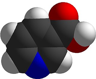
Niacin, also known as nicotinic acid, is an organic compound and a vitamer of vitamin B3, an essential human nutrient. It can be manufactured by plants and animals from the amino acid tryptophan. Niacin is obtained in the diet from a variety of whole and processed foods, with highest contents in fortified packaged foods, meat, poultry, red fish such as tuna and salmon, lesser amounts in nuts, legumes and seeds. Niacin as a dietary supplement is used to treat pellagra, a disease caused by niacin deficiency. Signs and symptoms of pellagra include skin and mouth lesions, anemia, headaches, and tiredness. Many countries mandate its addition to wheat flour or other food grains, thereby reducing the risk of pellagra.

<span class="mw-page-title-main">Pellagra</span> Human disease caused by a lack of vitamin niacin

Pellagra is a disease caused by a lack of the vitamin niacin (vitamin B3). Symptoms include inflamed skin, diarrhea, dementia, and sores in the mouth. Areas of the skin exposed to either sunlight or friction are typically affected first. Over time affected skin may become darker, stiffen, peel, or bleed.

<span class="mw-page-title-main">Cystinuria</span> Amino acid metabolic disorder involving cystine stones forming in the kidneys, ureter, and bladder

Cystinuria is an inherited autosomal recessive disease characterized by high concentrations of the amino acid cystine in the urine, leading to the formation of cystine stones in the kidneys, ureters, and bladder. It is a type of aminoaciduria. "Cystine", not "cysteine," is implicated in this disease; the former is a dimer of the latter.

<span class="mw-page-title-main">Cystinosis</span> Lysosomal storage disease

Cystinosis is a lysosomal storage disease characterized by the abnormal accumulation of cystine, the oxidized dimer of the amino acid cysteine. It is a genetic disorder that follows an autosomal recessive inheritance pattern. It is a rare autosomal recessive disorder resulting from accumulation of free cystine in lysosomes, eventually leading to intracellular crystal formation throughout the body. Cystinosis is the most common cause of Fanconi syndrome in the pediatric age group. Fanconi syndrome occurs when the function of cells in renal tubules is impaired, leading to abnormal amounts of carbohydrates and amino acids in the urine, excessive urination, and low blood levels of potassium and phosphates.

<span class="mw-page-title-main">Gitelman syndrome</span> Medical condition

Gitelman syndrome (GS) is an autosomal recessive kidney tubule disorder characterized by low blood levels of potassium and magnesium, decreased excretion of calcium in the urine, and elevated blood pH. It is the most frequent hereditary salt-losing tubulopathy. Gitelman syndrome is caused by disease-causing variants on both alleles of the SLC12A3 gene. The SLC12A3 gene encodes the thiazide-sensitive sodium-chloride cotransporter, which can be found in the distal convoluted tubule of the kidney.

Glutaric acidemia type 1 (GA1) is an inherited disorder in which the body is unable to completely break down the amino acids lysine, hydroxylysine and tryptophan. Excessive levels of their intermediate breakdown products can accumulate and cause damage to the brain, but particularly the basal ganglia, which are regions that help regulate movement. GA1 causes secondary carnitine deficiency, as glutaric acid, like other organic acids, is detoxified by carnitine. Mental retardation may occur.

<span class="mw-page-title-main">Lysinuric protein intolerance</span> Medical condition

Lysinuric protein intolerance (LPI) is an autosomal recessive metabolic disorder affecting amino acid transport.

<span class="mw-page-title-main">Indican</span> Chemical compound

Indican is a colourless organic compound, soluble in water, naturally occurring in Indigofera plants. It is a precursor of indigo dye.

<span class="mw-page-title-main">Renal tubular acidosis</span> Medical condition

Renal tubular acidosis (RTA) is a medical condition that involves an accumulation of acid in the body due to a failure of the kidneys to appropriately acidify the urine. In renal physiology, when blood is filtered by the kidney, the filtrate passes through the tubules of the nephron, allowing for exchange of salts, acid equivalents, and other solutes before it drains into the bladder as urine. The metabolic acidosis that results from RTA may be caused either by insufficient secretion of hydrogen ions into the latter portions of the nephron or by failure to reabsorb sufficient bicarbonate ions from the filtrate in the early portion of the nephron. Although a metabolic acidosis also occurs in those with chronic kidney disease, the term RTA is reserved for individuals with poor urinary acidification in otherwise well-functioning kidneys. Several different types of RTA exist, which all have different syndromes and different causes. RTA is usually an incidental finding based on routine blood draws that show abnormal results. Clinically, patients may present with vague symptoms such as dehydration, mental status changes, or delayed growth in adolescents.

<span class="mw-page-title-main">Blue diaper syndrome</span> Medical condition

Blue diaper syndrome is a rare, autosomal recessive or X linked recessive metabolic disorder characterized in infants by bluish urine-stained diapers. It is also known as Drummond's syndrome, and hypercalcemia.

<span class="mw-page-title-main">Aminoaciduria</span> Medical condition

Aminoaciduria occurs when the urine contains abnormally high amounts of amino acids. In the healthy kidney, the glomeruli filter all amino acids out of the blood, and the renal tubules then reabsorb over 95% of the filtered amino acids back into the blood.

<span class="mw-page-title-main">Dent's disease</span> Medical condition

Dent's disease is a rare X-linked recessive inherited condition that affects the proximal renal tubules of the kidney. It is one cause of Fanconi syndrome, and is characterized by tubular proteinuria, excess calcium in the urine, formation of calcium kidney stones, nephrocalcinosis, and chronic kidney failure.

b(0,+)-type amino acid transporter 1 Protein-coding gene in the species Homo sapiens

b(0,+)-type amino acid transporter 1, also known as b(0,+)AT1, is a protein which in humans is encoded by the SLC7A9 gene.

<span class="mw-page-title-main">Neutral and basic amino acid transport protein rBAT</span> Protein-coding gene in the species Homo sapiens

Neutral and basic amino acid transport protein rBAT is a protein that in humans is encoded by the SLC3A1 gene.

<span class="mw-page-title-main">Sodium-dependent neutral amino acid transporter B(0)AT1</span> Protein-coding gene in the species Homo sapiens

Sodium-dependent neutral amino acid transporter B(0)AT1 is a protein that in humans is encoded by the SLC6A19 gene.

<span class="mw-page-title-main">Hypertryptophanemia</span> Medical condition

Hypertryptophanemia is a rare autosomal recessive metabolic disorder that results in a massive buildup of the amino acid tryptophan in the blood, with associated symptoms and tryptophanuria.

<span class="mw-page-title-main">Iminoglycinuria</span> Medical condition

Iminoglycinuria is an autosomal recessive disorder of renal tubular transport affecting reabsorption of the amino acid glycine, and the imino acids proline and hydroxyproline. This results in excess urinary excretion of all three acids.
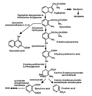
Dicarboxylic aminoaciduria is a rare form of aminoaciduria which is an autosomal recessive disorder of urinary glutamate and aspartate due to genetic errors related to transport of these amino acids. Mutations resulting in a lack of expression of the SLC1A1 gene, a member of the solute carrier family, are found to cause development of dicarboxylic aminoaciduria in humans. SLC1A1 encodes for EAAT3 which is found in the neurons, intestine, kidney, lung, and heart. EAAT3 is part of a family of high affinity glutamate transporters which transport both glutamate and aspartate across the plasma membrane.

Vitamin B<sub>3</sub> Class of chemically related vitamers

Vitamin B3, colloquially referred to as niacin, is a vitamin family that includes three forms, or vitamers: niacin (nicotinic acid), nicotinamide (niacinamide), and nicotinamide riboside. All three forms of vitamin B3 are converted within the body to nicotinamide adenine dinucleotide (NAD). NAD is required for human life and people are unable to make it within their bodies without either vitamin B3 or tryptophan. Nicotinamide riboside was identified as a form of vitamin B3 in 2004.

Proton-coupled amino acid transporters belong to the SLC26A5 family; they are protein receptors whose main function is the transmembrane movement of amino acids and their derivatives. This family of receptors is most commonly found within the luminal surface of the small intestine as well as in some lysosomes. The solute carrier family (SLC) of genes includes roughly 400 membrane proteins that are characterized by 66 families in total. The SLC36 family of genes maps to chromosome 11. The diversity of these receptors is vast, with the ability to transport both charged and uncharged amino acids along with their derivatives. In research and practice, SLC36A1/2 are both targets for drug-based delivery systems for a wide range of disorders.

References

  1. Rapini, Ronald P.; Bolognia, Jean L.; Jorizzo, Joseph L. (2007). Dermatology: 2-Volume Set. St. Louis: Mosby. ISBN   978-1-4160-2999-1.
  2. Online Mendelian Inheritance in Man (OMIM): 234500
  3. Kleta R, Romeo E, Ristic Z, Ohura T, Stuart C, Arcos-Burgos M, Dave MH, Wagner CA, Camargo SR, Inoue S, Matsuura N, Helip-Wooley A, Bockenhauer D, Warth R, Bernardini I, Visser G, Eggermann T, Lee P, Chairoungdua A, Jutabha P, Babu E, Nilwarangkoon S, Anzai N, Kanai Y, Verrey F, Gahl WA, Koizumi A (September 2004). "Mutations in SLC6A19, encoding B0AT1, cause Hartnup disorder". Nature Genetics. 36 (9): 999–1002. doi:10.1038/ng1405. PMID   15286787. S2CID   155361.
  4. James, William D.; Berger, Timothy G.; et al. (2006). Andrews' Diseases of the Skin: clinical Dermatology. Saunders Elsevier. ISBN   978-0-7216-2921-6.
  5. Seow HF, Brer S, Brer A, Bailey CG, Potter SJ, Cavanaugh JA, Rasko JE (September 2004). "Hartnup disorder is caused by mutations in the gene encoding the neutral amino acid transporter SLC6A19". Nature Genetics. 36 (9): 1003–7. doi: 10.1038/ng1406 . PMID   15286788.
  6. 1 2 LaRosa, CJ (January 2020). "Hartnup Disease" . Retrieved 6 July 2020.
  7. Milne, M.D., Crawford, M.A., Girao, C.B. and Loughridge, L. (1961) The metabolic disorder of the Hartnup disease. Q. J. Med. 29: 407-421
  8. 1 2 Sekulovic, LJ (February 2017). "Hartnup Disease" . Retrieved 6 July 2020.