Methylmalonyl-CoA mutase deficiency

Last updated
Methylmalonyl-CoA mutase deficiency
Other namesMCM Deficiency [1]
Methylmalonyl-CoA mutase
Protein MUT PDB 2XIJ.png
Rendering based on PDB 2XIJ .
Identifiers
Symbol MMUT
Alt. symbolsMCM, MUT
NCBI gene 4594
HGNC 7526
OMIM 609058
RefSeq NP_000246
UniProt P22033
Other data
EC number 5.4.99.2
Locus Chr. 6 p21
Search for
Structures Swiss-model
Domains InterPro
methylmalonyl-CoA mutase
Identifiers
EC no. 5.4.99.2
CAS no. 9023-90-9
Databases
IntEnz IntEnz view
BRENDA BRENDA entry
ExPASy NiceZyme view
KEGG KEGG entry
MetaCyc metabolic pathway
PRIAM profile
PDB structures RCSB PDB PDBe PDBsum
Gene Ontology AmiGO / QuickGO
Search
PMC articles
PubMed articles
NCBI proteins

Methylmalonyl-CoA mutase is a mitochondrial homodimer apoenzyme (EC. 5. 4.99.2) that focuses on the catalysis of methylmalonyl CoA to succinyl CoA. The enzyme is bound to adenosylcobalamin, a hormonal derivative of vitamin B12 in order to function. Methylmalonyl-CoA mutase deficiency [2] is caused by genetic defect in the MUT [3] gene responsible for encoding the enzyme. Deficiency in this enzyme accounts for 60% of the cases of methylmalonic acidemia. [4]

Contents

Symptoms

People with methylmalonyl CoA mutase deficiency exhibit many symptoms similar to other diseases involving inborn errors of metabolism.

Newborn babies experience with vomiting, acidosis, hyperammonemia, hepatomegaly (enlarged livers), hyperglycinemia (high glycine levels), and hypoglycemia (low blood sugar). Later, cases of thrombocytopenia and neutropenia can occur.

In some cases intellectual and developmental disabilities, such as autism, were noted with increased frequency in populations with methylmalonyl-CoA mutase deficiency. [5]

Causes

Although methylmalonic acidemia has a variety of causes, both genetic and dietary, methylmalonyl CoA mutase deficiency is an autosomal recessive genetic disorder. Patients with the deficiency either have a complete gene lesion, designated as mut0 or a partial mutation in the form of a frameshift designated as mut-. This frameshift affects the folding of the enzyme rendering its binding domain less effective. [6] Patients with a complete deletion have an inactivation of methylmalonyl CoA mutase and exhibit the most severe symptoms of the deficiency, while patients with a partial mutations have a wide range of symptoms. Over 49 different mutations [7] have been discovered for the MUT gene, yet only two appear in any discernible frequency.

Enzymatic activity

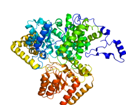
Methylmalonyl-CoA mutase catalyzes the isomerization of methylmalonyl-CoA to succinyl-CoA, and uses a B12 derived prosthetic group, adenosylcobalamin, in order to accomplish this transfer. The enzyme is a homodimer, located in the mitochondrial matrix. The enzyme is 750 amino acids long, with the a metal ligand binding region to bind to the Cobalt region of adenosylcobalamin. [8]

The enzyme works by cleaving the adenosylcobalamin C-Co(III) bond, giving the carbon and cobalt (III) atoms each an electron. The cobalt then fluctuates between its two oxidation states: Co(II) and Co(III). In this way the adenosylcobalamin works as a reversible free radical generator. The cobalt therefore donates an electron back to the methylmalonyl-CoA backbone in order to transfer the coenzyme A group. [8]

MUT.png

Metabolic activity

Methylmalonyl-CoA mutase is essential to the degradation pathways of many molecules including amino acids, and odd-chain fatty acids. Methylmalonyl-CoA mutase links the propionyl-CoA degradation byproduct of these macromolecules to the tricarboxylic acid cycle. [8]

For amino acid metabolism, methylmalonyl-CoA mutase works in the degradation pathways of isoleucine, threonine, valine, and methionine. These amino acids are degraded into propanoyl-CoA which is then further degraded into (S)-methylmalonyl-CoA. This substrate must be further metabolized by a very similar enzyme, methylmalonyl-CoA epimerase, which converts the (S) form of methylmalonyl-CoA into the (R) form. This is finally transformed using methylmalonyl-CoA mutase. L-methionine is also metabolized through a longer superpathway (see Figure 2). After transformation to L-homocystein, it is combined with L-serine to make L-cystathione, which is hydrolyzed by cystathione gamma lyase to create 2-oxobutanoate. This substrate is transformed to propionyl-CoA and undergoes the same metabolism previously described for propionyl-CoA. [9]

The cholesterol superpathway follows the degradation of cholesterol down to various substrates, however only a couple of these biotransformed molecules see propionyl-CoA as a byproduct. The conversion of 3,24-dioxocholest-4-en-26-oyl-CoA to 2-oxochol-4-en-24-oyl-CoA sees the release of a propionyl-CoA molecule. Additionally, the conversion of 3-oxo-23,24-bisnorchol-4-en-17-ol-22-oyl-CoA to androst-4-ene-3,17-dione release of a propionyl-CoA molecule. Finally, the degradation of (S)-4-hydroxy-2-oxohexanoate to pyruvate and propanal, in turn releases a propionyl-CoA substrate after the propanal is converted. All these propionyl-CoA substrates are converted to succinyl-CoA following the methylmalonyl pathway For amino acid metabolism, methylmalonyl-CoA mutase works in the degradation pathways of isoleucine, threonine, valine, and methionine. These amino acids are degraded into propanoyl-CoA which is then further degraded into (S)-methylmalonyl-CoA. This substrate must be further metabolized by a very similar enzyme, methylmalonyl-CoA epimerase, which converts the (S) form of methylmalonyl-CoA into the (R) form. This is finally transformed using methylmalonyl-CoA mutase. L-methionine is also metabolized through a longer superpathway (see Figure 2). After transformation to L-homocystein, it is combined with L-serine to make L-cystathione, which is hydrolyzed by cystathione gamma lyase to create 2-oxobutanoate. This substrate is transformed to propanoyl-CoA and undergoes the same metabolism previously described for propanoyl-CoA. [9]

Odd chain fatty acids are also metabolized through the methylmalonyl pathway. The degradation of odd chain fatty acids releases Acetyl-CoA and propionyl-CoA. Propionyl-CoA is then converted to succinyl-CoA, and both succinyl-CoA and propionyl-CoA are interjected into the tricarboxylic acid cycle for continued production of reductant. [10]

Metabolic pathology

Urea cycle colored. Urea cycle 2.png
Urea cycle colored.

The final product of methylmalonyl-CoA mutase activity is succinyl-CoA which is a tricarboxylic acid cycle substrate. A side effect of excess methylmalonyl-CoA is an interruption of the enzymes responsible for other transformations earlier in the metabolism of propionyl-CoA, leading to propanoic acidemia as well. Excess methylmalonyl-CoA leads to oxidative stress by inhibiting the methylation pathway and formation of glutathione, which is dependent on that pathway. This disrupts the biosynthesis of myelin, urea, and glucose. [11] Specifically, excess methylmalonyl-CoA places oxidative stress on the mitochondrial enzymes involved in the urea cycle (such as ammonia-dependent-carbamoyl-phosphate synthase or CPS1), and inhibits its mechanism of action. [12] The combination of inhibited urea synthesis and poor protein metabolism, as well as a weakly replenished tricarboxylic acid cycle contribute to the symptoms of methylmalonic acidemia.

Diagnosis

Several tests can be done to discover the dysfunction of methylmalonyl-CoA mutase. Ammonia test, blood count, CT scan, MRI scan, electrolyte levels, genetic testing, methylmalonic acid blood test, and blood plasma amino acid tests all can be conducted to determine deficiency.

Treatment

There is no treatment for complete lesion of the mut0 gene, though several treatments can help those with slight genetic dysfunction. Liver and kidney transplants, and a low-protein diet all help regulate the effects of the diseases. [13]

Prognosis

Infant mortality is high for patients diagnosed with early onset; mortality can occur within less than 2 months, while children diagnosed with late-onset syndrome seem to have higher rates of survival. [14] Patients with a complete lesion of mut0 have not only the poorest outcome of those with methylaonyl-CoA mutase deficiency, but also of all individuals with any form of methylmalonic acidemia.

See also

Related Research Articles

<span class="mw-page-title-main">Methionine</span> Sulfur-containing amino acid

Methionine is an essential amino acid in humans.

<span class="mw-page-title-main">Isoleucine</span> Chemical compound

Isoleucine (symbol Ile or I) is an α-amino acid that is used in the biosynthesis of proteins. It contains an α-amino group (which is in the protonated −NH+3 form under biological conditions), an α-carboxylic acid group (which is in the deprotonated −COO form under biological conditions), and a hydrocarbon side chain with a branch (a central carbon atom bound to three other carbon atoms). It is classified as a non-polar, uncharged (at physiological pH), branched-chain, aliphatic amino acid. It is essential in humans, meaning the body cannot synthesize it. Essential amino acids are necessary in our diet. In plants isoleucine can be synthesized from threonine and methionine. In plants and bacteria, isoleucine is synthesized from pyruvate employing leucine biosynthesis enzymes. It is encoded by the codons AUU, AUC, and AUA.

<span class="mw-page-title-main">Methylmalonic acidemia</span> Medical condition

Methylmalonic acidemia, also called methylmalonic aciduria, is an autosomal recessive metabolic disorder that disrupts normal amino acid metabolism. It is a classical type of organic acidemia. The result of this condition is the inability to properly digest specific fats and proteins, which in turn leads to a buildup of a toxic level of methylmalonic acid in the blood.

Propionic acidemia, also known as propionic aciduria or propionyl-CoA carboxylase deficiency, is a rare autosomal recessive metabolic disorder, classified as a branched-chain organic acidemia.

Succinyl-coenzyme A, abbreviated as succinyl-CoA or SucCoA, is a thioester of succinic acid and coenzyme A.

<span class="mw-page-title-main">Methylmalonyl-CoA mutase</span> Mammalian protein found in Homo sapiens

Methylmalonyl-CoA mutase (EC 5.4.99.2, MCM), mitochondrial, also known as methylmalonyl-CoA isomerase, is a protein that in humans is encoded by the MUT gene. This vitamin B12-dependent enzyme catalyzes the isomerization of methylmalonyl-CoA to succinyl-CoA in humans. Mutations in MUT gene may lead to various types of methylmalonic aciduria.

<span class="mw-page-title-main">Malonyl-CoA decarboxylase</span> Class of enzymes

Malonyl-CoA decarboxylase, is found in bacteria and humans and has important roles in regulating fatty acid metabolism and food intake, and it is an attractive target for drug discovery. It is an enzyme associated with Malonyl-CoA decarboxylase deficiency. In humans, it is encoded by the MLYCD gene.

Propionyl-CoA is a coenzyme A derivative of propionic acid. It is composed of a 24 total carbon chain and its production and metabolic fate depend on which organism it is present in. Several different pathways can lead to its production, such as through the catabolism of specific amino acids or the oxidation of odd-chain fatty acids. It later can be broken down by propionyl-CoA carboxylase or through the methylcitrate cycle. In different organisms, however, propionyl-CoA can be sequestered into controlled regions, to alleviate its potential toxicity through accumulation. Genetic deficiencies regarding the production and breakdown of propionyl-CoA also have great clinical and human significance.

<span class="mw-page-title-main">Propionyl-CoA carboxylase</span>

Propionyl-CoA carboxylase (EC 6.4.1.3, PCC) catalyses the carboxylation reaction of propionyl-CoA in the mitochondrial matrix. PCC has been classified both as a ligase and a lyase. The enzyme is biotin-dependent. The product of the reaction is (S)-methylmalonyl CoA.

<span class="mw-page-title-main">Methylmalonyl-CoA</span> Chemical compound

Methylmalonyl-CoA is the thioester consisting of coenzyme A linked to methylmalonic acid. It is an important intermediate in the biosynthesis of succinyl-CoA, which plays an essential role in the tricarboxylic acid cycle. The compound is sometimes referred to as "methylmalyl-CoA".

<span class="mw-page-title-main">Methylmalonyl CoA epimerase</span>

Methylmalonyl CoA epimerase is an enzyme involved in fatty acid catabolism that is encoded in human by the "MCEE" gene located on chromosome 2. It is routinely and incorrectly labeled as "methylmalonyl-CoA racemase". It is not a racemase because the CoA moiety has 5 other stereocenters.

α-Ketobutyric acid Chemical compound

α-Ketobutyric acid is an organic compound with the formula CH3CH2C(O)CO2H. It is a colorless solid that melts just above room temperature. Its conjugate base α-ketobutyrate is the predominant form found in nature (near neutral pH). It results from the lysis of cystathionine. It is also one of the degradation products of threonine, produced by the catabolism of the amino acid by threonine dehydratase. It is also produced by the degradation of homocysteine and the metabolism of methionine.

<span class="mw-page-title-main">Cyanocobalamin</span> Form of vitamin B-12

Cyanocobalamin is a form of vitamin B
12
used to treat and prevent vitamin B
12
deficiency
except in the presence of cyanide toxicity. The deficiency may occur in pernicious anemia, following surgical removal of the stomach, with fish tapeworm, or due to bowel cancer. It is less preferred than hydroxocobalamin for treating vitamin B
12
deficiency. Some study have shown that it has an antihypotensive effect. It is used by mouth, by injection into a muscle, or as a nasal spray.

<span class="mw-page-title-main">MMAB</span> Protein-coding gene in the species Homo sapiens

Cob(I)yrinic acid a,c-diamide adenosyltransferase, mitochondrial is an enzyme that in humans is encoded by the MMAB gene.

<span class="mw-page-title-main">MMAA</span> Protein-coding gene in the species Homo sapiens

Methylmalonic aciduria type A protein, mitochondrial also known as MMAA is a protein that in humans is encoded by the MMAA gene.

<span class="mw-page-title-main">Vitamin B12-binding domain</span> Type of protein domain

In molecular biology, the vitamin B12-binding domain is a protein domain which binds to cobalamin. It can bind two different forms of the cobalamin cofactor, with cobalt bonded either to a methyl group (methylcobalamin) or to 5'-deoxyadenosine (adenosylcobalamin). Cobalamin-binding domains are mainly found in two families of enzymes present in animals and prokaryotes, which perform distinct kinds of reactions at the cobalt-carbon bond. Enzymes that require methylcobalamin carry out methyl transfer reactions. Enzymes that require adenosylcobalamin catalyse reactions in which the first step is the cleavage of adenosylcobalamin to form cob(II)alamin and the 5'-deoxyadenosyl radical, and thus act as radical generators. In both types of enzymes the B12-binding domain uses a histidine to bind the cobalt atom of cobalamin cofactors. This histidine is embedded in a DXHXXG sequence, the most conserved primary sequence motif of the domain. Proteins containing the cobalamin-binding domain include:

<span class="mw-page-title-main">Cobalamin biosynthesis</span>

Cobalamin biosynthesis is the process by which bacteria and archea make cobalamin, vitamin B12. Many steps are involved in converting aminolevulinic acid via uroporphyrinogen III and adenosylcobyric acid to the final forms in which it is used by enzymes in both the producing organisms and other species, including humans who acquire it through their diet.

Odd-chain fatty acids are those fatty acids that contain an odd number of carbon atoms. In addition to being classified according to their saturation or unsaturation, fatty acids are also classified according to their odd or even numbers of constituent carbon atoms. With respect to natural abundance, most fatty acids are even chain, e.g. palmitic (C16) and stearic (C18). In terms of physical properties, odd and even fatty acids are similar, generally being colorless, soluble in alcohols, and often somewhat oily. The odd-chain fatty acids are biosynthesized and metabolized slightly differently from the even-chained relatives. In addition to the usual C12-C22 long chain fatty acids, some very long chain fatty acids (VLCFAs) are also known. Some of these VLCFAs are also of the odd-chain variety.

Combined malonic and methylmalonic aciduria (CMAMMA), also called combined malonic and methylmalonic acidemia is an inherited metabolic disease characterized by elevated levels of malonic acid and methylmalonic acid. Some researchers have hypothesized that CMAMMA might be one of the most common forms of methylmalonic acidemia, and possibly one of the most common inborn errors of metabolism. Due to being infrequently diagnosed, it most often goes undetected.

References

  1. "Methylmalonyl-Coenzyme A mutase deficiency". The Genetic and Rare Diseases Information Center. NIH. Retrieved 19 March 2019.
  2. Online Mendelian Inheritance in Man (OMIM): 251000
  3. "Genebase on MUT".
  4. Online Mendelian Inheritance in Man (OMIM): 251100
  5. Brismar J, Ozand PT (September 1994). "CT and MR of the brain in disorders of the propionate and methylmalonate metabolism". AJNR Am J Neuroradiol. 15 (8): 1459–73. PMC   8334421 . PMID   7985563.
  6. Kennedy DG, Cannavan A, Molloy A, O'Harte F, Taylor SM, Kennedy S, Blanchflower WJ (November 1990). "Methylmalonyl-CoA mutase (EC 5.4.99.2) and methionine synthetase (EC 2.1.1.13) in the tissues of cobalt-vitamin B12 deficient sheep". Br. J. Nutr. 64 (3): 721–32. doi: 10.1079/bjn19900074 . PMID   1979918.
  7. Acquaviva C, Benoist JF, Callebaut I, Guffon N, Ogier de Baulny H, Touati G, Aydin A, Porquet D, Elion J (August 2001). "N219Y, a new frequent mutation among mut(degree) forms of methylmalonic acidemia in Caucasian patients". Eur. J. Hum. Genet. 9 (8): 577–82. doi: 10.1038/sj.ejhg.5200675 . PMID   11528502.
  8. 1 2 3 Ledley FD, Rosenblatt DS (1997). "Mutations in mut methylmalonic acidemia: clinical and enzymatic correlations". Hum. Mutat. 9 (1): 1–6. doi:10.1002/(SICI)1098-1004(1997)9:1<1::AID-HUMU1>3.0.CO;2-E. PMID   8990001. S2CID   41661834.
  9. 1 2 Berg JM, Tymoczko JL, Stryer L (2002). "Section 22.3: Certain Fatty Acids Require Additional Steps for Degradation". Biochemistry (5th ed.).
  10. Jansen R, Kalousek F, Fenton WA, Rosenberg LE, Ledley FD (February 1989). "Cloning of full-length methylmalonyl-CoA mutase from a cDNA library using the polymerase chain reaction". Genomics. 4 (2): 198–205. doi:10.1016/0888-7543(89)90300-5. PMID   2567699.
  11. Yudkoff M, Siegel GJ, Agranoff BW, Albers RW (1999). "Organic Acid Metabolism". Basic Neurochemistry: Molecular, Cellular, and Medical Aspects (6th ed.).
  12. Hudak ML, Jones MD, Brusilow SW (November 1985). "Differentiation of transient hyperammonemia of the newborn and urea cycle enzyme defects by clinical presentation". J. Pediatr. 107 (5): 712–9. doi:10.1016/s0022-3476(85)80398-x. PMID   4056969.
  13. "Medline infomatics".[ dead link ]
  14. Kaplan P, Ficicioglu C, Mazur AT, Palmieri MJ, Berry GT (August 2006). "Liver transplantation is not curative for methylmalonic acidopathy caused by methylmalonyl-CoA mutase deficiency". Mol. Genet. Metab. 88 (4): 322–6. doi:10.1016/j.ymgme.2006.04.003. PMID   16750411.