Dermotherium

Last updated

Dermotherium
Temporal range: Late Eocene – Late Oligocene
Scientific classification OOjs UI icon edit-ltr.svg
Domain: Eukaryota
Kingdom: Animalia
Phylum: Chordata
Class: Mammalia
Order: Dermoptera
Family: Cynocephalidae
Genus: Dermotherium
Ducrocq et al., 1992
Type species
Dermotherium major
Ducrocq et al., 1992
other species
  • Dermotherium chimaeraMarivaux et al., 2006

Dermotherium is a genus of fossil mammals closely related to the living colugos, a small group of gliding mammals from Southeast Asia. Two species are recognized: D. major from the Late Eocene of Thailand, based on a single fragment of the lower jaw, and D. chimaera from the Late Oligocene of Thailand, known from three fragments of the lower jaw and two isolated upper molars. In addition, a single isolated upper molar from the Early Oligocene of Pakistan has been tentatively assigned to D. chimaera. All sites where fossils of Dermotherium have been found were probably forested environments and the fossil species were probably forest dwellers like living colugos, but whether they had the gliding adaptations of the living species is unknown.

Contents

Some features of the teeth differentiate Dermotherium from both living colugo species, but other features are shared with only one of the two. The third lower incisor, lower canine, and third lower premolar at least are pectinate or comblike, bearing longitudinal rows of tines or cusps, an unusual feature of colugos (the first two lower incisors are unknown in Dermotherium). The fourth lower premolar instead resembles the lower molars. The front part of these teeth, the trigonid, is broader in D. chimaera than in D. major, which is known only from the second and third lower molars. The two species also differ in the configuration of the inner back corner of the lower molars. The upper molars are triangular teeth bearing several distinct small cusps, particularly on the second upper molar, and with wrinkled enamel.

Taxonomy

A Sunda colugo (Galeopterus variegatus), a living relative of Dermotherium Colugo (Galeopterus variegatus, adult female), Central Catchment Area, Singapore - 20060618.jpg
A Sunda colugo (Galeopterus variegatus), a living relative of Dermotherium

Colugos are a small group of Southeast Asian gliding mammals closely related to the primates. [1] Their fossil record is exceptionally poor. Although Paleogene groups such as the Plagiomenidae are considered by some to be closely related to colugos, no fossils undoubtedly referable to the living colugo family, Cynocephalidae, had been reported until 1992. [2] In that year, Stéphane Ducrocq and colleagues described a jaw fragment from the Eocene of Thailand as a new genus and species of colugo, Dermotherium major. [3] In 2000, however, Brian Stafford and Frederick Szalay argued that Dermotherium might not be a colugo, since the fossil is so poorly preserved that few traits can be unambiguously recognized and some purported dermopteran features of the fossil are also seen in other placental groups. [4] Mary Silcox and colleagues reaffirmed the colugo affinities of Dermotherium in 2005 on the basis of detailed similarities in molar morphology. [5]

In 2006, Laurent Marivaux and colleagues described a second species of Dermotherium, D. chimaera, from material from the Oligocene of Thailand. They gave it the specific name chimaera (Latin for "chimera") because it shares characters with both the Philippine colugo (Cynocephalus volans) and Sunda colugo (Galeopterus variegatus), the two living colugo species. [6] In addition, they tentatively identified a fossil from the Oligocene of Pakistan as Dermotherium chimaera and regarded some fossils from the Eocene of Myanmar as indeterminate dermopterans. [7] According to a phylogenetic analysis carried out by Marivaux and colleagues, D. chimaera, D. major, and the Myanmar dermopteran are successive sister groups of the two living colugos. [8]

Description

Known material of Dermotherium includes a handful of jaw fragments and isolated teeth. Dermotherium major is known only from a fragment of the left lower jaw bearing the third lower molar (m3) and a poorly preserved second lower molar (m2). [9] The holotype of Dermotherium chimaera is a lower jaw fragment in which remnants of the deciduous third lower premolar are visible. X-ray microtomography reveals the unerupted lower third incisor (i3), canine (c1), third premolar (p3), and fourth premolar (p4) still inside the jaw. In addition, this species is known from two other jaw fragments, one bearing m1 and m2 and the other bearing m2 and m3, and two isolated molars, an upper first and second molar (M1 and M2). [10] The tentatively referred material of this species from Pakistan includes a single M2. [11]

The two species of Dermotherium were about as large as the Philippine colugo and larger than the Sunda colugo and differed from both in several characteristics of the dentition. [12] Not enough is known of the skeleton of Dermotherium to assess whether the animal already possessed the gliding adaptations of living colugos. [13] The two species are similar in size, but again differ in details of the dentition. [14] In two specimens of D. chimaera, the length and width of the m2 are 5 and 4.3 mm (0.20 and 0.17 in) and 5.4 and 4.8 mm (0.21 and 0.19 in) respectively; this tooth is 5.4 mm (0.21 in) long and 4.9 mm (0.19 in) wide in the only known specimen of Dermotherium major. The Pakistani M2 of Dermotherium is 4.3 mm (0.17 in) long and 6 mm (0.24 in) wide, compared to 4.7 and 6.6 mm (0.19 and 0.26 in) in the only known M2 of D. chimaera from Thailand. [15]

The lower jaw of Dermotherium major resembles that of living colugos in the presence of a strong angular process (a projecting piece of bone at the lower side of the back of the jawbone) and a retromolar space (a flat space behind the last molars). The coronoid process (a projecting piece of bone directly behind the molars) rises steeply, with its front wall virtually vertical. [9]

Lower dentition

The i3 of Dermotherium chimaera is an elongate tooth bearing six tines (narrow, high "fingers" as in a comb) arranged from front to back. The frontmost is larger, the next four are about equal in size, and the sixth is smaller. [16] The number of tines resembles that seen in the Sunda colugo, which has four to seven; the Philippine colugo has three to five. [17] The c1 is also an elongate, narrow tooth; at the front, it is slightly curved towards the midline of the jaw. On the buccal (outer) side of this tooth are six cusps, of which the third (counting from the front) is the largest. [18] The p3 is similarly elongate and rounded at the front, but it is broader at the back, forming a talonid (a "heel" of cusps at the back of a tribosphenic tooth). There are six cusps on the narrow anterior part of the tooth, and the fifth (identified as the protoconid) is by far the largest. On the buccal side of the talonid is a strong cusp, the hypoconid, with a crest, the cristid obliqua, descending from it towards the front. A second, smaller cusp, the hypoconulid, is present on the lingual side of the talonid, connected to the hypoconid by a postcristid. [19] The pectinate (comb-like) shape of the anterior teeth is a shared characteristic of the colugos and highly unusual among mammals. [20] Dermotherium chimaera resembles the Sunda colugo in that the c1 and p3 are also pectinate; in the Philippine colugo, these teeth are not pectinate. [21]

In Dermotherium chimaera, the p4 and m1 through m3 are similar to each other (and unlike the i3, canine, and p3) and appear to form a series of decreasing size from front to back. [21] In D. major, only m2 and m3 are known, but they are similar in morphology and m3 is smaller than m2. [22] In the Sunda colugo, however, the teeth get larger from p4 to m3. [11] The p4 and molars have a distinct trigonid (a triangular group of cusps at the front of a tribosphenic tooth) and talonid. The trigonid contains strong protoconid and metaconid cusps. The metaconid is stronger than the protoconid in the molars, but it is not clear whether this is the case in the p4. [23] In the living colugos, the protoconid is higher than the metaconid in both p4 and m1. [11] The trigonid is longer in the p4 than in the molars. [21] In D. chimaera, the trigonid is broader than in D. major. [6] A low crest, the paracristid, descends from the protoconid lingually and towards the front, forming the front margin of the trigonid; there is no distinct cusp (a paraconid) at the front of the trigonid on the p4, but this cusp is present in the molars. [23]

The talonid contains a hypoconid, hypoconulid, and entoconid and is much wider than the trigonid because the hypoconid is displaced buccally. A cristid obliqua descends from the hypoconid and reaches the protoconid. [23] Although a crest, the postmetacristid, descends from the back side of the metaconid, ending in a small cusp, the metastylid, it is separated from the entoconid by a notch. The living colugos lack such a strong postmetacristid. [24] The hypoconulid is near the back lingual margin of the tooth, behind the entoconid. In D. major, this cusp is further to the back than in both D. chimaera and the Sunda colugo, while the two cusps are merged in the Philippine colugo. [11] In D. chimaera, a low crest, the post-hypoconulid cristid, reaches from the hypoconulid to the back lingual corner of the tooth, where a small cuspule, the distocuspid, is located. A long crest, the postcristid, connects the distocuspid to the hypoconid along the posterior side of the tooth. [21] The Philippine colugo is similar, but in D. major, both the distocuspid and the post-hypoconulid cristid are absent, and the Sunda colugo has a weaker distocuspid and a postcristid that does not reach further lingually than the hypoconulid. [11]

Upper dentition

The upper molars of Dermotherium chimaera are triangular in overall shape and much broader than long, with the narrow end of the triangle pointing lingually. [25] The M2 from Pakistan that was tentatively placed in D. chimaera is slightly smaller than Thai fossils of the species, but otherwise very similar. [26] The crest in front of and behind the major cusps on the buccal side of the tooth, the paracone and metacone, are well-developed, together forming a long W-shaped ridge. [25] D. chimaera resembles the Philippine colugo in that the crests behind the paracone and in front of the metacone form an acute angle with each other, so that together they form a V; their shape rather resembles a U in the Sunda colugo. [11] A cingulum (shelf) is present on the buccal margin of the tooth, [27] but this cingulum is rather weak in the Pakistani fossil. [26] On the M2, smaller cusps, the paraconule and metaconule, are present on the lingual sides of the paracone and metacone, but on the M1 the paraconule is missing and the metaconule is small and ridgelike. [28] The small cusps are better developed in the Pakistani M2. [26] A third major cusp, the protocone, is present on the lingual side of both upper molars. [29] This crest is displaced towards the front in the living colugos, but less so in D. chimaera. [11] Two strong crests descend from the front and back faces of the protocone in a buccal direction. These crests end in small cusps (protoconules) that are directly lingual to the paracone and metacone. [29] The living colugos lack these protoconules. [11] The enamel is wrinkled on the flanks of the paracone and metacone and, in the Thai but not the Pakistani specimens, on the lingual side of the protocone. [30] Enamel wrinkling is also seen in some Philippine colugos. [27]

Range and ecology

Dermotherium major was found in a lignite pit known as Wai Lek in Krabi Province, southwestern Thailand. [31] It is part of the Krabi Basin fauna, which contains at least 40 mammalian genera, mostly artiodactyls but also including some primates, such as Siamopithecus , Wailekia , and Muangthanhinius . [32] This fauna has been dated to about 33 to 35 million years ago, during the Late Eocene. [33]

Fossils of Dermotherium chimaera come from Phetchaburi Province, further to the north in Thailand. The fossil deposits, located in the Cha Prong pit in a coal mine called Nong Ya Plong, are dated to the Late Oligocene. Other mammals found here include the rodent Fallomus ladakhensis , the rhinoceros Diceratherium sp. cf. D. lamilloquense, the carnivoran Chaprongictis phetchaburiensis , the lipotyphlan Siamosorex debonisi , and a pteropodid bat. [34]

Paali Nala, where the M2 tentatively assigned to Dermotherium chimaera was found, is in the Bugti Hills of Balochistan, western Pakistan. The fossil locality, placed stratigraphically in the Bugti Member of the Chitarwata Formation, is thought to be Early Oligocene in age. [11] The Paali Nala fauna also contains a diverse array of mammals, including many rodents, some primates, and rhinos. [35]

The specializations of dermopterans are such that they are dependent on a forested habitat, [36] and reconstructions of the paleoenvironments at Krabi, Nong Ya Plong, and Paali Nala suggest that all three fossil deposits developed in a humid, tropical forest environment. [37] Their extinction in the Indian subcontinent may have been caused by the development of a drier climate there during the Late Oligocene. [38]

Related Research Articles

<span class="mw-page-title-main">Molar (tooth)</span> Large tooth at the back of the mouth

The molars or molar teeth are large, flat teeth at the back of the mouth. They are more developed in mammals. They are used primarily to grind food during chewing. The name molar derives from Latin, molaris dens, meaning "millstone tooth", from mola, millstone and dens, tooth. Molars show a great deal of diversity in size and shape across mammal groups. The third molar of humans is sometimes vestigial.

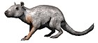
Ferugliotherium is a genus of fossil mammals in the family Ferugliotheriidae from the Campanian and/or Maastrichtian period of Argentina. It contains a single species, Ferugliotherium windhauseni, which was first described in 1986. Although originally interpreted on the basis of a single brachydont (low-crowned) molar as a member of Multituberculata, an extinct group of small, rodent-like mammals, it was recognized as related to the hypsodont (high-crowned) Sudamericidae following the discovery of additional material in the early 1990s. After a jaw of the sudamericid Sudamerica was described in 1999, these animals were no longer considered to be multituberculates and a few fossils that were previously considered to be Ferugliotherium were assigned to unspecified multituberculates instead. Since 2005, a relationship between gondwanatheres and multituberculates has again received support. A closely related animal, Trapalcotherium, was described in 2009 on the basis of a single tooth.

<span class="mw-page-title-main">Colugo</span> Family of mammals

Colugos are arboreal gliding mammals that are native to Southeast Asia. Their closest evolutionary relatives are primates. There are just two living species of colugos: the Sunda flying lemur and the Philippine flying lemur. These two species make up the entire family Cynocephalidae and order Dermoptera.

Tingamainus is a species of early placenta bearing mammal.

<i>Ambondro mahabo</i> Species of small mammal from the middle Jurassic of Madagascar

Ambondro mahabo is a mammal from the Middle Jurassic (Bathonian) Isalo III Formation of Madagascar. The only described species of the genus Ambondro, it is known from a fragmentary lower jaw with three teeth, interpreted as the last premolar and the first two molars. The premolar consists of a central cusp with one or two smaller cusps and a cingulum (shelf) on the inner, or lingual, side of the tooth. The molars also have such a lingual cingulum. They consist of two groups of cusps: a trigonid of three cusps at the front and a talonid with a main cusp, a smaller cusp, and a crest at the back. Features of the talonid suggest that Ambondro had tribosphenic molars, the basic arrangement of molar features also present in marsupial and placental mammals. It is the oldest known mammal with putatively tribosphenic teeth; at the time of its discovery it antedated the second oldest example by about 25 million years.

Many different terms have been proposed for features of the tooth crown in mammals.

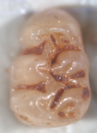
UA 8699 is a fossil mammalian tooth from the Cretaceous of Madagascar. A broken lower molar about 3.5 mm (0.14 in) long, it is from the Maastrichtian of the Maevarano Formation in northwestern Madagascar. Details of its crown morphology indicate that it is a boreosphenidan, a member of the group that includes living marsupials and placental mammals. David W. Krause, who first described the tooth in 2001, interpreted it as a marsupial on the basis of five shared characters, but in 2003 Averianov and others noted that all those are shared by zhelestid placentals and favored a close relationship between UA 8699 and the Spanish zhelestid Lainodon. Krause used the tooth as evidence that marsupials were present on the southern continents (Gondwana) as early as the late Cretaceous and Averianov and colleagues proposed that the tooth represented another example of faunal exchange between Africa and Europe at the time.

<i>Brachytarsomys mahajambaensis</i> Extinct species of rodent

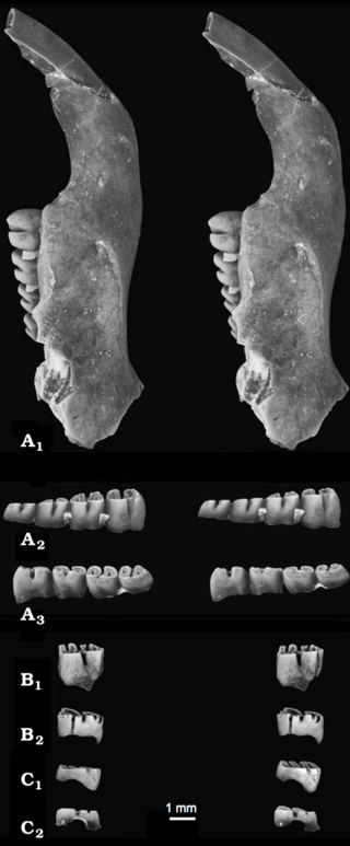
Brachytarsomys mahajambaensis is an extinct rodent from northwestern Madagascar. It is known from nine isolated molars found in several sites during fieldwork that started in 2001. First described in 2010, it is placed in the genus Brachytarsomys together with two larger living species, which may differ in some details of molar morphology. The presence of B. mahajambaensis, a rare element in the local rodent fauna, suggests that the region was previously more humid.

<i>Nesomys narindaensis</i> Extinct species of rodent

Nesomys narindaensis is an extinct rodent that lived in northwestern Madagascar. It is known from subfossil skull bones and isolated molars found in several sites during field work that started in 2001. First described in 2010, it is placed in the genus Nesomys together with three smaller living species, which may differ in some details of molar morphology. The presence of N. narindaensis, a rare element in the local rodent fauna, suggests that the region was previously more humid.

<i>Hipposideros besaoka</i> Extinct species of bat

Hipposideros besaoka is an extinct bat from Madagascar in the genus Hipposideros. It is known from numerous jaws and teeth, which were collected in a cave at Anjohibe in 1996 and described as a new species in 2007. The site where H. besaoka was found is at most 10,000 years old; other parts of the cave have yielded H. commersoni, a living species of Hipposideros from Madagascar, and some material that is distinct from both species. H. besaoka was larger than H. commersoni, making it the largest insectivorous bat of Madagascar, and had broader molars and a more robust lower jaw. As usual in Hipposideros, the second upper premolar is small and displaced from the toothrow, and the second lower premolar is large.

<i>Triaenops goodmani</i> Species of mammal

Triaenops goodmani is an extinct bat from Madagascar in the genus Triaenops. It is known from three lower jaws collected in a cave at Anjohibe in 1996, and described as a new species in 2007. The material is at most 10,000 years old. A bat humerus from the same site could not be identified as either T. goodmani or the living T. menamena. T. goodmani is identifiable as a member of Triaenops or the related genus Paratriaenops by a number of features of the teeth, such as the single-cusped, canine-like fourth premolar and the presence of a gap between the entoconid and hypoconulid cusps on the first two molars. T. goodmani is larger than the living species of Triaenops and Paratriaenops on Madagascar, and on the first molar the protoconid cusp is only slightly higher than the hypoconid, not much higher as in the other species.

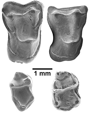
Lagrivea is a fossil genus of squirrel from the Middle Miocene of France. The single species, L. vireti, is known from three mandibles and two isolated teeth. All come from the fissure filling of La Grive L5, part of the La Grive-Saint-Alban complex in Saint-Alban-de-Roche, southeastern France. Lagrivea was a large tree squirrel with flat lower incisors and a large, triangular fourth lower premolar (p4). Each of the four cheekteeth bears a deep basin in the middle of the crown. The m3 is about rectangular in shape, but rounded at the back. Although m1 and m2 have two roots, m3 has three.

<i>Agathaeromys</i> Extinct genus of rodents

Agathaeromys is an extinct genus of oryzomyine rodents from the Pleistocene of Bonaire, Netherlands Antilles. Two species are known, which differ in size and some details of tooth morphology. The larger A. donovani, the type species, is known from hundreds of teeth that are probably 900,000 to 540,000 years old, found in four localities. A. praeuniversitatis, the smaller species, is known from 35 teeth found in a single fossil site, which is probably 540,000 to 230,000 years old.

<i>Pennatomys</i> Extinct oryzomyine rodent in the Lesser Antilles

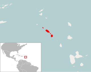
Pennatomys nivalis is an extinct oryzomyine rodent from the islands of Sint Eustatius, Saint Kitts, and Nevis in the Lesser Antilles. The only species in the genus Pennatomys, it is known from skeletal remains found in Amerindian archeological sites on all three islands, with dates ranging from 790–520 BCE to 900–1200 CE. No live specimens are known, but there are several historical records of rodents from Saint Kitts and Nevis that could conceivably refer to Pennatomys. The animal apparently belongs to a group within the tribe Oryzomyini that includes many other island-dwelling species.

Donodontidae is an extinct family of cladotherian mammals known from the Late Jurassic and Early Cretaceous of North Africa. When originally named in 1991, Donodontidae was a monotypic family containing a single species: Donodon perscriptoris. In 2022, four more species were designated and placed within the family: Donodon minor, Stylodens amerrukensis, Anoualestes incidens, and Amazighodon orbis. All five species are endemic to the Ksar Metlili Formation of Morocco, which is dated to the Tithonian and Berriasian. Donodontid fossils are restricted to postcanine teeth and associated jaw fragments.

<i>Afrasia djijidae</i> Eocene fossil primate from Myanmar

Afrasia djijidae is a fossil primate that lived in Myanmar approximately 37 million years ago, during the late middle Eocene. The only species in the genus Afrasia, it was a small primate, estimated to weigh around 100 grams (3.5 oz). Despite the significant geographic distance between them, Afrasia is thought to be closely related to Afrotarsius, an enigmatic fossil found in Libya and Egypt that dates to 38–39 million years ago. If this relationship is correct, it suggests that early simians dispersed from Asia to Africa during the middle Eocene and would add further support to the hypothesis that the first simians evolved in Asia, not Africa. Neither Afrasia nor Afrotarsius, which together form the family Afrotarsiidae, is considered ancestral to living simians, but they are part of a side branch or stem group known as eosimiiforms. Because they did not give rise to the stem simians that are known from the same deposits in Africa, early Asian simians are thought to have dispersed from Asia to Africa more than once prior to the late middle Eocene. Such dispersals from Asia to Africa also were seen around the same time in other mammalian groups, including hystricognathous rodents and anthracotheres.

Indraloris is a fossil primate from the Miocene of India and Pakistan in the family Sivaladapidae. Two species are now recognized: I. himalayensis from Haritalyangar, India and I. kamlialensis from the Pothohar Plateau, Pakistan. Other material from the Potwar Plateau may represent an additional, unnamed species. Body mass estimates range from about 2 kg (4.4 lb) for the smaller I. kamlialensis to over 4 kg (8.8 lb) for the larger I. himalayensis.

<i>Apeomyoides</i> Extinct genus of rodents

Apeomyoides savagei is a fossil rodent from the Miocene of the United States, the only species in the genus Apeomyoides. It is known from fragmentary jaws and isolated teeth from a site in the early Barstovian, around 15–16 million years ago, of Nevada. Together with other species from scattered localities in the United States, Japan, and Europe, Apeomyoides is classified in the subfamily Apeomyinae of the extinct rodent family Eomyidae. Apeomyines are a rare but widespread group that may have been adapted to a relatively dry habitat.

Sivaladapis is a genus of adapiform primate that lived in Asia during the middle Miocene.

Exiguodon is an extinct genus of hyainailourid hyaenodont mammal of the subfamily Hyainailourinae. Remains are known from early Miocene deposits in Kenya and Uganda, in East Africa.

References

  1. Stafford 2005; Marivaux et al. 2006, p. 396; Janečka et al. 2007.
  2. Marivaux et al. 2006, p. 396.
  3. Ducrocq et al. 1992, pp. 373–374.
  4. Stafford & Szalay 2000, p. 378.
  5. Silcox et al. 2005, p. 131.
  6. 1 2 Marivaux et al. 2006, p. 400.
  7. Marivaux et al. 2006, pp. 405–406.
  8. Marivaux et al. 2006, fig. 11.
  9. 1 2 Ducrocq et al. 1992, pp. 374–375.
  10. Marivaux et al. 2006, p. 400, fig. 3.
  11. 1 2 3 4 5 6 7 8 9 Marivaux et al. 2006, p. 405.
  12. Marivaux et al. 2006, pp. 398–399, 404.
  13. Marivaux et al. 2006, pp. 414–415.
  14. Marivaux et al. 2006, p. 400, table 1.
  15. Marivaux et al. 2006, table 1.
  16. Marivaux et al. 2006, p. 400, fig. 4.
  17. Marivaux et al. 2006, pp. 404–405.
  18. Marivaux et al. 2006, pp. 400–401, fig. 4.
  19. Marivaux et al. 2006, pp. 401–402, fig. 4.
  20. Ungar 2010, p. 190.
  21. 1 2 3 4 Marivaux et al. 2006, p. 403.
  22. Ducrocq et al. 1992, p. 375.
  23. 1 2 3 Marivaux et al. 2006, pp. 402–403, figs. 4, 5.
  24. Marivaux et al. 2006, pp. 403, 405.
  25. 1 2 Marivaux et al. 2006, p. 403, fig. 6.
  26. 1 2 3 Marivaux et al. 2006, p. 406.
  27. 1 2 Marivaux et al. 2006, p. 404.
  28. Marivaux et al. 2006, pp. 403–404, fig. 6.
  29. 1 2 Marivaux et al. 2006, p. 404, fig. 6.
  30. Marivaux et al. 2006, pp. 404, 406.
  31. Ducrocq et al. 1992, p. 373.
  32. Peigné et al. 2009, p. 991; Benammi et al. 2001, pp. 266–267; Marivaux et al. 2006, p. 425.
  33. Benammi et al. 2001, p. 265; Marivaux et al. 2006, p. 426.
  34. Marivaux et al. 2006, pp. 400, 414; Peigné et al. 2009, pp. 975–976.
  35. Welcomme et al. 2001, p. 398.
  36. Marivaux et al. 2006, pp. 412–413.
  37. Marivaux et al. 2006, p. 414; Marivaux et al. 2006, p. 426; Ducrocq et al. 1995, p. 67.
  38. Marivaux et al. 2006, p. 414.

Literature cited