Exophiala pisciphila

Last updated

Exophiala pisciphila
Scientific classification OOjs UI icon edit-ltr.svg
Domain: Eukaryota
Kingdom: Fungi
Division: Ascomycota
Class: Eurotiomycetes
Order: Chaetothyriales
Family: Herpotrichiellaceae
Genus: Exophiala
Species:
E. pisciphila
Binomial name
Exophiala pisciphila
McGinnis & Ajello (1974)

Exophiala pisciphila is a mesophilic black yeast and member of the dark septate endophytes. This saprotrophic fungus is found commonly in marine and soil environments. It is abundant in harsh environments like soil contaminated with heavy metals. E. pisciphila forms symbiotic relationships with various plants by colonizing on roots, conferring resistance to drought and heavy metal stress. It is an opportunistic pathogen that commonly causes infections in captive fish and amphibians, while rarely causing disease in humans. Secondary metabolites produced by this species have potential clinical antibiotic and antiretroviral applications.

Contents

History and taxonomy

In 1969, Nikola Fijan first described a systemic mycosis outbreak in channel catfish from a pond in Alabama and identified it as Exophiala salmonis. [1] In 1974, Michael McGinnis and Libero Ajello reevaluated the fungus and identified it as a new species Exophiala pisciphila. [2] The specific epithet pisciphila is a linguistic barbarism, combining the Latin word piscis meaning "fish" with the Greek suffix -philos (φίλος) meaning "loving". [3]

Habitat and ecology

Exophiala pisciphila is commonly found in soil, [4] plants [5] and water [6] in North America, Netherlands, United Kingdom, and Australia. [7] E. pisciphila occurs as a colonist or pathogen in cold-blooded vertebrates such as various commercially cultivated fish and amphibians. [8] It has low host specificity. [8] Captive fish are especially susceptible due to the confined space of aquariums and accumulation of fungal particles. [9] Decorative pieces, stones or contaminated food in aquariums can all be reservoirs of E. pisciphila. [9] This fungus has a high tolerance to certain metals ions and has been encountered in harsh environments such as heavy metal polluted soils. [10] When this fungus colonizes plant roots, it enhances plant tolerance to heavy metal ions. [11] Symbiotic relationships with host plants also allow for improved growth performance and plant survival rate in drought conditions. [12] [13]

Growth and morphology

Exophiala pisciphila is an exclusively asexual fungus that exhibits both filamentous and yeast-like growth. [14] Due to its variable growth forms and the dark pigmentation of its cell walls, it is considered a member of the descriptive grouping of similar fungi known as the black yeasts. [14] E. pisciphila forms slow growing colonies approximately 20–35 millimetres (0.79–1.38 in) in size which is similar to other species in the genus, E. salmonis and E. brunnea. [2] The texture of the colony is dry and fluffy due to the formation on aerial hyphae in mature colonies. [2] The upper surface is grey to green black in colour while the reverse surface tends to be black. [8]

Growth occurs on various media including malt extract agar (MA), oatmeal agar (OA), Sabourand's dextrose agar (SA), corn meal agar (CMA), Czapeck's solution agar, potato dextrose agar (PDA) and nutrient agar (NA). [15] Optimal growth occurs on PDA and MA with the most aerial hyphae forming dome shaped colonies. [14] [15] Media interpreted to be associated with less optimal growth result in the formation of flat colonies. [15] A distinguishing feature of this fungus from others in the genus is its ability to grow on L-arabinitol. [8]

Ideal growth conditions for E. pisciphila occur between 20–30 °C (68–86 °F), where maximum growth occurs at 37 °C (99 °F). [14] [2] This differentiates it from E. jeanselmei which has similar physiology otherwise. [14]

Reproduction for this species occurs asexually by conidiation which was observed to occur through various means in developing colonies. [8] The conidia are produced either by (1) pre-existing conidia, (2) mature hyphae or (3) the differentiation of the cell into a specialized conidium-producing cell called an annellide. [15] E. pisciphila have smooth-walled conidia with yellow-brown walls that characteristically differentiate into annelides. [4] Annelides are bottle-shaped cells that give rise to conidia from a point at the tip of the bottle-neck, as it were. In this way, annelides are similar to phialides but differ in that their necks incrementally elongate as each successive conidium is borne. The cell walls of this species contain the brown pigment melanin which is both a pathogenicity factor and a mechanism of enhancing cell survival during periods of stress. [16] The developing colonies also produce aerial hyphae that appear as hyphal strands that intertwine in a rope-like fashion. [15] The formation of aerial hyphae has been suggested as a means of enhancing survival during harsh growth conditions. [15] E. salmonis has single-celled conidia that are smaller than those of the otherwise morphologically the similar species, E. brunnea. [8]

Pathology

Unlike closely related species such as E. jeanselmei and E. dermatitidis , E. pisciphila rarely causes disease in humans primarily due to its inability to tolerate human body temperature. [8] One case of human disease was reported in Brazil where a person undergoing immunosuppressive therapy for a liver transplant developed a skin infection. [17] The infection did not disseminate and resolved with therapy within a month. [17] Uncontrolled asthmatics may manifest hypersensitivity to E. pisciphila antigens. [18] This fungus is pathogenic to an array of aquatic animals most notably freshwater and seawater fish in which infection is associated with the development of skin lesions and nodules on visceral organs. [4] It can cause deadly infections in Atlantic salmon where the hyphae invade the brain causing chronic inflammation. [19] These infections are associated with abnormal swimming behaviours, depression and darkening of skin. [20] Non-salmonid fish such as smooth dogfish, [16] channel catfish, [19] American sole, [19] Cardinal tetra, [21] cod, [4] triggerfish, [4] Japanese flounder, [8] King George whiting, [8] American plaice are also susceptible. [8] Systemic, lethal infections have been described in captive sharks [16] including the zebra, [19] bonnethead [22] and hammerhead sharks. [22] Infections of sharks, rays and skates are typically associated with severe tissue damage especially necrosis of the spleen and gills. [22] Other cold-blooded animals such as turtles, crabs, sea horses and frogs can be affected. [8] E. pisciphila has been implicated as a minor egg pathogen due to its ability to infect a small number of nematode larvae. [23] Isolates have been identified from tongue ulcers of various terrestrial animals such as horses and dogs. [7]

Uses

E. pisciphila produces Exophilin A, a secondary metabolite identified as a new antibiotic against Gram-positive bacteria. [24] [25] Another secondary metabolite produced by this species is a newly discovered polyketide compound 1-(3,5-dihydroxyphenyl)-4-hydroxypentan-2-one which may have antimicrobial activity. [26] [27] A novel fungal metabolite, Exophilic acid, has been isolated which acts as an inhibitor of HIV-1 integrase, an enzyme critical for replication and spread of HIV virus. This demonstrates its potential to be used for antiretroviral therapy. [28]

Related Research Articles

<i>Aspergillus terreus</i> Species of fungus

Aspergillus terreus, also known as Aspergillus terrestris, is a fungus (mold) found worldwide in soil. Although thought to be strictly asexual until recently, A. terreus is now known to be capable of sexual reproduction. This saprotrophic fungus is prevalent in warmer climates such as tropical and subtropical regions. Aside from being located in soil, A. terreus has also been found in habitats such as decomposing vegetation and dust. A. terreus is commonly used in industry to produce important organic acids, such as itaconic acid and cis-aconitic acid, as well as enzymes, like xylanase. It was also the initial source for the drug mevinolin (lovastatin), a drug for lowering serum cholesterol.

Acremonium strictum is an environmentally widespread saprotroph species found in soil, plant debris, and rotting mushrooms. Isolates have been collected in North and Central America, Asia, Europe and Egypt. A. strictum is an agent of hyalohyphomycosis and has been identified as an increasingly frequent human pathogen in immunosuppressed individuals, causing localized, disseminated and invasive infections. Although extremely rare, A. strictum can infect immunocompetent individuals, as well as neonates. Due to the growing number of infections caused by A. strictum in the past few years, the need for new medical techniques in the identification of the fungus as well as for the treatment of human infections has risen considerably.

<i>Acrophialophora fusispora</i> Species of ascomycete fungus found in soil, air and various plants

Acrophialophora fusispora is a poorly studied ascomycete fungus found in soil, air and various plants. A. fusispora is morphologically similar to the genera Paecilomyces and Masonia, but differ in the presence of pigmented conidiophores, verticillate phialides, and frequent sympodial proliferation. Moreover, A. fusispora is distinguished by its pigmented spindle-shaped conidia, covered with spiral bands. The fungus is naturally found in soils of tropical to temperate regions. The fungus has been identified as a plant and animal pathogen, and has recently been recognized as an emerging opportunistic human pathogen. A. fusispora infection in human is rare and has few documented clinical cases, but due to the rarity of the fungus and potential misidentification, the infections may be underdiagnosed. Clinical cases of A. fusispora include cases of keratitis, pulmonary colonization and infection, and cerebral infections. The fungus also has two documented cases of infection in dogs.

Nigrospora sphaerica is an airborne filamentous fungus in the phylum Ascomycota. It is found in soil, air, and plants as a leaf pathogen. It can occur as an endophyte where it produces antiviral and antifungal secondary metabolites. Sporulation of N. sphaerica causes its initial white coloured colonies to rapidly turn black. N. sphaerica is often confused with the closely related species N. oryzae due to their morphological similarities.

<i>Exophiala</i> Genus of fungi

Exophiala is a genus of anamorphic fungi in the family Herpotrichiellaceae. The widespread genus contains 28 species. The genus was formally described by J. W. Carmichael in 1966.

Exophiala jeanselmei is a saprotrophic fungus in the family Herpotrichiellaceae. Four varieties have been discovered: Exophiala jeanselmei var. heteromorpha, E. jeanselmei var. lecanii-corni, E. jeanselmei var. jeanselmei, and E. jeanselmei var. castellanii. Other species in the genus Exophiala such as E. dermatitidis and E. spinifera have been reported to have similar annellidic conidiogenesis and may therefore be difficult to differentiate.

Trichophyton concentricum is an anthropophilic dermatophyte believed to be an etiological agent of a type of skin mycosis in humans, evidenced by scaly cutaneous patches on the body known as tinea imbricata. This fungus has been found mainly in the Pacific Islands and South America.

<i>Exophiala dermatitidis</i> Species of fungus

Exophiala dermatitidis is a thermophilic black yeast, and a member of the Herpotrichiellaceae. While the species is only found at low abundance in nature, metabolically active strains are commonly isolated in saunas, steam baths, and dish washers. Exophiala dermatitidis only rarely causes infection in humans, however cases have been reported around the world. In East Asia, the species has caused lethal brain infections in young and otherwise healthy individuals. The fungus has been known to cause cutaneous and subcutaneous phaeohyphomycosis, and as a lung colonist in people with cystic fibrosis in Europe. In 2002, an outbreak of systemic E. dermatitidis infection occurred in women who had received contaminated steroid injections at North Carolina hospitals.

Exophiala hongkongensis is an ascomycete fungus species. Described as new to science in 2013, it was discovered on the toenail clipping of a patient with the fungal disease onychomycosis.

Dark septate endophytes (DSE) are a group of endophytic fungi characterized by their morphology of melanized, septate, hyphae. This group is likely paraphyletic, and contain conidial as well as sterile fungi that colonize roots intracellularly or intercellularly. Very little is known about the number of fungal taxa within this group, but all are in the Ascomycota. They are found in over 600 plant species and across 114 families of angiosperms and gymnosperms and co-occur with other types of mycorrhizal fungi. They have a wide global distribution and can be more abundant in stressed environments. Much of their taxonomy, physiology, and ecology are unknown.

Emmonsia parva is a filamentous, saprotrophic fungus and one of three species within the genus Emmonsia. The fungus is most known for its causal association with the lung disease, adiaspiromycosis which occurs most commonly in small mammals but is also seen in humans. The disease was first described from rodents in Arizona, and the first human case was reported in France in 1964. Since then, the disease has been reported from Honduras, Brazil, the Czech Republic, Russia, the United States of America and Guatemala. Infections in general are quite rare, especially in humans.

<i>Penicillium digitatum</i> Species of fungus

Penicillium digitatum is a mesophilic fungus found in the soil of citrus-producing areas. It is a major source of post-harvest decay in fruits and is responsible for the widespread post-harvest disease in Citrus fruit known as green rot or green mould. In nature, this necrotrophic wound pathogen grows in filaments and reproduces asexually through the production of conidiophores and conidia. However, P. digitatum can also be cultivated in the laboratory setting. Alongside its pathogenic life cycle, P. digitatum is also involved in other human, animal and plant interactions and is currently being used in the production of immunologically based mycological detection assays for the food industry.

<i>Trichothecium roseum</i> Species of fungus

Trichothecium roseum is a fungus in the division Ascomycota first reported in 1809. It is characterized by its flat and granular colonies which are initially white and develop to be light pink in color. This fungus reproduces asexually through the formation of conidia with no known sexual state. Trichothecium roseum is distinctive from other species of the genus Trichothecium in its characteristic zigzag patterned chained conidia. It is found in various countries worldwide and can grow in a variety of habitats ranging from leaf litter to fruit crops. Trichothecium roseum produces a wide variety of secondary metabolites including mycotoxins, such as roseotoxins and trichothecenes, which can infect and spoil a variety of fruit crops. It can act as both a secondary and opportunistic pathogen by causing pink rot on various fruits and vegetables and thus has an economical impact on the farming industry. Secondary metabolites of T. roseum, specifically Trichothecinol A, are being investigated as potential anti-metastatic drugs. Several agents including harpin, silicon oxide, and sodium silicate are potential inhibitors of T. roseum growth on fruit crops. Trichothecium roseum is mainly a plant pathogen and has yet to show a significant impact on human health.

<i>Candida tropicalis</i> Species of fungus

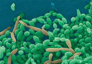
Candida tropicalis is a species of yeast in the genus Candida. It is a common pathogen in neutropenic hosts, in whom it may spread through the bloodstream to peripheral organs. For invasive disease, treatments include amphotericin B, echinocandins, or extended-spectrum triazole antifungals.

<i>Phialophora verrucosa</i> Species of fungus

Phialophora verrucosa is a pathogenic, dematiaceous fungus that is a common cause of chromoblastomycosis. It has also been reported to cause subcutaneous phaeohyphomycosis and mycetoma in very rare cases. In the natural environment, it can be found in rotting wood, soil, wasp nests, and plant debris. P. verrucosa is sometimes referred to as Phialophora americana, a closely related environmental species which, along with P. verrucosa, is also categorized in the P. carrionii clade.

<i>Metarhizium granulomatis</i> Species of fungus

Metarhizium granulomatis is a fungus in the family Clavicipitaceae associated with systemic mycosis in veiled chameleons. The genus Metarhizium is known to infect arthropods, and collectively are referred to green-spored asexual pathogenic fungi. This species grows near the roots of plants and has been reported as an agent of disease in captive veiled chameleons. The etymology of the species epithet, "granulomatis" refers to the ability of the fungus to cause granulomatous disease in susceptible reptiles.

Sarocladium kiliense is a saprobic fungus that is occasionally encountered as a opportunistic pathogen of humans, particularly immunocompromised and individuals. The fungus is frequently found in soil and has been linked with skin and systemic infections. This species is also known to cause disease in the green alga, Cladophora glomerata as well as various fruit and vegetable crops grown in warmer climates.

Myriodontium keratinophilum is a fungus widespread in nature, most abundantly found in keratin-rich environments such as feathers, nails and hair. Despite its ability to colonize keratinous surfaces of human body, the species has been known to be non-pathogenic in man and is phylogentically distant to other human pathogenic species, such as anthropophilic dermatophytes. However, its occasional isolation from clinical specimens along with its keratinolytic properties suggest the possibility it may contribute to disease.

Cladophialophora arxii is a black yeast shaped dematiaceous fungus that is able to cause serious phaeohyphomycotic infections. C. arxii was first discovered in 1995 in Germany from a 22-year-old female patient suffering multiple granulomatous tracheal tumours. It is a clinical strain that is typically found in humans and is also capable of acting as an opportunistic fungus of other vertebrates Human cases caused by C. arxii have been reported from all parts of the world such as Germany and Australia.

Curvularia geniculata is a fast-growing anamorphic fungus in the division Ascomycota, most commonly found in soil, especially in areas of warmer climates. The fungus is a pathogen, mainly causing plant and animal infections, and rarely causing human infections. C. geniculata is characterized by its curved conidia, which has a dark brown centre and pale tapered tips, and produces anti-fungal compounds called Curvularides A-E.

References

  1. Fijan, Nikola (1969). "Systemic Mycosis in Channel Catfish". Bulletin of the Wildlife Disease Association. 5 (2): 109–110. doi:10.7589/0090-3558-5.2.109. PMID   5816092. S2CID   20510874.
  2. 1 2 3 4 Mcginnis, M; Ajello, L (1974). "A New Species of Exophiala Isolated from Channel Catfish". Mycologia. 66 (3): 518–520. doi:10.1080/00275514.1974.12019633. PMID   4858287.
  3. "Online Etymology Dictionary". www.etymonline.com.
  4. 1 2 3 4 5 Brady, B (1975). "CMI Descriptions of Pathogenic Fungi and Bacteria No. 744". Bulletin of the Wildlife Disease Association. 75 (2): 105–106.
  5. Zhan, Fangdong; He, Yongmei; Li, Tao; Yang, Yun-ya; Toor, Gurpal S.; Zhao, Zhiwei (17 October 2014). "Tolerance and Antioxidant Response of a Dark Septate Endophyte (DSE), Exophiala pisciphila, to Cadmium Stress". Bulletin of Environmental Contamination and Toxicology. 94 (1): 96–102. doi:10.1007/s00128-014-1401-8. PMID   25323040. S2CID   22294797.
  6. Wang, L.; Yokoyama, K.; Miyaji, M.; Nishimura, K. (1 December 2001). "Identification, Classification, and Phylogeny of the Pathogenic Species Exophiala jeanselmei and Related Species by Mitochondrial Cytochrome b Gene Analysis". Journal of Clinical Microbiology. 39 (12): 4462–4467. doi:10.1128/JCM.39.12.4462-4467.2001. PMC   88566 . PMID   11724862.
  7. 1 2 Hoog, G.S. de; Hermanides-Nijhof, E.J. (1977). "The black yeasts and allied Hyphomycetes". Studies in Mycology. 15.
  8. 1 2 3 4 5 6 7 8 9 10 11 de Hoog, G.S.; Vicente, V.A.; Najafzadeh, M.J.; Harrak, M.J.; Badali, H.; Seyedmousavi, S. (2011-12-31). "Waterborne Exophiala species causing disease in cold-blooded animals". Persoonia. 27 (1): 46–72. doi:10.3767/003158511x614258. ISSN   0031-5850. PMC   3251318 . PMID   22403476.
  9. 1 2 Řehulka, J; Kubátová, A; Hubka, V (March 2018). "Swim bladder mycosis in pretty tetra (Hemigrammus pulcher) caused by Exophiala pisciphila and Phaeophleospora hymenocallidicola, and experimental verification of pathogenicity". Journal of Fish Diseases. 41 (3): 487–500. doi:10.1111/jfd.12750. PMID   29159880.
  10. Zhan, Fangdong; He, Yongmei; Li, Yuan; Li, Tao; Yang, Yun-Ya; Toor, Gurpal S.; Zhao, Zhiwei (14 July 2015). "Subcellular distribution and chemical forms of cadmium in a dark septate endophyte (DSE), Exophiala pisciphila". Environmental Science and Pollution Research. 22 (22): 17897–17905. doi:10.1007/s11356-015-5012-7. PMID   26165995. S2CID   22794201.
  11. Li, T.; Liu, M.J.; Zhang, X.T.; Zhang, H.B.; Sha, T.; Zhao, Z.W. (February 2011). "Improved tolerance of maize (Zea mays L.) to heavy metals by colonization of a dark septate endophyte (DSE) Exophiala pisciphila". Science of the Total Environment. 409 (6): 1069–1074. Bibcode:2011ScTEn.409.1069L. doi:10.1016/j.scitotenv.2010.12.012. PMID   21195456.
  12. Druzhinina, Irina S.; Kubicek, Christian P. (2016). Environmental and Microbial Relationships. Springer. ISBN   9783319295329.
  13. Zhang, Q; Gong, M; Yuan, J (2017). "Dark Septate Endophyte Improves Drought Tolerance in Sorghum". International Journal of Agriculture and Biology. 19 (1): 53. doi: 10.17957/ijab/15.0241 .
  14. 1 2 3 4 5 Kwon-Chung, K. June; Bennett, Joan E. (1992). Medical mycology. Philadelphia: Lea & Febiger. ISBN   978-0812114638.
  15. 1 2 3 4 5 6 Cheung, P; Gaskins, J (1986). "Exophilia psciphila: A study of its development". Mycopathologia. 93 (3): 173–184. doi:10.1007/BF00443521. PMID   3713799. S2CID   22725393.
  16. 1 2 3 Zhan, Fangdong; He, Yongmei; Zu, Yanqun; Li, Tao; Zhao, Zhiwei (13 March 2011). "Characterization of melanin isolated from a dark septate endophyte (DSE), Exophiala pisciphila". World Journal of Microbiology and Biotechnology. 27 (10): 2483–2489. doi:10.1007/s11274-011-0712-8. S2CID   85195084.
  17. 1 2 Sughayer, Maher; DeGirolami, Paola C.; Khettry, Urmila; Korzeniowski, Denise; Grumney, Anne; Pasarell, Lester; McGinnis, Michael R. (1991-05-01). "Human Infection Caused by Exophiala pisciphila: Case Report and Review". Clinical Infectious Diseases. 13 (3): 379–382. doi:10.1093/clinids/13.3.379. ISSN   1537-6591. PMID   1866539.
  18. Kebbe, Jad; Mador, M. Jeffery (6 March 2016). "Exophiala pisciphila : a novel cause of allergic bronchopulmonary mycosis". Journal of Thoracic Disease. 8 (7): E538–E541. doi: 10.21037/jtd.2016.05.77 . ISSN   2077-6624. PMC   4958854 . PMID   27499992.
  19. 1 2 3 4 Hurst, Christon J. (2016). The Rasputin effect : when commensals and symbionts become parasitic. Springer. p. 112. ISBN   978-3319281704.
  20. Buller, Nicky B (2014). Bacteria and fungi from fish and other aquatic animals : a practical identification manual (2 ed.). CABI. ISBN   978-1845938055.
  21. Řehulka, J; Kolařík, M; Hubka, V (August 2017). "Disseminated infection due to E. pisciphila in Cardinal tetra". Journal of Fish Diseases. 40 (8): 1015–1024. doi:10.1111/jfd.12577. PMID   27982440.
  22. 1 2 3 Marancik, David P. (2011). "Disseminated fungal infection in two species of captive sharks". Journal of Zoo and Wildlife Medicine. 42 (4): 686–694. doi:10.1638/2010-0175.1. PMID   22204064. S2CID   34699522.
  23. Poinar, George O. (2018). Diseases Of Nematodes. CRC Press. ISBN   9781351088367.
  24. Doshida, Junko (1996). "Exophilin A, a New Antibiotic from a Marine Microorganism Exophiala pisciphila". The Journal of Antibiotics. 49 (11): 1105–1109. doi: 10.7164/antibiotics.49.1105 . PMID   8982339.
  25. Bartonn, Sir Derek; Nakanishi, Kōji; Mori, Kenji; Meth-Cohn, Otto (1999). Comprehensive natural products chemistry (1st ed.). Elsevier. p. 601. ISBN   978-0080431604.
  26. Wang, Cui-Cui; Liu, Hai-Zhou; Liu, Ming; Zhang, Yu-Yan; Li, Tian-Tian; Lin, Xiu-Kun (30 March 2011). "Cytotoxic Metabolites from the Soil-Derived Fungus Exophiala pisciphila". Molecules. 16 (4): 2796–2801. doi: 10.3390/molecules16042796 . PMC   6260601 . PMID   21455093.
  27. Tidgewell, Kevin; Clark, Benjamin R.; Gerwick, William H. (2010). The Natural Products Chemistry of Cyanobacteria. Comprehensive Natural Products II. pp. 141–188. doi:10.1016/b978-008045382-8.00041-1. ISBN   9780080453828.
  28. Ondeyka, John G; Deborah, Zink (2003). "Isolation, Structure and HIV-1 Integrase Inhibitory Activity of Exophillic Acid, a Novel Fungal Metabolite from Exophiala pisciphila". Journal of Antibiotics. 56 (12): 1018–1023. doi: 10.7164/antibiotics.56.1018 . PMID   15015729.