Ganglionic eminence

Last updated
Ganglionic eminence
Interneuron-radial glial interactions in the developing cerebral cortex.png
Interneurons (green) migrate tangentially from the ganglionic eminence to the cerebral cortex. The tangentially migrating interneurons travel perpendicular to the radial glial cells (red). The radially migrating interneurons travel parallel to the radial glial cells.
Anatomical terminology

The ganglionic eminence (GE) is a transitory structure in the development of the nervous system that guides cell and axon migration. [1] It is present in the embryonic and fetal stages of neural development found between the thalamus and caudate nucleus. [1]

Contents

The eminence is divided into three regions of the ventral ventricular zone of the telencephalon (a lateral, medial and caudal eminence), where they facilitate tangential cell migration during embryonic development. Tangential migration does not involve interactions with radial glial cells; instead the interneurons migrate perpendicularly through the radial glial cells to reach their final location. The characteristics and function of the cells that follow the tangential migration pathway seem to be closely related to the location and precise timing of their production, [2] and the GEs contribute significantly to building up the GABAergic cortical cell population. [1] [3] [4] Another structure that the GEs contribute to is the basal ganglia. [5] The GEs also guide the axons growing from the thalamus into the cortex and vice versa. [1]

In humans, the GEs disappear by one year of age. [1] During development, neuronal migration continues until the extinction of the germ layer, at which point the remnants from the germ layer make up the eminences. [1]

Categorization

Coronal section in the forebrain of an embryonic mouse at 12.5 days of gestation (preplate stage), showing the lateral and medial ganglionic eminences (LGE, MGE) from which GABAergic interneurons tangentially migrate to the cortical anlage (left, yellow). Glutamatergic neurons destined for the cortex are generated locally in the cortical ventricular zone and migrate radially (right, red). Ganglionic eminence mice E12.5 doi 10.1186 1747-5333-1-16-1.jpg
Coronal section in the forebrain of an embryonic mouse at 12.5 days of gestation (preplate stage), showing the lateral and medial ganglionic eminences (LGE, MGE) from which GABAergic interneurons tangentially migrate to the cortical anlage (left, yellow). Glutamatergic neurons destined for the cortex are generated locally in the cortical ventricular zone and migrate radially (right, red).

Ganglionic eminences are categorized into three groups based on their location within the subventricular zone:

A sulcus separates the medial and lateral ganglionic eminences. The expression of Nkx2-1, Gsx2, and Pax6 is required to determine the independent progenitor cell populations in the LGE and MGE. Interactions between these three genes define the boundaries between the different progenitor zones and mutations of these genes can cause abnormal expansion around the MGE, LGE, ventral pallium (VP), and anterior entopeduncular region (AEP). The cells of the GEs are quite homogenous, with the MGE, LGE, and CGE all having small, dark, irregular nuclei and moderately dense cytoplasm, however, each eminence can be identified by the type of progeny that it produces. [6] See the individual GE sections below for more information on the different types of progeny produced.

Additionally, the subventricular zone is the starting point of multiple streams of tangentially migrating interneurons that express Dlx genes. There are three main tangential migration pathways that have been identified in this region:

  1. the latero-caudal migration (subpallial telencephalon to cortex)
  2. the medio-rostral migration (subpallial basal telencephalon to the olfactory bulb)
  3. the latero-caudal migration (basal telencephalon to the striatum)

These pathways are temporally and spatially distinct, and produce a variety of GABAergic, and non-GABAergic interneurons. One example of GABAergic interneurons that the GEs guide are parvalbumin-containing interneurons in the neocortex. Some examples of non-GABAergic interneurons that the GEs guide are dopaminergic interneurons in the olfactory bulb, and cholinergic interneurons in the striatum. Cells migrating along these pathways move at different rates. Some molecules that have been implicated in controlling the rate of the unidirectional movement of cells derived from the GEs are hepatocyte growth factor/scattered factor (HGF/SF), and various neurotrophic factors. [2]

Medial ganglionic eminence (MGE)

The primary purpose of the MGE during development is to produce GABAergic stellate cells and direct their migration to the neocortex. [6] The precursors of most GABAergic interneurons in the cerebral cortex migrate from the subcortical progenitor zone. More specifically, performing a mechanical transection of the migratory route from the MGE to the neocortex causes a 33% decrease in GABAergic interneurons in the neocortex. [6] The MGE also produces some of the neurons and glia of the basal ganglia and hippocampus. [6] [7] The MGE may also be a source of Cajal-Retzius cells, but this remains controversial. [6] Early in embryonic development, the interneurons in the cortex stem primarily from the MGE [8] and the AEP. In vitro experiments show that MGE cells migrate more than 300 μm per day, three times faster than the migration of LGE cells. [2] See more about the time frame and function of MGE in comparison to the LGE in the following section.

Lateral ganglionic eminence (LGE)

Compared to the early temporal frame of development in the MGE, the LGE aids in the tangential migration of cells later in the mid-embryogenic stage. Unlike the MGE, which guides most cell migration into the cortex during this stage, the LGE contributes less to cell migration to the cortex, and instead guides many cells to the olfactory bulbs. In fact, the migration to the olfactory bulb is led by the LGE into adulthood. The route that newly generated neurons take from the anterior subventricular zone to the olfactory bulb is called the rostral migratory stream. During the late stages of embryonic development, both the LGE and MGE guide cell migration to the cortex, specifically the proliferative regions of the cortex. [2] Some studies have found that the LGE also contributes cells to the neocortex, but this remains an issue of debate. [6] In vitro, cells migrating from the LGE travel at a rate of 100 μm per day, slower than the MGE cells. [2]

Caudal ganglionic eminence (CGE)

The caudal ganglionic eminence is another subcortical structure that is essential to the generation of cortical interneurons. It is located next to the lateral ventricle, posterior to where the LGE and MGE fuse. [6] The CGE is a fusion of the rostral medial and lateral ganglionic eminence, which begins at the mid to caudal thalamus. There are two molecular domains that exist within the CGE and closely resemble extensions of the caudal MGE and LGE. [9] The CGE is distinct from the LGE and MGE in gene expression patterns and progeny produced. Unlike the cells from the MGE, the cells from the CGE were rarely parvalbumin-containing neurons. It seems that the majority of cells from the CGE were GABAergic interneurons, but depending on where they are located, CGE-derived cells are very diverse. CGE-derived cells include GABAergic interneurons, spiny interneurons, mossy cells, pyramidal and granule neurons, and even oligodendrocyte and astrocyte glial cells. [6]

Cell migration

Cells in the ganglionic eminence migrate tangentially to neocortex, giving rise to interneurons. A variety of molecular mechanisms cooperate to direct this process. Embryonic interneuronal migration to the cerebral cortex is mediated by an array of motogenic growth factors in the MGE, repulsive factors in the striatum and LGE, permissive factors in migratory corridors in the ganglionic eminence, and attractive factors in the cortex itself. [3] Cells in the LGE migrate to the striatal domain (caudate nucleus and putamen) and parts of the septum and amygdala. MGE cells follow a migratory path to the globus pallidus and part of the septum. The CGE gives rise to interneurons in the nucleus accumbens, the bed nucleus of the stria terminals, the hippocampus, and specific nuclei in the amygdala. This directed migration is induced by differences in gene expression between these subpallial domains. [4] An array of genes are involved in the differentiation and specification of interneurons and oligodendrocytes, including: Dlx1, Dlx2, Gsh1, Mash1, Gsh2, Nkx2.1, Nkx5.1, Isl1, Six3 and Vax1. [4]

Molecular mechanisms for directed migration

The induced migration of cells from the ganglionic eminence during development is directed by a variety of motogenic factors, molecules that increase cell motility, and chemotactic molecules. The motogenic factor HGF/SF enhances cell motility and directs cells away from subpallial regions and demarcates the routes followed by migrating cells. Neurotrophins, such as BDNF, are a family of motogenic factors involved in directing migration. The cerebral cortex provides chemoattractant molecules (for example NRG1 type I and II in the cortex) while subpallial areas produce chemorepulsive molecules (for example Slit) to direct cell migration. Additionally, some permissive factors (such as NRG1 type III) in the migratory corridors are necessary for this process to occur. [3] [4]

The neurotransmitters GABA and 5-HT have been implicated in the migration as well. High GABA concentrations have been seen to cause random cell movement ("random walk migration"), while low concentrations promote directed migration. 5-HT has been tied to the process of incorporating interneurons into the cortical plate, as well as in the differentiation into subpopulations of interneurons. [4]

Associated disorders

The migration of cells from the ventricular zone to their intended destination and the success of their differentiation can be interrupted in many different ways, including interference with mechanical motors or an alteration of molecular signals that initiate movement, lead the cell in migration, and terminate its migration. The function of the molecules that affect migration are not confined to cell movement, overlapping considerably with the events associated with neurogenesis. As a result, neuronal migration syndromes are difficult to classify. The largest class of neuronal migration syndromes is lissencephaly. This includes a spectrum of simplified cortex ranging from agyria (a total absence of cortical convolutions) to pachygyria (broadened gyri) with unusually thick cortex.

Mis-migration of neurons can also result in bilateral periventricular nodular heterotopia, a disease recognized by neuronal heterotopia lining the lateral ventricles. Zellweger Syndrome is characterized by a cortical dysplasia similar to polymicrogyria of cerebral and cerebellar cortex, occasionally with pachygyria surrounding the Sylvian fissure, and focal/subependymal heterotopia. Kallmann syndrome is recognized by anosmia associated with mental retardation, hypogonadism, and the failure of the olfactory bulb to develop.

Disorders of axonal projection and assembly are rarely pure, but closely related to neuronal migration genes. This notably includes agenesis of the corpus callosum.

Disturbances in the genesis of neural elements can result in cortical dysplasia. Examples include ectopic neurogenesis, microencephaly, and altered cell survival resulting in areas of hyperplasia, reduced apoptosis, and heterotopia. [10]

Further research

Further research could be done on the migration of cells from the basal ganglia to the neocortex. The molecular mechanisms in control of this are still not completely clarified. The number of known mutations that could interfere with neuronal migration is rapidly growing, and will continue to do so as further research is performed. The complexity of molecular steps needed to correctly place cells in a system as complicated as the brain is impressive, and as more pieces to this intricate puzzle arise, it will be easier to come up with strategies to remedy disorders associated with neuronal migration, and to potentially repair damage caused by trauma, stroke, maldevelopment, and aging. [10]

Related Research Articles

<span class="mw-page-title-main">Cerebral cortex</span> Outer layer of the cerebrum of the mammalian brain

The cerebral cortex, also known as the cerebral mantle, is the outer layer of neural tissue of the cerebrum of the brain in humans and other mammals. The cerebral cortex mostly consists of the six-layered neocortex, with just 10% consisting of the allocortex. It is separated into two cortices, by the longitudinal fissure that divides the cerebrum into the left and right cerebral hemispheres. The two hemispheres are joined beneath the cortex by the corpus callosum. The cerebral cortex is the largest site of neural integration in the central nervous system. It plays a key role in attention, perception, awareness, thought, memory, language, and consciousness. The cerebral cortex is part of the brain responsible for cognition.

The development of the nervous system, or neural development (neurodevelopment), refers to the processes that generate, shape, and reshape the nervous system of animals, from the earliest stages of embryonic development to adulthood. The field of neural development draws on both neuroscience and developmental biology to describe and provide insight into the cellular and molecular mechanisms by which complex nervous systems develop, from nematodes and fruit flies to mammals.

An apical dendrite is a dendrite that emerges from the apex of a pyramidal cell. Apical dendrites are one of two primary categories of dendrites, and they distinguish the pyramidal cells from spiny stellate cells in the cortices. Pyramidal cells are found in the prefrontal cortex, the hippocampus, the entorhinal cortex, the olfactory cortex, and other areas. Dendrite arbors formed by apical dendrites are the means by which synaptic inputs into a cell are integrated. The apical dendrites in these regions contribute significantly to memory, learning, and sensory associations by modulating the excitatory and inhibitory signals received by the pyramidal cells.

<span class="mw-page-title-main">Stellate cell</span>

Stellate cells are neurons in the central nervous system, named for their star-like shape formed by dendritic processes radiating from the cell body. Many stellate cells are GABAergic and are located in the molecular layer of the cerebellum. Stellate cells are derived from dividing progenitor cells in the white matter of postnatal cerebellum. Dendritic trees can vary between neurons. There are two types of dendritic trees in the cerebral cortex, which include pyramidal cells, which are pyramid shaped and stellate cells which are star shaped. Dendrites can also aid neuron classification. Dendrites with spines are classified as spiny, those without spines are classified as aspinous. Stellate cells can be spiny or aspinous, while pyramidal cells are always spiny. Most common stellate cells are the inhibitory interneurons found within the upper half of the molecular layer in the cerebellum. Cerebellar stellate cells synapse onto the dendritic trees of Purkinje cells and send inhibitory signals. Stellate neurons are sometimes found in other locations in the central nervous system; cortical spiny stellate cells are found in layer IVC of the primary visual cortex. In the somatosensory barrel cortex of mice and rats, glutamatergic (excitatory) spiny stellate cells are organized in the barrels of layer 4. They receive excitatory synaptic fibres from the thalamus and process feed forward excitation to 2/3 layer of the primary visual cortex to pyramidal cells. Cortical spiny stellate cells have a 'regular' firing pattern. Stellate cells are chromophobes, that is cells that does not stain readily, and thus appears relatively pale under the microscope.

<span class="mw-page-title-main">Rostral migratory stream</span> One path neural stem cells take to reach the olfactory bulb


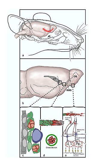
The rostral migratory stream (RMS) is a specialized migratory route found in the brain of some animals along which neuronal precursors that originated in the subventricular zone (SVZ) of the brain migrate to reach the main olfactory bulb (OB). The importance of the RMS lies in its ability to refine and even change an animal's sensitivity to smells, which explains its importance and larger size in the rodent brain as compared to the human brain, as our olfactory sense is not as developed. This pathway has been studied in the rodent, rabbit, and both the squirrel monkey and rhesus monkey. When the neurons reach the OB they differentiate into GABAergic interneurons as they are integrated into either the granule cell layer or periglomerular layer.

Pachygyria is a congenital malformation of the cerebral hemisphere. It results in unusually thick convolutions of the cerebral cortex. Typically, children have developmental delay and seizures, the onset and severity depending on the severity of the cortical malformation. Infantile spasms are common in affected children, as is intractable epilepsy.

<span class="mw-page-title-main">Olfactory tubercle</span> Area at the bottom of the forebrain

The olfactory tubercle (OT), also known as the tuberculum olfactorium, is a multi-sensory processing center that is contained within the olfactory cortex and ventral striatum and plays a role in reward cognition. The OT has also been shown to play a role in locomotor and attentional behaviors, particularly in relation to social and sensory responsiveness, and it may be necessary for behavioral flexibility. The OT is interconnected with numerous brain regions, especially the sensory, arousal, and reward centers, thus making it a potentially critical interface between processing of sensory information and the subsequent behavioral responses.

<span class="mw-page-title-main">Islands of Calleja</span> Group of neural granule cells

The islands of Calleja are a group of neural granule cells located within the ventral striatum in the brains of most animals. This region of the brain is part of the limbic system, where it aids in the reinforcing effects of reward-like activities. Within most species, the islands are specifically located within the olfactory tubercle; however, in primates, these islands are located within the nucleus accumbens, the reward center of the brain, since the olfactory tubercle has practically disappeared in the brains of primates. Both of these structures have been implicated in the processing of incentives as well as addictions to drugs. Projections to and from the islands supplement this knowledge with their involvement in the reward pathways for both cocaine and amphetamines.

<span class="mw-page-title-main">Radial glial cell</span> Bipolar-shaped progenitor cells of all neurons in the cerebral cortex and some glia

Radial glial cells, or radial glial progenitor cells (RGPs), are bipolar-shaped progenitor cells that are responsible for producing all of the neurons in the cerebral cortex. RGPs also produce certain lineages of glia, including astrocytes and oligodendrocytes. Their cell bodies (somata) reside in the embryonic ventricular zone, which lies next to the developing ventricular system.

<span class="mw-page-title-main">Subventricular zone</span> Region outside each lateral ventricle of the brain

The subventricular zone (SVZ) is a region situated on the outside wall of each lateral ventricle of the vertebrate brain. It is present in both the embryonic and adult brain. In embryonic life, the SVZ refers to a secondary proliferative zone containing neural progenitor cells, which divide to produce neurons in the process of neurogenesis. The primary neural stem cells of the brain and spinal cord, termed radial glial cells, instead reside in the ventricular zone (VZ).

<span class="mw-page-title-main">Paleocortex</span> Region within the telencephalon in the vertebrate brain

In anatomy of animals, the paleocortex, or paleopallium, is a region within the telencephalon in the vertebrate brain. This type of cortical tissue consists of three cortical laminae. In comparison, the neocortex has six layers and the archicortex has three or four layers. Because the number of laminae that compose a type of cortical tissue seems to be directly proportional to both the information-processing capabilities of that tissue and its phylogenetic age, paleocortex is thought to be an intermediate between the archicortex and the neocortex in both aspects.

<span class="mw-page-title-main">EMX1</span> Protein-coding gene in the species Homo sapiens

Homeobox protein EMX1 is a protein that in humans is encoded by the EMX1 gene. The transcribed EMX1 gene is a member of the EMX family of transcription factors. The EMX1 gene, along with its family members, are expressed in the developing cerebrum. EMX1 plays a role in specification of positional identity, the proliferation of neural stem cells, differentiation of layer-specific neuronal phenotypes and commitment to a neuronal or glial cell fate.

<span class="mw-page-title-main">Pallium (neuroanatomy)</span> Layers of grey and white matter that cover the upper surface of the cerebrum in vertebrates

In neuroanatomy, pallium refers to the layers of grey and white matter that cover the upper surface of the cerebrum in vertebrates. The non-pallial part of the telencephalon builds the subpallium. In basal vertebrates the pallium is a relatively simple three-layered structure, encompassing 3–4 histogenetically distinct domains, plus the olfactory bulb.

<span class="mw-page-title-main">TBR1</span> Protein-coding gene in Homo sapiens

T-box, brain, 1 is a transcription factor protein important in vertebrate embryo development. It is encoded by the TBR1 gene. This gene is also known by several other names: T-Brain 1, TBR-1, TES-56, and MGC141978. TBR1 is a member of the TBR1 subfamily of T-box family transcription factors, which share a common DNA-binding domain. Other members of the TBR1 subfamily include EOMES and TBX21. TBR1 is involved in the differentiation and migration of neurons and is required for normal brain development. TBR1 interacts with various genes and proteins in order to regulate cortical development, specifically within layer VI of the developing six-layered human cortex. Studies show that TBR1 may play a role in major neurological diseases such as Alzheimer's disease (AD), Parkinson's disease (PD) and autism spectrum disorder (ASD).

Guidepost cells are cells which assist in the subcellular organization of both neural axon growth and migration. They act as intermediate targets for long and complex axonal growths by creating short and easy pathways, leading axon growth cones towards their target area.

The development of the cerebral cortex, known as corticogenesis is the process during which the cerebral cortex of the brain is formed as part of the development of the nervous system of mammals including its development in humans. The cortex is the outer layer of the brain and is composed of up to six layers. Neurons formed in the ventricular zone migrate to their final locations in one of the six layers of the cortex. The process occurs from embryonic day 10 to 17 in mice and between gestational weeks seven to 18 in humans.

Cajal–Retzius cells are a heterogeneous population of morphologically and molecularly distinct reelin-producing cell types in the marginal zone/layer I of the developmental cerebral cortex and in the immature hippocampus of different species and at different times during embryogenesis and postnatal life.

Neurogenesis is the process by which nervous system cells, the neurons, are produced by neural stem cells (NSCs). In short, it is brain growth in relation to its organization. This occurs in all species of animals except the porifera (sponges) and placozoans. Types of NSCs include neuroepithelial cells (NECs), radial glial cells (RGCs), basal progenitors (BPs), intermediate neuronal precursors (INPs), subventricular zone astrocytes, and subgranular zone radial astrocytes, among others.

<span class="mw-page-title-main">Radial unit hypothesis</span> Conceptual theory of cerebral cortex development

The Radial Unit Hypothesis (RUH) is a conceptual theory of cerebral cortex development, first described by Pasko Rakic. The RUH states that the cerebral cortex develops during embryogenesis as an array of interacting cortical columns, or 'radial units', each of which originates from a transient stem cell layer called the ventricular zone, which contains neural stem cells known as radial glial cells.

Neurogliaform cells (NGF) are inhibitory (GABAergic) interneurons found in the cortex and the hippocampus. NGF cells represent approximately 10% of the total hippocampal inhibitory interneuron population.

References

  1. 1 2 3 4 5 6 Encha-Razavi & Sonigo. (2003). Features of the developing brain. Child's Nervous System. pp. 426–428
  2. 1 2 3 4 5 Marín, O; Rubenstein, JL (November 2001). "A long, remarkable journey: tangential migration in the telencephalon". Nature Reviews. Neuroscience. 2 (11): 780–90. doi:10.1038/35097509. PMID   11715055. S2CID   5604192.
  3. 1 2 3 Ghashghaei, HT; Lai, C; Anton, ES (February 2007). "Neuronal migration in the adult brain: are we there yet?". Nature Reviews. Neuroscience. 8 (2): 141–51. doi:10.1038/nrn2074. PMID   17237805. S2CID   9322780.
  4. 1 2 3 4 5 Hernández-Miranda, Parnavelas, & Chiara. (2010). Molecules & mechanisms involved in the generation and migration of cortical interneurons. ASN Neuro, 2(2). pp. 75-86.
  5. Purves, D., Augustine, G., Fitzpatrick, D., Hall, W., LaMantia, A.S., McNamara, J., and White, L. (2008). Neuroscience. 4th ed. Sinauer Associates. pp. 555–8. ISBN   978-0-87893-697-7.{{cite book}}: CS1 maint: multiple names: authors list (link)
  6. 1 2 3 4 5 6 7 8 9 Brazel, CY; Romanko, MJ; Rothstein, RP; Levison, SW (January 2003). "Roles of the mammalian subventricular zone in brain development". Progress in Neurobiology. 69 (1): 49–69. doi:10.1016/s0301-0082(03)00002-9. PMID   12637172. S2CID   24001139.
  7. Sanes, Reh, & Harris. (2012). Development of the Nervous System. 3rd ed. Academic Press. pp. 62–63. ISBN   978-0-12-374539-2.
  8. Lavdas, Grigoriou, Pachnis, & Parnavelas. (1999). The medial ganglionic eminence gives rise to a population of early neurons in the developing cerebral cortex. The Journal of Neuroscience, 99(19). pp. 7881–7888.
  9. Wonders, CP; Anderson, SA (September 2006). "The origin and specification of cortical interneurons". Nature Reviews. Neuroscience. 7 (9): 687–96. doi:10.1038/nrn1954. PMID   16883309. S2CID   3329713.
  10. 1 2 Ross, M. E., & Walsh, C. A. (2001). Human brain malformations and their lessons for neuronal migration. Annual Review of Neuroscience, 24(1), 1041–1070.