Related Research Articles

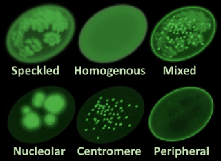
Antinuclear antibodies are autoantibodies that bind to contents of the cell nucleus. In normal individuals, the immune system produces antibodies to foreign proteins (antigens) but not to human proteins (autoantigens). In some cases, antibodies to human antigens are produced; these are known as autoantibodies.

Eosinophilic is the staining of tissues, cells, or organelles after they have been washed with eosin, a dye.

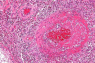
Fibrinoid necrosis is a specific pattern of irreversible, uncontrolled cell death that occurs when antigen-antibody complexes are deposited in the walls of blood vessels along with fibrin. It is common in the immune-mediated vasculitides which are a result of type III hypersensitivity. When stained with hematoxylin and eosin, they appear brightly eosinophilic and smudged.

A malar rash, also called butterfly rash, is a medical sign consisting of a characteristic form of facial rash. It is often seen in lupus erythematosus. More rarely, it is also seen in other diseases, such as pellagra, dermatomyositis, and Bloom syndrome.

Lupus nephritis is an inflammation of the kidneys caused by systemic lupus erythematosus (SLE), an autoimmune disease. It is a type of glomerulonephritis in which the glomeruli become inflamed. Since it is a result of SLE, this type of glomerulonephritis is said to be secondary, and has a different pattern and outcome from conditions with a primary cause originating in the kidney. The diagnosis of lupus nephritis depends on blood tests, urinalysis, X-rays, ultrasound scans of the kidneys, and a kidney biopsy. On urinalysis, a nephritic picture is found and red blood cell casts, red blood cells and proteinuria is found.

Hematoxylin and eosin stain is one of the principal tissue stains used in histology. It is the most widely used stain in medical diagnosis and is often the gold standard. For example, when a pathologist looks at a biopsy of a suspected cancer, the histological section is likely to be stained with H&E.

Belimumab, sold under the brand name Benlysta, is a human monoclonal antibody that inhibits B-cell activating factor (BAFF), also known as B-lymphocyte stimulator (BLyS). It is approved in the United States and Canada, and the European Union to treat systemic lupus erythematosus and lupus nephritis.

Anti-double stranded DNA (Anti-dsDNA) antibodies are a group of anti-nuclear antibodies (ANA) the target antigen of which is double stranded DNA. Blood tests such as enzyme-linked immunosorbent assay (ELISA) and immunofluorescence are routinely performed to detect anti-dsDNA antibodies in diagnostic laboratories. They are highly diagnostic of systemic lupus erythematosus (SLE) and are implicated in the pathogenesis of lupus nephritis.
Chilblain lupus erythematosus was initially described by Hutchinson in 1888 as an uncommon manifestation of chronic cutaneous lupus erythematosus. Chilblain lupus erythematosus is characterized by a rash that primarily affects acral surfaces that are frequently exposed to cold temperatures, such as the toes, fingers, ears, and nose. The rash is defined by oedematous skin, nodules, and tender plaques with a purple discoloration.
Tumid lupus erythematosus is a rare, but distinctive entity in which patients present with edematous erythematous plaque.
Lupus erythematosus panniculitis presents with subcutaneous nodules that are commonly firm, sharply defined and nontender.

Lupus, technically known as systemic lupus erythematosus (SLE), is an autoimmune disease in which the body's immune system mistakenly attacks healthy tissue in many parts of the body. Symptoms vary among people and may be mild to severe. Common symptoms include painful and swollen joints, fever, chest pain, hair loss, mouth ulcers, swollen lymph nodes, feeling tired, and a red rash which is most commonly on the face. Often there are periods of illness, called flares, and periods of remission during which there are few symptoms.
Anti-histone antibodies are autoantibodies that are a subset of the anti-nuclear antibody family, which specifically target histone protein subunits or histone complexes. They were first reported by Henry Kunkel, H.R. Holman, and H.R.G. Dreicher in their studies of cellular causes of lupus erythematosus in 1959–60. Today, anti-histone antibodies are still used as a marker for systemic lupus erythematosus, but are also implicated in other autoimmune diseases like Sjögren syndrome, dermatomyositis, or rheumatoid arthritis. Anti-histone antibodies can be used as a marker for drug-induced lupus.

A lupus erythematosus cell, also known as Hargraves cell, is a neutrophil or macrophage that has phagocytized (engulfed) the denatured nuclear material of another cell. The denatured material is an absorbed hematoxylin body.

Anti-SSA autoantibodies are a type of anti-nuclear autoantibodies that are associated with many autoimmune diseases, such as systemic lupus erythematosus (SLE), SS/SLE overlap syndrome, subacute cutaneous lupus erythematosus (SCLE), neonatal lupus and primary biliary cirrhosis. They are often present in Sjögren's syndrome (SS). Additionally, Anti-Ro/SSA can be found in other autoimmune diseases such as systemic sclerosis (SSc), polymyositis/dermatomyositis (PM/DM), rheumatoid arthritis (RA), and mixed connective tissue disease (MCTD), and are also associated with heart arrhythmia.

Lupus vasculitis is one of the secondary vasculitides that occurs in approximately 50% of patients with systemic lupus erythematosus (SLE).
Neuropsychiatric systemic lupus erythematosus or NPSLE refers to the neurological and psychiatric manifestations of systemic lupus erythematosus. SLE is a disease in which the immune system attacks the body's own cells and tissues. It can affect various organs or systems of the body. It is estimated that over half of people with SLE have neuropsychiatric involvement.

Robert George Lahita is an American physician, internist and rheumatologist, best known for his research into systemic lupus erythematosus. and other autoimmune diseases. He is the author of more than 16 books and 150 scientific publications in the field of autoimmunity and immuno-endocrinology and a media consultant on health-related issues. He currently serves as Director of the Institute of Autoimmune and Rheumatic Diseases at St. Joseph's Healthcare System, specializing in autoimmunity, rheumatology, and treatment of diseases of joints, muscle, bones and tendons including arthritis, back pain, muscle strains, common athletic injuries and collagen diseases.
Cell-Bound Complement Activation Products (CB-CAPs) or complement split products, refers to complement activation fragments, C4d, that are bound covalently to somatic cells, as a result of activation of the classical complement pathway. They appear potentially useful for the diagnosis of systemic lupus erythematosus as of 2015.

Dr. George C. Tsokos, MD is a Greek-American rheumatologist who serves as a Professor of Medicine at Harvard Medical School and the Chief of the Division of Rheumatology and Clinical Immunology at the Beth Israel Deaconess Medical Center, Boston. He is widely recognized as one of the foremost leaders of modern lupus research with landmark discoveries that have brought understanding of lupus to new levels, shedding light on how the disease develops and progresses over time.
References
- ↑ Chan JK (2014). "The wonderful colors of the hematoxylin-eosin stain in diagnostic surgical pathology". Int. J. Surg. Pathol. 22 (1): 12–32. doi:10.1177/1066896913517939. PMID 24406626. S2CID 26847314.
- 1 2 Hematoxylin body. Medical Dictionary
- ↑ Moreland, LW. Rheumatology and Immunology Therapy Springer Berlin Heidelberg 2004 page 384 ISBN 9783540206255 doi:10.1007/3-540-29662-X_1191
- ↑ Cibas ES, Ducatman BS. Cytology: Diagnostic Principles and Clinical Correlates. Elsevier Health Sciences. 2013. Pages 132-133 ISBN 9781455750795
- ↑ Hollander JL. Arthritis and allied conditions: a textbook of rheumatology. 1966 Page 100 and 784
- ↑ Wallace DJ, Hahn B. (editors). Dubois' Lupus Erythematosus. Lippincott Williams & Wilkins, 2007. ISBN 9780781793940
- ↑ Robbins & Cotran Pathologic Basis of Disease. Vinay Kumar V, Abbas AK, Aster JC. Elsevier Health Sciences, 2014. Edition 9, revised. pages 222 and 225. ISBN 9780323296359