Histoplasma capsulatum

Last updated

Histoplasma capsulatum
Histopathology of histoplasma, HE stain.png
Histopathology of Histoplasma capsulatum, H&E stain, showing organisms surrounded by halos, in a granuloma of epithelioid histiocytes.
Scientific classification
Kingdom:
Division:
Class:
Order:
Family:
Genus:
Species:
H. capsulatum
Binomial name
Histoplasma capsulatum
Darling (1906)

Histoplasma capsulatum is a species of dimorphic fungus. Its sexual form is called Ajellomyces capsulatus. It can cause pulmonary and disseminated histoplasmosis.

Contents

H. capsulatum is "distributed worldwide, except in Antarctica, but most often associated with river valleys" [1] and occurs chiefly in the "Central and Eastern United States" [2] followed by "Central and South America, and other areas of the world". [2] It is most prevalent in the Ohio and Mississippi River valleys. It was discovered by Samuel Taylor Darling in 1906.

Growth and morphology

H. capsulatum is an ascomycetous fungus closely related to Blastomyces dermatitidis . It is potentially sexual, and its sexual state, Ajellomyces capsulatus, can readily be produced in culture, though it has not been directly observed in nature. H. capsulatum groups with B. dermatitidis and the South American pathogen Paracoccidioides brasiliensis in the recently recognized fungal family Ajellomycetaceae. [3] [4] It is dimorphic and switches from a mould-like (filamentous) growth form in the natural habitat to a small, budding yeast form in the warm-blooded animal host.

Like B. dermatitidis, H. capsulatum has two mating types, "+" and "–". The great majority of North American isolates belongs to a single genetic type, [5] [6] but a study of multiple genes suggests a recombining, sexual population. [6] A recent analysis has suggested that the prevalent North American genetic type and a less common type should be considered separate phylogenetic species, distinct from H. capsulatum isolates obtained in Central and South America and other parts of the world. These entities are temporarily designated NAm1 (the rare type, which includes a famous experimental isolate designated "the Downs strain") and NAm2 (the common type). [6] As yet, no well-established clinical or geographic distinction is seen between these two genetic groups.

In its asexual form, the fungus grows as a colonial microfungus strongly similar in macromorphology to B. dermatitidis. A microscopic examination shows a marked distinction: H. capsulatum produces two types of conidia, globose macroconidia, 8–15 µm, with distinctive tuberculate or finger-like cell wall ornamentation, and ovoid microconidia, 2–4 µm, which appear smooth or finely roughened. Whether either of these conidial types is the principal infectious particle is unclear. They form on individual short stalks and readily become airborne when the colony is disturbed. Ascomata of the sexual state are 80–250 µm, and are very similar in appearance and anatomy to those described above for B. dermatitidis. The ascospores are similarly minute, averaging 1.5 µm.

The budding yeast cells formed in infected tissues are small (about 2–4 µm) and are characteristically seen forming in clusters within phagocytic cells, including histiocytes and other macrophages, as well as monocytes. An African phylogenetic species, H. duboisii , often forms larger yeast cells to 15 µm.

Geographic distribution

H. capsulatum is "distributed worldwide, except in Antarctica, but most often associated with river valleys" [1] and occurs chiefly in the "Central and Eastern United States" [2] followed by "Central and South America, and other areas of the world" [2] It is most prevalent in the Ohio and Mississippi river valleys.

The enzootic and endemic zones of H. capsulatum can be roughly divided into core areas, where the fungus occurs widely in soil or on vegetation contaminated by bird droppings or equivalent organic inputs, and peripheral areas, where the fungus occurs relatively rarely in association with soil, but is still found abundantly in heavy accumulations of bat or bird guano in enclosed spaces such as caves, buildings, and hollow trees. The principal core area for this species includes the valleys of the Mississippi, Ohio, and Potomac Rivers in the USA, as well as a wide span of adjacent areas extending from Kansas, Illinois, Indiana, and Ohio in the north to Mississippi, Louisiana, and Texas in the south. [7] [8] [9] In some areas, such as Kansas City, skin testing with the histoplasmin antigen preparation shows that 80–90% of the resident population have an antibody reaction to H. capsulatum, probably indicating prior subclinical infection. [7] Northern U.S. states such as Minnesota, Michigan, New York and Vermont are peripheral areas for histoplasmosis, but have scattered counties where 5–19% of lifetime residents show exposure to H. capsulatum. One New York county, St. Lawrence county (across the St. Lawrence River from the CornwallPrestonBrockville area of Ontario, Canada) shows exposures over 20%. [7]

The distribution of H. capsulatum in Canada is not as well documented as in the US. The St. Lawrence Valley is probably the best known endemic region based both on case reports and on a number of skin test reaction studies that were done between 1945 and 1970. The Montreal area is a particularly well documented endemic focus, not just in the agricultural regions surrounding the city [10] but also within the city itself. [11] The Mount Royal area in central Montreal, especially the north and east sides of Mt. Royal Park, showed exposure rates between 20 and 50% in schoolchildren [11] and locally lifetime-resident university students. [12] A particularly high rate of 79.3% exposure was shown in St. Thomas, Ontario, south of London, Ontario, after 7 local residents had died of histoplasmosis in 1957. [13] Based on numerous small regional studies, histoplasmin skin test reactors form ca. 10–50 % of the population in much of southern Ontario and in Quebec’s St. Lawrence Valley, ca. 5% in southern Manitoba and some northerly parts of Quebec (e.g., Abitibi-Témiscamingue), and ca. 1% in Nova Scotia. [12] [13] [14] [15] [16] Exposure of aboriginal Canadians occurs remarkably far north in Quebec, [17] [18] but has not been reported in similar boreal biogeoclimatic zones in many other parts of Canada. Recently and remarkably, a cluster of four indigenously acquired cases of histoplasmosis was shown to be associated with a golf course in suburban Edmonton, Alberta. [19] Examination suggested that local soil was the source.

Spectrum of disease

Histoplasmosis is usually a subclinical infection that does not come to the attention of the person involved. The organism tends to remain alive in the scattered pulmonary calcifications; therefore, some cases are detected by emergence of serious infection when a patient becomes immunocompromised, perhaps decades later. Frank cases are most often seen as acute pulmonary histoplasmosis, a disease that resembles acute pneumonia but is usually self-limited. [7] [20] It is most often seen in children newly exposed to H. capsulatum or in heavily exposed individuals. Erythematous skin conditions arising from antigen reactions may complicate the disease, as may myalgias, arthralgias, and rarely, arthritic conditions. Emphysema sufferers may contract chronic cavitary pulmonary histoplasmosis as a disease complication; eventually the cavity formed may be occupied by an Aspergillus fungus ball (aspergilloma), potentially leading to massive hemoptysis. [20] Another uncommon form of histoplasmosis is a slowly progressing condition known as granulomatous mediastinitis, in which the lymph nodes in the mediastinal cavity between the lungs become inflamed and ultimately necrotic; the swollen nodes or draining fluid may ultimately affect the bronchi, the superior vena cava, the esophagus or the pericardium. A particularly dangerous condition is mediastinal fibrosis, in which a subset of individuals with granulomatous mediastinitis develop an uncontrolled fibrotic reaction that may press on the lungs or the bronchi, or may cause right heart failure. There are a number of other rare pulmonary manifestations of histoplasmosis.

Histoplasmosis, like blastomycosis, may disseminate haematogenously to infect internal organs and tissues, but it does so in a very low proportion of cases, and half or more of these dissemination cases involve immunocompromisation. Unlike blastomycosis, histoplasmosis is a recognized AIDS-defining illness in people with HIV infection; disseminated histoplasmosis affects approximately 5% of AIDS patients with CD4+ cell counts <150 cells/µL in highly endemic areas. [21] The incidence of this condition dropped significantly after introduction of current anti-HIV therapies. [20] Other conditions very uncommonly associated with H. capsulatum include endocarditis and peritonitis. [7] [22]

Ecology and epidemiology

H. capsulatum appears to be strongly associated with the droppings of certain bird species as well as bats. [7] A mixture of these droppings and certain soil types is particularly conducive to proliferation. In highly endemic areas there is a strong association with soil under and around chicken houses, and with areas where soil or vegetation has become heavily contaminated with faecal material deposited by flocking birds such as starlings and blackbirds. Bird roosting areas that are Histoplasma-free appear to be lower in nitrogen, phosphorus, organic matter and moisture than contaminated roosting areas. [7] The guano of gulls and other colonially nesting water-associated birds is rarely connected to histoplasmosis. [23] Bat dwellings, including caves, attics and hollow trees, are classic H. capsulatum habitats. [7] [22]

Histoplasmosis outbreaks are typically associated with cleaning guano accumulations or clearing guano-covered vegetation, or with exploration of bat caves. In addition, however, outbreaks may be associated with wind-blown dust liberated by construction projects in endemic areas: a classic outbreak is one associated with intense construction activity, including subway construction, in Montreal in 1963. [24]

As with blastomycosis, a good understanding of the precise ecological affinities of H. capsulatum is greatly complicated by the difficulty of isolating the fungus directly from nature. Again, the mouse passage procedure originally devised by Emmons [25] must be used. A direct PCR technique for detection of H. capsulatum in soil has been published. [26] H. capsulatum appears particularly likely to cause clinical disease in young children, persons working in sites contaminated by conducive bird or bat droppings, persons exposed to construction dust raised from contaminated sites, immunocompromised patients, and emphysema sufferers. Elimination of the agent from contaminated soils typically involves the use of toxic fumigants with limited success. [27]

Etymology

In 1905, Samuel Taylor Darling serendipitously identified a protozoan-like microorganism in an autopsy specimen while trying to understand malaria, which was prevalent during the construction of the Panama Canal. He named this microorganism Histoplasma capsulatum because it invaded the cytoplasm (plasma) of histiocyte-like cells (Histo) and had a refractive halo mimicking a capsule (capsulatum), a misnomer. [28]

Additional images

See also

Related Research Articles

<span class="mw-page-title-main">Histoplasmosis</span> Human disease

Histoplasmosis is a fungal infection caused by Histoplasma capsulatum. Symptoms of this infection vary greatly, but the disease affects primarily the lungs. Occasionally, other organs are affected; called disseminated histoplasmosis, it can be fatal if left untreated.

<i>Talaromyces marneffei</i> Species of fungus

Talaromyces marneffei, formerly called Penicillium marneffei, was identified in 1956. The organism is endemic to southeast Asia where it is an important cause of opportunistic infections in those with HIV/AIDS-related immunodeficiency. Incidence of T. marneffei infections has increased due to a rise in HIV infection rates in the region.

<span class="mw-page-title-main">Granuloma</span> Aggregation of macrophages in response to chronic inflammation

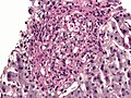
A granuloma is an aggregation of macrophages that forms in response to chronic inflammation. This occurs when the immune system attempts to isolate foreign substances that it is otherwise unable to eliminate. Such substances include infectious organisms including bacteria and fungi, as well as other materials such as foreign objects, keratin, and suture fragments.

<span class="mw-page-title-main">Blastomycosis</span> Medical condition

Blastomycosis, also known as Gilchrist's disease, is a fungal infection, typically of the lungs, which can spread to brain, stomach, intestine and skin, where it appears as crusting purplish warty plaques with a roundish bumpy edge and central depression. Only about half of people with the disease have symptoms, which can include fever, cough, night sweats, muscle pains, weight loss, chest pain, and feeling tired. Symptoms usually develop between three weeks and three months after breathing in the spores. In 25% to 40% of cases, the infection also spreads to other parts of the body, such as the skin, bones or central nervous system. Although blastomycosis is especially dangerous for those with weak immune systems, most people diagnosed with blastomycosis have healthy immune systems.

<span class="mw-page-title-main">Ziehl–Neelsen stain</span> Bacteriological technique

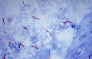
The Ziehl-Neelsen stain, also known as the acid-fast stain, is a bacteriological staining technique used in cytopathology and microbiology to identify acid-fast bacteria under microscopy, particularly members of the Mycobacterium genus. This staining method was initially introduced by Paul Ehrlich (1854–1915) and subsequently modified by the German bacteriologists Franz Ziehl (1859–1926) and Friedrich Neelsen (1854–1898) during the late 19th century.

<span class="mw-page-title-main">Opportunistic infection</span> Infection caused by pathogens that take advantage of an opportunity not normally available

An opportunistic infection is an infection caused by pathogens that take advantage of an opportunity not normally available. These opportunities can stem from a variety of sources, such as a weakened immune system, an altered microbiome, or breached integumentary barriers. Many of these pathogens do not necessarily cause disease in a healthy host that has a non-compromised immune system, and can, in some cases, act as commensals until the balance of the immune system is disrupted. Opportunistic infections can also be attributed to pathogens which cause mild illness in healthy individuals but lead to more serious illness when given the opportunity to take advantage of an immunocompromised host.

<span class="mw-page-title-main">Presumed ocular histoplasmosis syndrome</span> Medical condition

Presumed ocular histoplasmosis syndrome (POHS) is a syndrome affecting the eye, which is characterized by peripheral atrophic chorioretinal scars, atrophy or scarring adjacent to the optic disc and maculopathy.

<i>Histoplasma</i> Genus of fungi


Histoplasma is a genus of fungi in the order Onygenales. Species are known human pathogens producing yeast-like states under pathogenic conditions. They are the causative agents of histoplasmosis in humans and epizootic lymphangitis in horses.

<i>Blastomyces dermatitidis</i> Species of fungus

Blastomyces dermatitidis is a dimorphic fungus that causes blastomycosis, an invasive and often serious fungal infection found occasionally in humans and other animals. It lives in soil and wet, decaying wood, often in an area close to a waterway such as a lake, river or stream. Indoor growth may also occur, for example, in accumulated debris in damp sheds or shacks. The fungus is endemic to parts of eastern North America, particularly boreal northern Ontario, southeastern Manitoba, Quebec south of the St. Lawrence River, parts of the U.S. Appalachian mountains and interconnected eastern mountain chains, the west bank of Lake Michigan, the state of Wisconsin, and the entire Mississippi Valley including the valleys of some major tributaries such as the Ohio River. In addition, it occurs rarely in Africa both north and south of the Sahara Desert, as well as in the Arabian Peninsula and the Indian subcontinent. Though it has never been directly observed growing in nature, it is thought to grow there as a cottony white mold, similar to the growth seen in artificial culture at 25 °C (77 °F). In an infected human or animal, however, it converts in growth form and becomes a large-celled budding yeast. Blastomycosis is generally readily treatable with systemic antifungal drugs once it is correctly diagnosed; however, delayed diagnosis is very common except in highly endemic areas.

<span class="mw-page-title-main">Dimorphic fungus</span>

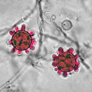
Dimorphic fungi are fungi that can exist in the form of both mold and yeast. This is usually brought about by change in temperature and the fungi are also described as thermally dimorphic fungi. An example is Talaromyces marneffei, a human pathogen that grows as a mold at room temperature, and as a yeast at human body temperature.

<span class="mw-page-title-main">Guano</span> Excrement of seabirds or bats

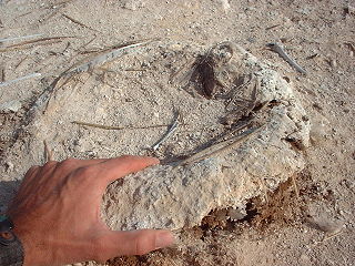
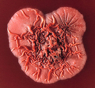
Guano is the accumulated excrement of seabirds or bats. Guano is a highly effective fertilizer due to the high content of nitrogen, phosphate, and potassium, all key nutrients essential for plant growth. Guano was also, to a lesser extent, sought for the production of gunpowder and other explosive materials.

Pathogenic fungi are fungi that cause disease in humans or other organisms. Although fungi are eukaryotic, many pathogenic fungi are microorganisms. Approximately 300 fungi are known to be pathogenic to humans; their study is called "medical mycology". Fungal infections kill more people than either tuberculosis or malaria—about 2 million people per year.

<span class="mw-page-title-main">Fungal meningitis</span> Meningitis caused by a fungal infection

Fungal meningitis refers to meningitis caused by a fungal infection.

African histoplasmosis is a fungal infection caused by Histoplasma capsulatumvar. duboisii, or Histoplama duboisii (Hcd). Disease has been most often reported in Uganda, Nigeria, Zaire and Senegal, as Hcd is exclusive to Africa. In human disease it manifests differently than histoplasmosis, most often involving the skin and bones and rarely involving the lungs. Also unlike Hcc, Hcd has been reported to rarely present in those with HIV, likely due to underreporting. However, this along with the differences in Hcc and Hcd have been disputed.

Ajellomyces is a genus of fungi in the division Ascomycota, in the family Ajellomycetaceae. The genus contains two species, which have a widespread distribution, especially in tropical areas. The species Ajellomyces capsulatus is significant to human health as the causative agent of histoplasmosis. This species is more usually referred to as Histoplasma capsulatum, with the designation Ajellomyces capsulatus referring to the ascomycetous perfect stage.

Emmonsia parva is a filamentous, saprotrophic fungus and one of three species within the genus Emmonsia. The fungus is most known for its causal association with the lung disease, adiaspiromycosis which occurs most commonly in small mammals but is also seen in humans. The disease was first described from rodents in Arizona, and the first human case was reported in France in 1964. Since then, the disease has been reported from Honduras, Brazil, the Czech Republic, Russia, the United States of America and Guatemala. Infections in general are quite rare, especially in humans.

Histoplasma duboisii is a saprotrophic fungus responsible for the invasive infection known as African histoplasmosis. This species is a close relative of Histoplasma capsulatum, the agent of classical histoplasmosis, and the two occur in similar habitats. Histoplasma duboisii is restricted to continental Africa and Madagascar, although scattered reports have arisen from other places usually in individuals with an African travel history. Like, H. capsulatum, H. duboisii is dimorphic – growing as a filamentous fungus at ambient temperature and a yeast at body temperature. It differs morphologically from H. capsulatum by the typical production of a large-celled yeast form. Both agents cause similar forms of disease, although H. duboisii predominantly causes cutaneous and subcutaneous disease in humans and non-human primates. The agent responds to many antifungal drug therapies used to treat serious fungal diseases.

Emmonsiosis, also known as emergomycosis, is a systemic fungal infection that can affect the lungs, generally always affects the skin and can become widespread. The lesions in the skin look like small red bumps and patches with a dip, ulcer and dead tissue in the centre.

<span class="mw-page-title-main">Chester Wilson Emmons</span> American mycologist (1900–1985)

Chester Wilson Emmons was an American scientist, who researched fungi that cause diseases. He was the first mycologist at the National Institutes of Health (NIH), where for 31 years he served as head of its Medical Mycology Section.

<span class="mw-page-title-main">Libero Ajello</span> American mycologist (1916–2004)

Libero Ajello was an American mycologist. He cofounded and was first president of the International Society of Human and Animal Mycology (ISHAM). He was the head of the Division of Mycotic Diseases at the Communicable Disease Center (CDC), and editor of the ISHAM Journal Medical Mycology for several years. He was one of the first researchers to investigate histoplasmosis and coccidioidomycosis in the United States and made valuable contributions to the comprehensive field of veterinary and human fungal disease diagnosis.

References

  1. 1 2 Chiller, Tom M. (2016), "Histoplasmosis", CDC Yellow Book: CDC Health Information for International Travel.
  2. 1 2 3 4 CDC (2014), Fungal Diseases > Global fungal diseases > Preventing Deaths from Histoplasmosis.
  3. Untereiner, W.A.; Scott, J.A.; Naveau, F.A.; Bachewich, J. (2002). "Phylogeny of Ajellomyces, Polytolypa and Spiromastix (Onygenaceae) inferred from rDNA sequence and non-molecular data". Studies in Mycology. 47: 25–35.
  4. Untereiner, WA; Scott, JA; Naveau, FA; Sigler, L; Bachewich, J; Angus, A (2004). "The Ajellomycetaceae, a new family of vertebrate-associated Onygenales". Mycologia. 96 (4): 812–821. doi:10.2307/3762114. JSTOR   3762114. PMID   21148901.
  5. Karimi, K; Wheat, LJ; Connolly, P; Cloud, G; Hajjeh, R; Wheat, E; Alves, K; Lacaz Cd Cda, S; Keath, E (1 December 2002). "Differences in histoplasmosis in patients with acquired immunodeficiency syndrome in the United States and Brazil". The Journal of Infectious Diseases. 186 (11): 1655–1660. doi: 10.1086/345724 . PMID   12447743.
  6. 1 2 3 Kasuga, T; White, TJ; Koenig, G; McEwen, J; Restrepo, A; Castañeda, E; Da Silva Lacaz, C; Heins-Vaccari, EM; De Freitas, RS; Zancopé-Oliveira, RM; Qin, Z; Negroni, R; Carter, DA; Mikami, Y; Tamura, M; Taylor, ML; Miller, GF; Poonwan, N; Taylor, JW (2003). "Phylogeography of the fungal pathogen Histoplasma capsulatum". Molecular Ecology. 12 (12): 3383–401. doi:10.1046/j.1365-294x.2003.01995.x. PMID   14629354. S2CID   13060796.
  7. 1 2 3 4 5 6 7 8 Kwon-Chung, K. June; Bennett, Joan E. (1992). Medical mycology. Philadelphia: Lea & Febiger. ISBN   978-0812114638.
  8. Chamany, S; Mirza, SA; Fleming, JW; Howell, JF; Lenhart, SW; Mortimer, VD; Phelan, MA; Lindsley, MD; Iqbal, NJ; Wheat, LJ; Brandt, ME; Warnock, DW; Hajjeh, RA (2004). "A large histoplasmosis outbreak among high school students in Indiana, 2001". The Pediatric Infectious Disease Journal. 23 (10): 909–14. doi:10.1097/01.inf.0000141738.60845.da. PMID   15602189. S2CID   23475233.
  9. Stobierski, MG; Hospedales, CJ; Hall, WN; Robinson-Dunn, B; Hoch, D; Sheill, DA (1996). "Outbreak of histoplasmosis among employees in a paper factory--Michigan, 1993". Journal of Clinical Microbiology. 34 (5): 1220–1223. doi:10.1128/JCM.34.5.1220-1223.1996. PMC   228985 . PMID   8727906.
  10. Guy, R; Roy, O (1949). "Histoplasmin sensitivity; preliminary observations in a group of school children in the Province of Quebec". Canadian Journal of Public Health. 40 (2): 68–71. PMID   18113223.
  11. 1 2 Leznoff, A; Frank, H; Taussig, A; Brandt, JL (1969). "The focal distribution of histoplasmosis in Montreal". Canadian Journal of Public Health. 60 (8): 321–5. PMID   5806145.
  12. 1 2 MacEachern, EJ; McDonald, JC (1971). "Histoplasmin sensitivity in McGill University students". Canadian Journal of Public Health. 62 (5): 415–422. PMID   5137623.
  13. 1 2 Haggar, R.A; Brown, E.L.; Toplack, N.J. (1957). "Histoplasmosis in South Western Ontario". Canadian Medical Association Journal. 77 (9): 855–861. PMC   1824134 . PMID   13472567.
  14. Rostocka, M; Hiltz, JE (1966). "Histoplasmin skin sensitivity in Nova Scotia". Canadian Journal of Public Health. 57 (9): 413–418. PMID   5981243.
  15. Hoff, B.; Fogle, B. (1970). "Case report. Histoplasmosis in a dog". Canadian Veterinary Journal. 11 (7): 145–148. PMC   1695089 . PMID   5464271.
  16. Mochi, A; Edwards, PQ (1952). "Geographical distribution of histoplasmosis and histoplasmin sensitivity". Bulletin of the World Health Organization. 5 (3): 259–291. PMC   2554039 . PMID   14935779.
  17. Guy, R; Panisset, M; Frappier, A (1949). "Histoplasmin sensitivity; a brief study of the incidence of hypersensitivity to histoplasmin in an Indian tribe of northern Quebec". Canadian Journal of Public Health. 40 (7): 306–309. PMID   18133141.
  18. Schaefer, O (1966). "Pulmonary miliary calcification and histoplasmin sensitivity in Canadian eskimos". Canadian Journal of Public Health. 57 (9): 410–418. PMID   5977447.
  19. Anderson, Heather; Honish, Lance; Taylor, Geoff; Johnson, Marcia; Tovstiuk, Chrystyna; Fanning, Anne; Tyrrell, Gregory; Rennie, Robert; Jaipaul, Joy; Sand, Crystal; Probert, Steven (2006). "Histoplasmosis Cluster, Golf Course, Canada". Emerging Infectious Diseases. 12 (1): 163–165. doi:10.3201/eid1201.051083. PMC   3291405 . PMID   16494738.
  20. 1 2 3 Kauffman, C. A. (2007). "Histoplasmosis: a Clinical and Laboratory Update". Clinical Microbiology Reviews. 20 (1): 115–132. doi:10.1128/CMR.00027-06. PMC   1797635 . PMID   17223625.
  21. Hajjeh, RA (1995). "Disseminated histoplasmosis in persons infected with human immunodeficiency virus". Clinical Infectious Diseases. 21 Suppl 1: S108–10. doi:10.1093/clinids/21.supplement_1.s108. PMID   8547497.
  22. 1 2 Rippon, John Willard (1988). Medical mycology: the pathogenic fungi and the pathogenic actinomycetes (3rd ed.). Philadelphia, PA: Saunders. ISBN   978-0721624440.
  23. Waldman, RJ; England, AC; Tauxe, R; Kline, T; Weeks, RJ; Ajello, L; Kaufman, L; Wentworth, B; Fraser, DW (1983). "A winter outbreak of acute histoplasmosis in northern Michigan". American Journal of Epidemiology. 117 (1): 68–75. doi:10.1093/oxfordjournals.aje.a113517. PMID   6823954.
  24. Leznoff, A; Frank, H; Telner, P; Rosensweig, J; Brandt, JL (1964). "Histoplasmosis in Montreal during the fall of 1963, with observations on erythema multiforme". Canadian Medical Association Journal. 91 (22): 1154–1160. PMC   1928451 . PMID   14226089.
  25. Emmons, C.W. (1961). "Isolation of Histoplasma capsulatum from soil in Washington, D.C." Public Health Reports. 76 (591–596): 591–5. doi:10.2307/4591218. JSTOR   4591218. PMC   1929641 . PMID   13696714.
  26. Reid, TM; Schafer, MP (1999). "Direct detection of Histoplasma capsulatum in soil suspensions by two-stage PCR". Molecular and Cellular Probes. 13 (4): 269–273. doi:10.1006/mcpr.1999.0247. PMID   10441199.
  27. Ajello, L.; Weeks, R.J. (1983). "Soil decontamination and other control measures". In DiSalvo, Arthur F. (ed.). Occupational mycoses. Philadelphia, Pa.: Lea and Febiger. pp.  229–238. ISBN   9780812108859.
  28. Mahajan, Monika (2021). "Etymologia: Histoplasma capsulatum". Emerging Infectious Diseases. 27 (3): 969. doi: 10.3201/eid2703.et2703 . PMC   7920646 . Citing public domain text from the CDC