Horseradish peroxidase

Last updated
Horseradish peroxidase
HRP-xray.png
Horseradish peroxidase C1 [1]
Identifiers
Organism Armoracia rusticana
SymbolPeroxidase C1A
Alt. symbolsPRXC1A
PDB 1W4W More structures
UniProt P00433
Other data
EC number 1.11.1.7

The enzyme horseradish peroxidase (HRP), found in the roots of horseradish, is used extensively in biochemistry applications. It is a metalloenzyme with many isoforms, of which the most studied type is C. It catalyzes the oxidation of various organic substrates by hydrogen peroxide.

Contents

Structure

The structure of the enzyme was first solved by X-ray crystallography in 1997 [2] and has since been solved several times with various substrates. [3] It is a large alpha-helical glycoprotein which binds heme as a redox cofactor.

Substrates

Alone, the HRP enzyme, or conjugates thereof, is of little value; its presence must be made visible using a substrate that, when oxidized by HRP using hydrogen peroxide as the oxidizing agent, yields a characteristic color change that is detectable by spectrophotometric methods. [4] [5]

Numerous substrates for horseradish peroxidase have been described and commercialized to exploit the desirable features of HRP. These substrates fall into several distinct categories. HRP catalyzes the conversion of chromogenic substrates (e.g., TMB, DAB, ABTS) into colored products, and produces light when acting on chemiluminescent substrates (e.g. Enhanced Chemiluminescence by luminol).[ citation needed ]

Some of the most common HRP chromogenic substrates. Luminol is the exception, as it is not a chromophore and light is generated after the HRP-catalyzed reaction. Structures of HRP substrates.png
Some of the most common HRP chromogenic substrates. Luminol is the exception, as it is not a chromophore and light is generated after the HRP-catalyzed reaction.

Applications

Horseradish peroxidase is a 44,173.9-dalton glycoprotein with 6 lysine residues which can be conjugated to a labeled molecule. It produces a coloured, fluorimetric, [6] or luminescent derivative of the labeled molecule when incubated with a proper substrate, allowing it to be detected and quantified. HRP is often used in conjugates (molecules that have been joined genetically or chemically) to determine the presence of a molecular target. For example, an antibody conjugated to HRP may be used to detect a small amount of a specific protein in a western blot. Here, the antibody provides the specificity to locate the protein of interest, and the HRP enzyme, in the presence of a substrate, produces a detectable signal. [7] Horseradish peroxidase is also commonly used in techniques such as ELISA and Immunohistochemistry due to its monomeric nature and the ease with which it produces coloured products. Peroxidase, a heme-containing oxidoreductase, is a commercially important enzyme which catalyses the reductive cleavage of hydrogen peroxide by an electron donor.

Horseradish peroxidase is ideal in many respects for these applications because it is smaller, more stable, and less expensive than other popular alternatives such as alkaline phosphatase. It also has a high turnover rate that allows generation of strong signals in a relatively short time span. [8] High concentrations of phosphate severely decrease stability of horseradish peroxidase. In addition to biomedical applications, horseradish peroxidase is one of the enzymes with important environmental applications. This enzyme is suitable for the removal of hydroxylated aromatic compounds (HACs) that are considered to be primary pollutants in a wide variety of industrial wastewater. [9]

Moreover, "In recent years the technique of marking neurons with the enzyme horseradish peroxidase has become a major tool. In its brief history, this method has probably been used by more neurobiologists than have used the Golgi stain since its discovery in 1870." [10]

Enhanced chemiluminescence (ECL)

Horseradish peroxidase catalyses the oxidation of luminol to 3-aminophthalate via several intermediates. The reaction is accompanied by emission of low-intensity light at 428 nm. However, in the presence of certain chemicals, the light emitted is enhanced up to 1000-fold, making the light easier to detect and increasing the sensitivity of the reaction. The enhancement of light emission is called enhanced chemiluminescence (ECL). Several enhancers can be used such as the commonly known modified phenols (mainly iodo-phenol). However, there are several substrates on the market that use other enhancers which result in luminescence signals up to 13 times greater than phenol-enhanced substrates. [11] The intensity of light is a measure of the number of enzyme molecules reacting and thus of the amount of hybrid. ECL is simple to set up and is sensitive, detecting about 0.5 pg nucleic acid in Southern blots and in northern blots. Detection by chemiluminescent substrates has several advantages over chromogenic substrates. The sensitivity is 10- to 100-fold greater, and quantifying of light emission is possible over a wide dynamic range, whereas that for coloured precipitates is much more limited, about one order of magnitude less. Stripping filters are much easier when chemiluminescent substrates are used.[ citation needed ]

HRP mimics

Many materials have been explored to mimic natural HRP. For example, iron oxide nanoparticles and hemin-containing complexes have been used to mimic HRP. [12] These HRP-like artificial enzymes have been used for many applications, ranging from biomarker detection and tumor immunostaining to antibiofouling.

See also

Related Research Articles

Northern blot Molecular biology technique

The northern blot, or RNA blot, is a technique used in molecular biology research to study gene expression by detection of RNA in a sample.

Peroxidase Peroxide decomposing Biocatalyst

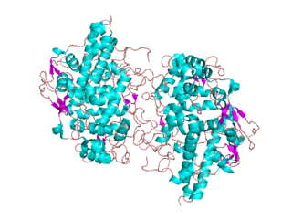
Peroxidases or peroxide reductases are a large group of enzymes which play a role in various biological processes. They are named after the fact that they commonly break up peroxides.

ELISA Method to detect an antigen using an antibody and enzyme

The enzyme-linked immunosorbent assay (ELISA) is a commonly used analytical biochemistry assay, first described by Eva Engvall and Peter Perlmann in 1971. The assay uses a solid-phase type of enzyme immunoassay (EIA) to detect the presence of a ligand in a liquid sample using antibodies directed against the protein to be measured. ELISA has been used as a diagnostic tool in medicine, plant pathology, and biotechnology, as well as a quality control check in various industries.

Western blot Analytical technique used in molecular biology

The western blot, or western blotting, is a widely used analytical technique in molecular biology and immunogenetics to detect specific proteins in a sample of tissue homogenate or extract.

Chemiluminescence Emission of light as a result of a chemical reaction

Chemiluminescence is the emission of light (luminescence) as the result of a chemical reaction. There may also be limited emission of heat. Given reactants A and B, with an excited intermediate ,

Luminol Chemical compound

Luminol (C8H7N3O2) is a chemical that exhibits chemiluminescence, with a blue glow, when mixed with an appropriate oxidizing agent. Luminol is a white-to-pale-yellow crystalline solid that is soluble in most polar organic solvents, but insoluble in water.

Immunostaining

In biochemistry, immunostaining is any use of an antibody-based method to detect a specific protein in a sample. The term "immunostaining" was originally used to refer to the immunohistochemical staining of tissue sections, as first described by Albert Coons in 1941. However, immunostaining now encompasses a broad range of techniques used in histology, cell biology, and molecular biology that use antibody-based staining methods.

Immunohistochemistry Common application of immunostaining

Immunohistochemistry (IHC) is the most common application of immunostaining. It involves the process of selectively identifying antigens (proteins) in cells of a tissue section by exploiting the principle of antibodies binding specifically to antigens in biological tissues. IHC takes its name from the roots "immuno", in reference to antibodies used in the procedure, and "histo", meaning tissue. Albert Coons conceptualized and first implemented the procedure in 1941.

Immunoassay Biochemical test for a protein or other molecule using an antibody

An immunoassay (IA) is a biochemical test that measures the presence or concentration of a macromolecule or a small molecule in a solution through the use of an antibody (usually) or an antigen (sometimes). The molecule detected by the immunoassay is often referred to as an "analyte" and is in many cases a protein, although it may be other kinds of molecules, of different sizes and types, as long as the proper antibodies that have the required properties for the assay are developed. Analytes in biological liquids such as serum or urine are frequently measured using immunoassays for medical and research purposes.

ABTS Chemical compound

In biochemistry, ABTS is a chemical compound used to observe the reaction kinetics of specific enzymes. A common use for it is in the enzyme-linked immunosorbent assay (ELISA) to detect the binding of molecules to each other.

Dot blot

A dot blot is a technique in molecular biology used to detect proteins. It represents a simplification of the western blot method, with the exception that the proteins to be detected are not first separated by electrophoresis. Instead, the sample is applied directly on a membrane in a single spot, and the blotting procedure is performed.

3,3,5,5-Tetramethylbenzidine Chemical compound

3,3′,5,5′-Tetramethylbenzidine or TMB is a chromogenic substrate used in staining procedures in immunohistochemistry as well as being a visualising reagent used in enzyme-linked immunosorbent assays (ELISA). TMB is a white solid that forms a pale blue-green liquid in solution with ethyl acetate. TMB is degraded by sunlight and by fluorescent lights.

Ascorbate peroxidase

Ascorbate peroxidase (or L-ascorbate peroxidase, APX) (EC 1.11.1.11) is an enzyme that catalyzes the chemical reaction

In enzymology, a manganese peroxidase (EC 1.11.1.13) is an enzyme that catalyzes the chemical reaction

Magnetic immunoassay (MIA) is a type of diagnostic immunoassay using magnetic beads as labels in lieu of conventional enzymes (ELISA), radioisotopes (RIA) or fluorescent moieties to detect a specified analyte. MIA involves the specific binding of an antibody to its antigen, where a magnetic label is conjugated to one element of the pair. The presence of magnetic beads is then detected by a magnetic reader (magnetometer) which measures the magnetic field change induced by the beads. The signal measured by the magnetometer is proportional to the analyte concentration in the initial sample.

A reverse phase protein lysate microarray (RPMA) is a protein microarray designed as a dot-blot platform that allows measurement of protein expression levels in a large number of biological samples simultaneously in a quantitative manner when high-quality antibodies are available.

Chromogenic in situ hybridization (CISH) is a cytogenetic technique that combines the chromogenic signal detection method of immunohistochemistry (IHC) techniques with in situ hybridization. It was developed around the year 2000 as an alternative to fluorescence in situ hybridization (FISH) for detection of HER-2/neu oncogene amplification. CISH is similar to FISH in that they are both in situ hybridization techniques used to detect the presence or absence of specific regions of DNA. However, CISH is much more practical in diagnostic laboratories because it uses bright-field microscopes rather than the more expensive and complicated fluorescence microscopes used in FISH.

Primary and secondary antibodies are two groups of antibodies that are classified based on whether they bind to antigens or proteins directly or target another (primary) antibody that, in turn, is bound to an antigen or protein.

Eclox, which stands for Enhanced ChemiLuminescence and OXyradical test for water quality analysis, is a rapid toxicity technology that has been shown to correlate with other established toxicity tests. Rapid toxicity testing is unable to identify specific contaminants or their concentrations and instead function as a screening tool to quickly determine whether water is potentially toxic.
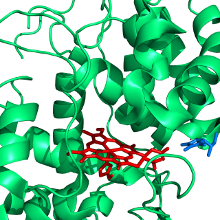
The enzyme-linked immune absorbent spot (ELISpot) is a type of assay that focuses on quantitatively measuring the frequency of cytokine secretion for a single cell. The ELISpot Assay is also a form of immunostaining since it is classified as a technique that uses antibodies to detect a protein analyte, with the word analyte referring to any biological or chemical substance being identified or measured.

References

  1. PDB: 1w4w ; Carlsson GH, Nicholls P, Svistunenko D, Berglund GI, Hajdu J (January 2005). "Complexes of horseradish peroxidase with formate, acetate, and carbon monoxide". Biochemistry. 44 (2): 635–42. doi:10.1021/bi0483211. PMID   15641789.
  2. PDB: 1ATJ ; Gajhede M, Schuller DJ, Henriksen A, Smith AT, Poulos TL (December 1997). "Crystal structure of horseradish peroxidase C at 2.15 A resolution". Nature Structural Biology. 4 (12): 1032–8. doi:10.1038/nsb1297-1032. PMID   9406554. S2CID   8535268.
  3. "Peroxidase C1A Related PDB sequences". UniPDB. European Bioinformatics Institute.
  4. Veitch NC (February 2004). "Horseradish peroxidase: a modern view of a classic enzyme". Phytochemistry. 65 (3): 249–59. doi:10.1016/j.phytochem.2003.10.022. PMID   14751298.
  5. Akkara JA, Senecal KJ, Kaplan DL (October 1991). "Synthesis and characterization of polymers produced by horseradish peroxidase in dioxane". Journal of Polymer Science. 29 (11): 1561–74. Bibcode:1991JPoSA..29.1561A. doi: 10.1002/pola.1991.080291105 .
  6. Acharya AP, Nafisi PM, Gardner A, MacKay JL, Kundu K, Kumar S, Murthy N (2013). "A fluorescent peroxidase probe increases the sensitivity of commercial ELISAs by two orders of magnitude". Chem Commun. 49 (88): 10379–10381. doi:10.1039/c3cc44783a. PMC   4011665 . PMID   24071916.
  7. Chau YP, Lu KS (1995). "Investigation of the blood-ganglion barrier properties in rat sympathetic ganglia by using lanthanum ion and horseradish peroxidase as tracers". Acta Anatomica. 153 (2): 135–44. doi:10.1159/000313647. PMID   8560966.
  8. Beyzavi K, Hampton S, Kwasowski P, Fickling S, Marks V, Clift R (March 1987). "Comparison of horseradish peroxidase and alkaline phosphatase-labelled antibodies in enzyme immunoassays". Annals of Clinical Biochemistry. 24 ( Pt 2) (2): 145–52. doi:10.1177/000456328702400204. PMID   3035992. S2CID   40978557.
  9. Ghasempur S, Torabi SF, Ranaei-Siadat SO, Jalali-Heravi M, Ghaemi N, Khajeh K (October 2007). "Optimization of peroxidase-catalyzed oxidative coupling process for phenol removal from wastewater using response surface methodology". Environmental Science & Technology. 41 (20): 7073–9. Bibcode:2007EnST...41.7073G. doi:10.1021/es070626q. PMID   17993150.
  10. Lichtman JW, Purves D (1985). "Cell marking with horseradish peroxidase". Principles of neural development. Sunderland, Mass: Sinauer Associates. p.  114. ISBN   978-0-87893-744-8.
  11. High Intensity HRP-Chemiluminescence ELISA Substrate Archived 2016-04-08 at the Wayback Machine . Haemoscan.com (2016-02-11). Retrieved on 2016-03-29.
  12. Wei H, Wang E (July 2013). "Nanomaterials with enzyme-like characteristics (nanozymes): next-generation artificial enzymes". Chemical Society Reviews. 42 (14): 6060–93. doi:10.1039/C3CS35486E. PMID   23740388.