This page is based on this
Wikipedia article Text is available under the
CC BY-SA 4.0 license; additional terms may apply.
Images, videos and audio are available under their respective licenses.

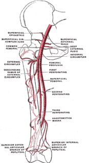
The femoral artery is a large artery in the thigh and the main arterial supply to the thigh and leg. It enters the thigh from behind the inguinal ligament as the continuation of the external iliac artery.

The external iliac veins are large veins that connect the femoral veins to the common iliac veins. Their origin is at the inferior margin of the inguinal ligaments and they terminate when they join the internal iliac veins.

The external iliac arteries are two major arteries which bifurcate off the common iliac arteries anterior to the sacroiliac joint of the pelvis. They proceed anterior and inferior along the medial border of the psoas major muscles. They exit the pelvic girdle posterior and inferior to the inguinal ligament about one third laterally from the insertion point of the inguinal ligament on the pubic tubercle at which point they are referred to as the femoral arteries. The external iliac artery is usually the artery used to attach the renal artery to the recipient of a kidney transplant.

The common iliac arteries are two large arteries that originate from the aortic bifurcation at the level of the fourth lumbar vertebra. They end in front of the sacroiliac joint, one on either side, and each bifurcates into the external and internal iliac arteries.

The external oblique muscle is the largest and the outermost of the three flat muscles of the lateral anterior abdomen.
The internal iliac artery is the main artery of the pelvis.

The iliolumbar artery is the first branch of the posterior trunk of the internal iliac artery.
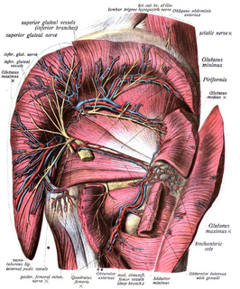
The inferior gluteal veins, or venæ comitantes of the inferior gluteal artery, begin on the upper part of the back of the thigh, where they anastomose with the medial femoral circumflex and first perforating veins.

The rectal venous plexus surrounds the rectum, and communicates in front with the vesical venous plexus in the male, and the vaginal venous plexus in the female.
The cruciate anastomosis is a circulatory anastomosis in the upper thigh of the inferior gluteal artery, the lateral and medial circumflex femoral arteries, and the first perforating artery of the profunda femoris artery. Also, the anastomotic branch of the posterior branch of the obturator artery. The cruciate anastomosis is clinically relevant because if there is a blockage between the femoral artery and external iliac artery, blood can reach the popliteal artery by means of the anastomosis. The route of blood is through the internal iliac, to the inferior gluteal artery, to a perforating branch of the deep femoral artery, to the lateral circumflex femoral artery, then to its descending branch into the superior lateral genicular artery and thus into the popliteal artery.

The internal iliac vein begins near the upper part of the greater sciatic foramen, passes upward behind and slightly medial to the internal iliac artery and, at the brim of the pelvis, joins with the external iliac vein to form the common iliac vein.

The deep circumflex iliac artery is an artery in the pelvis that travels along the iliac crest of the pelvic bone.
The superficial iliac circumflex artery, the smallest of the cutaneous branches of the femoral artery, arises close to the superficial epigastric artery, and, piercing the fascia lata, runs lateralward, parallel with the inguinal ligament, as far as the crest of the ilium.

The superficial external pudendal artery is one of the three pudendal arteries. It arises from the medial side of the femoral artery, close to the superficial epigastric artery and superficial iliac circumflex artery.
In the course of the round ligament of liver, small veins (paraumbilical) are found which establish an anastomosis between the veins of the anterior abdominal wall and the hepatic portal, hypogastric, and iliac veins.

The following outline is provided as an overview of and topical guide to human anatomy:
Iliac circumflex or Circumflex iliac can refer to:

