LIG3

Last updated
LIG3
PDB 1imo EBI.jpg
Available structures
PDB Ortholog search: PDBe RCSB
Identifiers
Aliases LIG3 , LIG2, DNA ligase 3, LIG3alpha, MTDPS20
External IDs OMIM: 600940 MGI: 109152 HomoloGene: 32109 GeneCards: LIG3
Orthologs
SpeciesHumanMouse
Entrez
Ensembl
UniProt
RefSeq (mRNA)

NM_002311
NM_013975

NM_001291245
NM_001291246
NM_001291247
NM_010716

RefSeq (protein)

NP_002302
NP_039269

n/a

Location (UCSC) Chr 17: 34.98 – 35.01 Mb Chr 11: 82.67 – 82.7 Mb
PubMed search [3] [4]
Wikidata
View/Edit Human View/Edit Mouse

DNA ligase 3 is an enzyme that, in humans, is encoded by the LIG3 gene. [5] [6] The human LIG3 gene encodes ATP-dependent DNA ligases that seal interruptions in the phosphodiester backbone of duplex DNA.

Contents

There are three families of ATP-dependent DNA ligases in eukaryotes. [7] These enzymes utilize the same three step reaction mechanism; (i) formation of a covalent enzyme-adenylate intermediate; (ii) transfer of the adenylate group to the 5' phosphate terminus of a DNA nick; (iii) phosphodiester bond formation. Unlike LIG1 and LIG4 family members that are found in almost all eukaryotes, LIG3 family members are less widely distributed. [8] The LIG3 gene encodes several distinct DNA ligase species by alternative translation initiation and alternative splicing mechanisms that are described below.

Structure, DNA binding and catalytic activities

Eukaryotic ATP-dependent DNA ligases have related catalytic region that contains three domains, a DNA binding domain, an adenylation domain and an oligonucleotide / oligosaccharide binding-fold domain. When these enzymes engage a nick in duplex DNA, these domains encircle the DNA duplex with each one making contact with the DNA. The structure of the catalytic region of DNA ligase III complexed with a nicked DNA has been determined by X-ray crystallography and is remarkably similar to that formed by the catalytic region of human DNA ligase I bound to nicked DNA. [9] A unique feature of the DNA ligases encoded by the LIG3 gene is an N-terminal zinc finger that resembles the two zinc fingers at the N-terminus of poly (ADP-ribose) polymerase 1 (PARP1). [10] As with the PARP1 zinc fingers, the DNA ligase III zinc finger is involved in binding to DNA strand breaks. [10] [11] [12] Within the DNA ligase III polypeptide, the zinc finger co-operates with the DNA binding domain to form a DNA binding module. [13] In addition, the adenylation domain and an oligonucleotide/oligosaccharide binding-fold domain form a second DNA binding module. [13] In the jackknife model proposed by the Ellenberger laboratory, [13] the zinc finger-DNA binding domain module serves as a strand break sensor that binds to DNA single strand interruptions irrespective of the nature of the strand break termini. If these breaks are ligatable, they are transferred to the adenylation domain-oligonucleotide/oligosaccharide binding-fold domain module that binds specifically to ligatable nicks. Compared with DNA ligases I and IV, DNA ligase III is the most active enzyme in the intermolecular joining of DNA duplexes. [14] This activity is predominantly dependent upon the DNA ligase III zinc finger suggesting that the two DNA binding modules of DNA ligase III may be able to simultaneously engage duplex DNA ends. [9] [13]

Alternative splicing

The alternative translation initiation and splicing mechanisms alter the amino- and carboxy-terminal sequences that flank the DNA ligase III catalytic region. [15] [16] In the alternative splicing mechanism, the exon encoding a C-terminal breast cancer susceptibility protein 1 C-terminal (BRCT) domain at the C-terminus of DNA ligase III-alpha is replaced by a short positively charged sequence that acts as a nuclear localization signal, generating DNA ligase III-beta. This alternatively spliced variant has, to date, only been detected in male germs cells. [16] Based on its expression pattern during spermatogenesis, it appears likely that DNA ligase IIIbeta is involved in meiotic recombination and/or DNA repair in haploid sperm but this has not been definitively demonstrated. Although an internal ATG is the preferred site for translation initiation within the DNA ligase III open reading frame, translation initiations does also occur at the first ATG within the open reading frame, resulting in the synthesis of a polypeptide with an N-terminal mitochondrial targeting sequence. [15] [17] [18]

Cellular function

As mentioned above, DNA ligase III-alpha mRNA encodes nuclear and mitochondrial versions of DNA ligase III-alpha. Nuclear DNA ligase III-alpha exists and functions in a stable complex with the DNA repair protein XRCC1. [19] [20] These proteins interact via their C-terminal BRCT domains. [16] [21] XRCC1 has no enzymatic activity but instead appears to acts as a scaffold protein by interacting with a large number of proteins involved in base excision and single-strand break repair. The participation of XRCC1 in these pathways is consistent with the phenotype of xrcc1 cells. [19] In contrast to nuclear DNA ligase III-alpha, mitochondrial DNA ligase III-alpha functions independently of XRCC1, which is not found in mitochondria. [22] It appears that nuclear DNA ligase III-alpha forms a complex with XRCC1 in the cytoplasm and the subsequent nuclear targeting of the resultant complex is directed by the XRCC1 nuclear localization signal. [23] While mitochondrial DNA ligase III-alpha also interacts with XRCC1, it is likely that the activity of the mitochondrial targeting sequence of DNA ligase III-alpha is greater than the activity of the XRCC1 nuclear localization signal and that the DNA ligase III-alpha/XRCC1 complex is disrupted when mitochondrial DNA ligase III-alpha passes through the mitochondrial membrane.

Since the LIG3 gene encodes the only DNA ligase in mitochondria, inactivation of the LIG3 gene results in loss of mitochondrial DNA that in turn leads to loss of mitochondrial function. [24] [25] [26] Fibroblasts with inactivated Lig3 gene can be propagated in the media supplemented with uridine and pyruvate. However, these cells lack mtDNA. [27] Physiological levels of mitochondrial DNA ligase III appear excessive, and cells with 100-fold reduced mitochondrial content of mitochondrial DNA ligase III-alpha maintain normal mtDNA copy number. [27] The essential role of DNA ligase III-alpha in mitochondrial DNA metabolism can be fulfilled by other DNA ligases, including the NAD-dependent DNA ligase of E. coli , if they are targeted to mitochondria. [24] [26] Thus, viable cells that lack nuclear DNA ligase III-alpha can be generated. While DNA ligase I is the predominant enzyme that joins Okazaki fragments during DNA replication, it is now evident that the DNA ligase III-alpha/XRCC1 complex enables cells that either lack or have reduced DNA ligase I activity to complete DNA replication. [24] [26] [28] [29] Given the biochemical and cell biology studies linking the DNA ligase III-alpha/XRCC1 complex with excision repair and the repair of DNA single strand breaks, [30] [31] [32] [33] it was surprising that the cells lacking nuclear DNA ligase III-alpha did not exhibit significantly increased sensitivity to DNA damaging agent. [24] [26] These studies suggest that there is significant functional redundancy between DNA ligase I and DNA ligase III-alpha in these nuclear DNA repair pathways. In mammalian cells, most DNA double strand breaks are repaired by DNA ligase IV-dependent non-homologous end joining (NHEJ). [34] DNA ligase III-alpha participates in a minor alternative NHEJ pathway that generates chromosomal translocations. [35] [36] Unlike the other nuclear DNA repair functions, it appears that the role of DNA ligase III-alpha in alternative NHEJ is independent of XRCC1. [37]

Clinical significance

Unlike the LIG1 and LIG4 genes, [38] [39] [40] [41] inherited mutations in the LIG3 gene have not been identified in the human population. DNA ligase III-alpha has, however, been indirectly implicated in cancer and neurodegenerative diseases. In cancer, DNA ligase III-alpha is frequently overexpressed and this serves as a biomarker to identify cells that are more dependent upon the alternative NHEJ pathway for the repair of DNA double strand breaks. [42] [43] [44] [45] Although the increased activity of the alternative NHEJ pathway causes genomic instability that drives disease progression, it also constitutes a novel target for the development of cancer cell-specific therapeutic strategies. [43] [44] Several genes encoding proteins that interact directly with DNA ligase III-alpha or indirectly via interactions with XRCC1 have been identified as being mutated in inherited neurodegenerative diseases. [46] [47] [48] [49] [50] Thus, it appears that DNA transactions involving DNA ligase III-alpha play an important role in maintaining the viability of neuronal cells.

LIG3 has a role in microhomology-mediated end joining (MMEJ) repair of double strand breaks. It is one of 6 enzymes required for this error prone DNA repair pathway. [51] LIG3 is upregulated in chronic myeloid leukemia, [45] multiple myeloma, [52] and breast cancer. [43]

Cancers are very often deficient in expression of one or more DNA repair genes, but over-expression of a DNA repair gene is less usual in cancer. For instance, at least 36 DNA repair enzymes, when mutationally defective in germ line cells, cause increased risk of cancer (hereditary cancer syndromes).[ citation needed ] (Also see DNA repair-deficiency disorder.) Similarly, at least 12 DNA repair genes have frequently been found to be epigenetically repressed in one or more cancers.[ citation needed ] (See also Epigenetically reduced DNA repair and cancer.) Ordinarily, deficient expression of a DNA repair enzyme results in increased un-repaired DNA damages which, through replication errors (translesion synthesis), lead to mutations and cancer. However, LIG3 mediated MMEJ repair is highly inaccurate, so in this case, over-expression, rather than under-expression, apparently leads to cancer.

Notes

Related Research Articles

<span class="mw-page-title-main">DNA ligase</span> Class of enzymes

DNA ligase is a type of enzyme that facilitates the joining of DNA strands together by catalyzing the formation of a phosphodiester bond. It plays a role in repairing single-strand breaks in duplex DNA in living organisms, but some forms may specifically repair double-strand breaks. Single-strand breaks are repaired by DNA ligase using the complementary strand of the double helix as a template, with DNA ligase creating the final phosphodiester bond to fully repair the DNA.

<span class="mw-page-title-main">DNA polymerase</span> Form of DNA replication

A DNA polymerase is a member of a family of enzymes that catalyze the synthesis of DNA molecules from nucleoside triphosphates, the molecular precursors of DNA. These enzymes are essential for DNA replication and usually work in groups to create two identical DNA duplexes from a single original DNA duplex. During this process, DNA polymerase "reads" the existing DNA strands to create two new strands that match the existing ones. These enzymes catalyze the chemical reaction

DNA glycosylases are a family of enzymes involved in base excision repair, classified under EC number EC 3.2.2. Base excision repair is the mechanism by which damaged bases in DNA are removed and replaced. DNA glycosylases catalyze the first step of this process. They remove the damaged nitrogenous base while leaving the sugar-phosphate backbone intact, creating an apurinic/apyrimidinic site, commonly referred to as an AP site. This is accomplished by flipping the damaged base out of the double helix followed by cleavage of the N-glycosidic bond.

<span class="mw-page-title-main">Poly (ADP-ribose) polymerase</span> Family of proteins

Poly (ADP-ribose) polymerase (PARP) is a family of proteins involved in a number of cellular processes such as DNA repair, genomic stability, and programmed cell death.

<span class="mw-page-title-main">Chromosome 2</span> Human chromosome

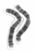
Chromosome 2 is one of the twenty-three pairs of chromosomes in humans. People normally have two copies of this chromosome. Chromosome 2 is the second-largest human chromosome, spanning more than 242 million base pairs and representing almost eight percent of the total DNA in human cells.

<span class="mw-page-title-main">MUTYH</span> Protein-coding gene in the species Homo sapiens

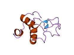
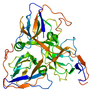
MUTYH is a human gene that encodes a DNA glycosylase, MUTYH glycosylase. It is involved in oxidative DNA damage repair and is part of the base excision repair pathway. The enzyme excises adenine bases from the DNA backbone at sites where adenine is inappropriately paired with guanine, cytosine, or 8-oxo-7,8-dihydroguanine, a common form of oxidative DNA damage.

<span class="mw-page-title-main">Proliferating cell nuclear antigen</span> Mammalian protein found in Homo sapiens

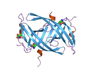
Proliferating cell nuclear antigen (PCNA) is a DNA clamp that acts as a processivity factor for DNA polymerase δ in eukaryotic cells and is essential for replication. PCNA is a homotrimer and achieves its processivity by encircling the DNA, where it acts as a scaffold to recruit proteins involved in DNA replication, DNA repair, chromatin remodeling and epigenetics.

<span class="mw-page-title-main">Protein inhibitor of activated STAT</span>

Protein inhibitor of activated STAT (PIAS), also known as E3 SUMO-protein ligase PIAS, is a protein that regulates transcription in mammals. PIAS proteins act as transcriptional co-regulators with at least 60 different proteins in order to either activate or repress transcription. The transcription factors STAT, NF-κB, p73, and p53 are among the many proteins that PIAS interacts with.

<span class="mw-page-title-main">XRCC1</span> Protein

DNA repair protein XRCC1, also known as X-ray repair cross-complementing protein 1, is a protein that in humans is encoded by the XRCC1 gene. XRCC1 is involved in DNA repair, where it complexes with DNA ligase III.

<span class="mw-page-title-main">DNA repair protein XRCC4</span> Protein-coding gene in the species Homo sapiens

DNA repair protein XRCC4 also known as X-ray repair cross-complementing protein 4 or XRCC4 is a protein that in humans is encoded by the XRCC4 gene. In addition to humans, the XRCC4 protein is also expressed in many other metazoans, fungi and in plants. The X-ray repair cross-complementing protein 4 is one of several core proteins involved in the non-homologous end joining (NHEJ) pathway to repair DNA double strand breaks (DSBs).

<span class="mw-page-title-main">MRE11A</span> Protein-coding gene in the species Homo sapiens

Double-strand break repair protein MRE11 is an enzyme that in humans is encoded by the MRE11 gene. The gene has been designated MRE11A to distinguish it from the pseudogene MRE11B that is nowadays named MRE11P1.

<span class="mw-page-title-main">LIG4</span> Protein-coding gene in the species Homo sapiens

DNA ligase 4 is an enzyme that in humans is encoded by the LIG4 gene.

<span class="mw-page-title-main">LIG1</span> Protein-coding gene in the species Homo sapiens

DNA ligase 1 is an enzyme that in humans is encoded by the LIG1 gene. DNA ligase I is the only known eukaryotic DNA ligase involved in both DNA replication and repair, making it the most studied of the ligases.

<span class="mw-page-title-main">Flap structure-specific endonuclease 1</span> Protein-coding gene in the species Homo sapiens

Flap endonuclease 1 is an enzyme that in humans is encoded by the FEN1 gene.

<span class="mw-page-title-main">Aprataxin</span> Protein-coding gene in the species Homo sapiens

Aprataxin is a protein that in humans is encoded by the APTX gene.

<span class="mw-page-title-main">ERCC4</span> Protein-coding gene in the species Homo sapiens

ERCC4 is a protein designated as DNA repair endonuclease XPF that in humans is encoded by the ERCC4 gene. Together with ERCC1, ERCC4 forms the ERCC1-XPF enzyme complex that participates in DNA repair and DNA recombination.

<span class="mw-page-title-main">DUT (gene)</span> Protein-coding gene in the species Homo sapiens

DUTP pyrophosphatase, also known as DUT, is an enzyme which in humans is encoded by the DUT gene on chromosome 15.

<span class="mw-page-title-main">PNKP</span> Protein-coding gene in the species Homo sapiens

Bifunctional polynucleotide phosphatase/kinase is an enzyme that in humans is encoded by the PNKP gene. A detailed structural study of the crystallized mouse protein examined both the 5´-polynucleotide kinase and 3’-polynucleotide phosphatase activities. Additional features of the peptide sequence include a forkhead association (FHA) domain, ATP binding site and nuclear and mitochondrial localization sequences.

<span class="mw-page-title-main">PARP2</span> Protein-coding gene in the species Homo sapiens

Poly [ADP-ribose] polymerase 2 is an enzyme that in humans is encoded by the PARP2 gene. It is one of the PARP family of enzymes.

<span class="mw-page-title-main">Single-stranded binding protein</span>

Single-stranded binding proteins (SSBs) are a class of proteins that have been identified in both viruses and organisms from bacteria to humans.

References

  1. 1 2 3 GRCh38: Ensembl release 89: ENSG00000005156 - Ensembl, May 2017
  2. 1 2 3 GRCm38: Ensembl release 89: ENSMUSG00000020697 - Ensembl, May 2017
  3. "Human PubMed Reference:". National Center for Biotechnology Information, U.S. National Library of Medicine.
  4. "Mouse PubMed Reference:". National Center for Biotechnology Information, U.S. National Library of Medicine.
  5. "Entrez Gene: Ligase III, DNA, ATP-dependent" . Retrieved 2012-03-12.
  6. Tomkinson AE, Sallmyr A (December 2013). "Structure and function of the DNA ligases encoded by the mammalian LIG3 gene". Gene. 531 (2): 150–7. doi:10.1016/j.gene.2013.08.061. PMC   3881560 . PMID   24013086.
  7. Ellenberger T, Tomkinson AE (2008). "Eukaryotic DNA ligases: structural and functional insights". Annu. Rev. Biochem. 77: 313–38. doi:10.1146/annurev.biochem.77.061306.123941. PMC   2933818 . PMID   18518823.
  8. Simsek D, Jasin M (November 2011). "DNA ligase III: a spotty presence in eukaryotes, but an essential function where tested". Cell Cycle. 10 (21): 3636–44. doi:10.4161/cc.10.21.18094. PMC   3266004 . PMID   22041657.
  9. 1 2 Cotner-Gohara E, Kim IK, Hammel M, Tainer JA, Tomkinson AE, Ellenberger T (July 2010). "Human DNA ligase III recognizes DNA ends by dynamic switching between two DNA-bound states". Biochemistry. 49 (29): 6165–76. doi:10.1021/bi100503w. PMC   2922849 . PMID   20518483.
  10. 1 2 Mackey ZB, Niedergang C, Murcia JM, Leppard J, Au K, Chen J, de Murcia G, Tomkinson AE (July 1999). "DNA ligase III is recruited to DNA strand breaks by a zinc finger motif homologous to that of poly(ADP-ribose) polymerase. Identification of two functionally distinct DNA binding regions within DNA ligase III". Journal of Biological Chemistry. 274 (31): 21679–87. doi: 10.1074/jbc.274.31.21679 . PMID   10419478.
  11. Leppard JB, Dong Z, Mackey ZB, Tomkinson AE (August 2003). "Physical and functional interaction between DNA ligase IIIalpha and poly(ADP-Ribose) polymerase 1 in DNA single-strand break repair". Molecular and Cellular Biology. 23 (16): 5919–27. doi:10.1128/MCB.23.16.5919-5927.2003. PMC   166336 . PMID   12897160.
  12. Taylor RM, Whitehouse CJ, Caldecott KW (September 2000). "The DNA ligase III zinc finger stimulates binding to DNA secondary structure and promotes end joining". Nucleic Acids Res. 28 (18): 3558–63. doi:10.1093/nar/28.18.3558. PMC   110727 . PMID   10982876.
  13. 1 2 3 4 Cotner-Gohara E, Kim IK, Tomkinson AE, Ellenberger T (April 2008). "Two DNA-binding and nick recognition modules in human DNA ligase III". Journal of Biological Chemistry. 283 (16): 10764–72. doi: 10.1074/jbc.M708175200 . PMC   2447648 . PMID   18238776.
  14. Chen L, Trujillo K, Sung P, Tomkinson AE (August 2000). "Interactions of the DNA ligase IV-XRCC4 complex with DNA ends and the DNA-dependent protein kinase". Journal of Biological Chemistry. 275 (34): 26196–205. doi: 10.1074/jbc.M000491200 . PMID   10854421.
  15. 1 2 Lakshmipathy U, Campbell C (May 1999). "The human DNA ligase III gene encodes nuclear and mitochondrial proteins". Molecular and Cellular Biology. 19 (5): 3869–76. doi:10.1128/MCB.19.5.3869. PMC   84244 . PMID   10207110.
  16. 1 2 3 Mackey ZB, Ramos W, Levin DS, Walter CA, McCarrey JR, Tomkinson AE (February 1997). "An alternative splicing event which occurs in mouse pachytene spermatocytes generates a form of DNA ligase III with distinct biochemical properties that may function in meiotic recombination". Molecular and Cellular Biology. 17 (2): 989–98. doi:10.1128/MCB.17.2.989. PMC   231824 . PMID   9001252.
  17. Wei YF, Robins P, Carter K, Caldecott K, Pappin DJ, Yu GL, Wang RP, Shell BK, Nash RA, Schär P (June 1995). "Molecular cloning and expression of human cDNAs encoding a novel DNA ligase IV and DNA ligase III, an enzyme active in DNA repair and recombination". Molecular and Cellular Biology. 15 (6): 3206–16. doi:10.1128/mcb.15.6.3206. PMC   230553 . PMID   7760816.
  18. Chen J, Tomkinson AE, Ramos W, Mackey ZB, Danehower S, Walter CA, Schultz RA, Besterman JM, Husain I (October 1995). "Mammalian DNA ligase III: molecular cloning, chromosomal localization, and expression in spermatocytes undergoing meiotic recombination". Molecular and Cellular Biology. 15 (10): 5412–22. doi:10.1128/MCB.15.10.5412. PMC   230791 . PMID   7565692.
  19. 1 2 Caldecott KW, McKeown CK, Tucker JD, Ljungquist S, Thompson LH (January 1994). "An interaction between the mammalian DNA repair protein XRCC1 and DNA ligase III". Molecular and Cellular Biology. 14 (1): 68–76. doi:10.1128/MCB.14.1.68. PMC   358357 . PMID   8264637.
  20. Caldecott KW, Tucker JD, Stanker LH, Thompson LH (December 1995). "Characterization of the XRCC1-DNA ligase III complex in vitro and its absence from mutant hamster cells". Nucleic Acids Res. 23 (23): 4836–43. doi:10.1093/nar/23.23.4836. PMC   307472 . PMID   8532526.
  21. Nash RA, Caldecott KW, Barnes DE, Lindahl T (April 1997). "XRCC1 protein interacts with one of two distinct forms of DNA ligase III". Biochemistry. 36 (17): 5207–11. doi:10.1021/bi962281m. PMID   9136882.
  22. Lakshmipathy U, Campbell C (October 2000). "Mitochondrial DNA ligase III function is independent of Xrcc1". Nucleic Acids Res. 28 (20): 3880–6. doi:10.1093/nar/28.20.3880. PMC   110795 . PMID   11024166.
  23. Parsons JL, Dianova II, Finch D, Tait PS, Ström CE, Helleday T, Dianov GL (July 2010). "XRCC1 phosphorylation by CK2 is required for its stability and efficient DNA repair". DNA Repair (Amst.). 9 (7): 835–41. doi: 10.1016/j.dnarep.2010.04.008 . PMID   20471329.
  24. 1 2 3 4 Gao Y, Katyal S, Lee Y, Zhao J, Rehg JE, Russell HR, McKinnon PJ (March 2011). "DNA ligase III is critical for mtDNA integrity but not Xrcc1-mediated nuclear DNA repair". Nature. 471 (7337): 240–4. Bibcode:2011Natur.471..240G. doi:10.1038/nature09773. PMC   3079429 . PMID   21390131.
  25. Lakshmipathy U, Campbell C (February 2001). "Antisense-mediated decrease in DNA ligase III expression results in reduced mitochondrial DNA integrity". Nucleic Acids Res. 29 (3): 668–76. doi:10.1093/nar/29.3.668. PMC   30390 . PMID   11160888.
  26. 1 2 3 4 Simsek D, Furda A, Gao Y, Artus J, Brunet E, Hadjantonakis AK, Van Houten B, Shuman S, McKinnon PJ, Jasin M (March 2011). "Crucial role for DNA ligase III in mitochondria but not in Xrcc1-dependent repair". Nature. 471 (7337): 245–8. Bibcode:2011Natur.471..245S. doi:10.1038/nature09794. PMC   3261757 . PMID   21390132.
  27. 1 2 Shokolenko IN, Fayzulin RZ, Katyal S, McKinnon PJ, Alexeyev MF (Sep 13, 2013). "Mitochondrial DNA ligase is dispensable for the viability of cultured cells but essential for mtDNA maintenance". Journal of Biological Chemistry. 288 (37): 26594–605. doi: 10.1074/jbc.M113.472977 . PMC   3772206 . PMID   23884459.
  28. Arakawa H, Bednar T, Wang M, Paul K, Mladenov E, Bencsik-Theilen AA, Iliakis G (March 2012). "Functional redundancy between DNA ligases I and III in DNA replication in vertebrate cells". Nucleic Acids Research. 40 (6): 2599–610. doi:10.1093/nar/gkr1024. PMC   3315315 . PMID   22127868.
  29. Le Chalony C, Hoffschir F, Gauthier LR, Gross J, Biard DS, Boussin FD, Pennaneach V (September 2012). "Partial complementation of a DNA ligase I deficiency by DNA ligase III and its impact on cell survival and telomere stability in mammalian cells". Cellular and Molecular Life Sciences. 69 (17): 2933–49. doi:10.1007/s00018-012-0975-8. PMC   3417097 . PMID   22460582.
  30. Cappelli E, Taylor R, Cevasco M, Abbondandolo A, Caldecott K, Frosina G (September 1997). "Involvement of XRCC1 and DNA ligase III gene products in DNA base excision repair". Journal of Biological Chemistry. 272 (38): 23970–5. doi: 10.1074/jbc.272.38.23970 . PMID   9295348.
  31. Okano S, Lan L, Tomkinson AE, Yasui A (2005). "Translocation of XRCC1 and DNA ligase III-alpha from centrosomes to chromosomes in response to DNA damage in mitotic human cells". Nucleic Acids Res. 33 (1): 422–9. doi:10.1093/nar/gki190. PMC   546168 . PMID   15653642.
  32. Kubota Y, Nash RA, Klungland A, Schär P, Barnes DE, Lindahl T (December 1996). "Reconstitution of DNA base excision-repair with purified human proteins: interaction between DNA polymerase beta and the XRCC1 protein". EMBO Journal. 15 (23): 6662–70. doi:10.1002/j.1460-2075.1996.tb01056.x. PMC   452490 . PMID   8978692.
  33. Moser J, Kool H, Giakzidis I, Caldecott K, Mullenders LH, Fousteri MI (July 2007). "Sealing of chromosomal DNA nicks during nucleotide excision repair requires XRCC1 and DNA ligase III alpha in a cell-cycle-specific manner" (PDF). Molecular Cell. 27 (2): 311–23. doi:10.1016/j.molcel.2007.06.014. PMID   17643379. S2CID   6333969.
  34. Lieber MR (2010). "The mechanism of double-strand DNA break repair by the nonhomologous DNA end-joining pathway". Annu. Rev. Biochem. 79: 181–211. doi:10.1146/annurev.biochem.052308.093131. PMC   3079308 . PMID   20192759.
  35. Wang H, Rosidi B, Perrault R, Wang M, Zhang L, Windhofer F, Iliakis G (May 2005). "DNA ligase III as a candidate component of backup pathways of nonhomologous end joining". Cancer Res. 65 (10): 4020–30. doi: 10.1158/0008-5472.CAN-04-3055 . PMID   15899791.
  36. Simsek D, Brunet E, Wong SY, Katyal S, Gao Y, McKinnon PJ, Lou J, Zhang L, Li J, Rebar EJ, Gregory PD, Holmes MC, Jasin M (June 2011). Haber JE (ed.). "DNA ligase III promotes alternative nonhomologous end-joining during chromosomal translocation formation". PLOS Genet. 7 (6): e1002080. doi: 10.1371/journal.pgen.1002080 . PMC   3107202 . PMID   21655080.
  37. Boboila C, Oksenych V, Gostissa M, Wang JH, Zha S, Zhang Y, Chai H, Lee CS, Jankovic M, Saez LM, Nussenzweig MC, McKinnon PJ, Alt FW, Schwer B (February 2012). "Robust chromosomal DNA repair via alternative end-joining in the absence of X-ray repair cross-complementing protein 1 (XRCC1)". Proceedings of the National Academy of Sciences, USA. 109 (7): 2473–8. Bibcode:2012PNAS..109.2473B. doi: 10.1073/pnas.1121470109 . PMC   3289296 . PMID   22308491.
  38. Girard PM, Kysela B, Härer CJ, Doherty AJ, Jeggo PA (October 2004). "Analysis of DNA ligase IV mutations found in LIG4 syndrome patients: the impact of two linked polymorphisms". Human Molecular Genetics. 13 (20): 2369–76. doi: 10.1093/hmg/ddh274 . PMID   15333585.
  39. O'Driscoll M, Cerosaletti KM, Girard PM, Dai Y, Stumm M, Kysela B, Hirsch B, Gennery A, Palmer SE, Seidel J, Gatti RA, Varon R, Oettinger MA, Neitzel H, Jeggo PA, Concannon P (December 2001). "DNA ligase IV mutations identified in patients exhibiting developmental delay and immunodeficiency". Molecular Cell. 8 (6): 1175–85. doi: 10.1016/S1097-2765(01)00408-7 . PMID   11779494.
  40. Riballo E, Critchlow SE, Teo SH, Doherty AJ, Priestley A, Broughton B, Kysela B, Beamish H, Plowman N, Arlett CF, Lehmann AR, Jackson SP, Jeggo PA (July 1999). "Identification of a defect in DNA ligase IV in a radiosensitive leukaemia patient". Curr. Biol. 9 (13): 699–702. Bibcode:1999CBio....9..699R. doi: 10.1016/S0960-9822(99)80311-X . PMID   10395545. S2CID   17103936.
  41. Barnes DE, Tomkinson AE, Lehmann AR, Webster AD, Lindahl T (May 1992). "Mutations in the DNA ligase I gene of an individual with immunodeficiencies and cellular hypersensitivity to DNA-damaging agents". Cell. 69 (3): 495–503. doi:10.1016/0092-8674(92)90450-Q. PMID   1581963. S2CID   11736507.
  42. Chen X, Zhong S, Zhu X, Dziegielewska B, Ellenberger T, Wilson GM, MacKerell AD, Tomkinson AE (May 2008). "Rational design of human DNA ligase inhibitors that target cellular DNA replication and repair". Cancer Res. 68 (9): 3169–77. doi:10.1158/0008-5472.CAN-07-6636. PMC   2734474 . PMID   18451142.
  43. 1 2 3 Tobin LA, Robert C, Nagaria P, Chumsri S, Twaddell W, Ioffe OB, Greco GE, Brodie AH, Tomkinson AE, Rassool FV (2012). "Targeting abnormal DNA repair in therapy-resistant breast cancers". Molecular Cancer Research. 10 (1): 96–107. doi:10.1158/1541-7786.MCR-11-0255. PMC   3319138 . PMID   22112941.
  44. 1 2 Tobin LA, Robert C, Rapoport AP, Gojo I, Baer MR, Tomkinson AE, Rassool FV (April 2013). "Targeting abnormal DNA double-strand break repair in tyrosine kinase inhibitor-resistant chronic myeloid leukemias". Oncogene. 32 (14): 1784–93. doi:10.1038/onc.2012.203. PMC   3752989 . PMID   22641215.
  45. 1 2 Sallmyr A, Tomkinson AE, Rassool FV (August 2008). "Up-regulation of WRN and DNA ligase III-alpha in chronic myeloid leukemia: consequences for the repair of DNA double-strand breaks". Blood. 112 (4): 1413–23. doi:10.1182/blood-2007-07-104257. PMC   2967309 . PMID   18524993.
  46. Ahel I, Rass U, El-Khamisy SF, Katyal S, Clements PM, McKinnon PJ, Caldecott KW, West SC (October 2006). "The neurodegenerative disease protein aprataxin resolves abortive DNA ligation intermediates". Nature. 443 (7112): 713–6. Bibcode:2006Natur.443..713A. doi:10.1038/nature05164. PMID   16964241. S2CID   4431045.
  47. Date H, Onodera O, Tanaka H, Iwabuchi K, Uekawa K, Igarashi S, Koike R, Hiroi T, Yuasa T, Awaya Y, Sakai T, Takahashi T, Nagatomo H, Sekijima Y, Kawachi I, Takiyama Y, Nishizawa M, Fukuhara N, Saito K, Sugano S, Tsuji S (October 2001). "Early-onset ataxia with ocular motor apraxia and hypoalbuminemia is caused by mutations in a new HIT superfamily gene". Nature Genetics. 29 (2): 184–8. doi:10.1038/ng1001-184. PMID   11586299. S2CID   25665707.
  48. Moreira MC, Barbot C, Tachi N, Kozuka N, Uchida E, Gibson T, Mendonça P, Costa M, Barros J, Yanagisawa T, Watanabe M, Ikeda Y, Aoki M, Nagata T, Coutinho P, Sequeiros J, Koenig M (October 2001). "The gene mutated in ataxia-ocular apraxia 1 encodes the new HIT/Zn-finger protein aprataxin". Nature Genetics. 29 (2): 189–93. doi:10.1038/ng1001-189. PMID   11586300. S2CID   23001321.
  49. El-Khamisy SF, Saifi GM, Weinfeld M, Johansson F, Helleday T, Lupski JR, Caldecott KW (March 2005). "Defective DNA single-strand break repair in spinocerebellar ataxia with axonal neuropathy-1". Nature. 434 (7029): 108–13. Bibcode:2005Natur.434..108E. doi:10.1038/nature03314. PMID   15744309. S2CID   4423748.
  50. Shen J, Gilmore EC, Marshall CA, Haddadin M, Reynolds JJ, Eyaid W, Bodell A, Barry B, Gleason D, Allen K, Ganesh VS, Chang BS, Grix A, Hill RS, Topcu M, Caldecott KW, Barkovich AJ, Walsh CA (March 2010). "Mutations in PNKP cause microcephaly, seizures and defects in DNA repair". Nature Genetics. 42 (3): 245–9. doi:10.1038/ng.526. PMC   2835984 . PMID   20118933.
  51. Sharma S, Javadekar SM, Pandey M, Srivastava M, Kumari R, Raghavan SC (2015). "Homology and enzymatic requirements of microhomology-dependent alternative end joining". Cell Death Dis. 6 (3): e1697. doi:10.1038/cddis.2015.58. PMC   4385936 . PMID   25789972.
  52. Herrero AB, San Miguel J, Gutierrez NC (2015). "Deregulation of DNA double-strand break repair in multiple myeloma: implications for genome stability". PLOS ONE. 10 (3): e0121581. Bibcode:2015PLoSO..1021581H. doi: 10.1371/journal.pone.0121581 . PMC   4366222 . PMID   25790254.