Leghemoglobin reductase

Last updated
leghemoglobin reductase
Identifiers
EC no. 1.6.2.6
CAS no. 60440-35-9
Databases
IntEnz IntEnz view
BRENDA BRENDA entry
ExPASy NiceZyme view
KEGG KEGG entry
MetaCyc metabolic pathway
PRIAM profile
PDB structures RCSB PDB PDBe PDBsum
Gene Ontology AmiGO / QuickGO
Search
PMC articles
PubMed articles
NCBI proteins

In enzymology, a leghemoglobin reductase (EC 1.6.2.6) is an enzyme that catalyzes the chemical reaction

Contents

NAD(P)H + H+ + 2 ferrileghemoglobin NAD(P)+ + 2 ferroleghemoglobin

In other words, a leghemoglobin (or phytoglobin in general) with a Fe3+ is reduced to one with the ferrous ion, Fe2+.

This enzyme belongs to the family of oxidoreductases, specifically those acting on NADH or NADPH with a heme protein as acceptor. The systematic name of this enzyme class is NAD(P)H:ferrileghemoglobin oxidoreductase. This enzyme is also called ferric leghemoglobin reductase.

Role in legume nodules

Leghemoglobin (Lb) is a heme-containing protein that reversibly binds and transports O2 into the N2-fixing nodules of leguminous plants. [1] In order to function as an O2-carrier Lb must be in the ferrous oxidation state (Lb2+). Oxygenated Lb2+ (Lb2+O2) readily autoxidizes to ferric Lb (Lb3+) generating O2 in the presence of trace amounts of transition metals, chelators and toxic metabolites (such as nitrite, superoxide radical and peroxides), [2] however Lb2+ is the predominant form in nodules. [3] [4] Therefore, mechanisms exist in vivo for maintaining Lb in the functional ferrous status. [5]

History

Burris and Hass [6] were the first to propose that reduced pyridine nucleotides might function as reductants of Lb3+ in leguminous root nodules and in 1969 Appleby [7] reported that Lb3+ was reduced to Lb2+ by a suspension of bacteroids. In 1982 Kretovich and collaborators [8] purified an enzyme from lupine nodules which catalyzed the reduction of Lb3+ to Lb2+ using NADH as reductant. This enzyme (named by these authors as Legoglobin Reductase -LR) is similar to NADH:cytochrome b5 reductase (EC 1.6.2.2) from erythrocytes and bovine muscle. Lupin LR is a flavoprotein with a molecular mass of 60 kDa and its activity is specific for NADH. In 1984 Klucas and collaborators [9] purified a protein with ferric Lb reductase (FLbR) activity from soybean nodules. The activity of soybean FLbR was 90% in the nodule cytosol and 10% in the bacteroids. NADH was the best reductant for soybean FLbR, although NADPH also functioned at rates that were three-fold less than NADH. These investigations by Klucas and collaborators [9] also showed that the oxidation of NADH and reduction of Lb3+ was undetectable when O2 was removed from the reaction system, but all were restored upon re-addition of O2, which indicated that the FLbR activity is O2-dependent.

Pea DLDH.tif
Predicted structure of soy FLbR2.tif
Rice Phytogb red by FLbR2.tif

In legumes

Soybean FLbR is a flavoprotein with flavin adenine dinucleotide (FAD) as the prosthetic group and consists of two identical subunits, each having a molecular mass of 54 kDa. The Km and Kcat values of soybean FLbR for soybean Lb3+ reduction are 9.2 μM and 6.2 s−1, respectively (Kcat/Km = 674 M−1 s−1). The amino acid sequence of soybean FLbR is highly related to that of the flavin-nucleotide disulfide oxidoreductases, especially dihydrolipoamide dehydrogenase (DLDH) (EC 1.8.1.4) of the pyruvate dehydrogenase complex. The amino acid sequence of soybean FLbR contains a 30-residue signal peptide for translocation into the mitochondria as well as conserved regions for the FAD-binding site, NAD(P)H-binding site and disulfide active site characteristic of pea DLDH and other enzymes in the family of the pyridine nucleotide-disulfide oxidoreductases. [10]

The soybean genome contains at least two copies (named flbr1 and flbr2) of the flbr gene. [11] The amino acid sequence of soybean FLbR2 has considerable homology with soybean FLbR1 and pea leaf mitochondria DLDH and contains a 30-residue mitochondrial transit peptide. [12] FLbR sequences have also been detected and analyzed in legumes other than soybean. For example, the nucleotide sequence of a cowpea FLbR cDNA has 88 and 85% similarity with soybean FLbR and pea DLDH, respectively. The Km and Kcat values of cowpea FLbR for cowpea Lb3+ reduction are 10.4 μM and 3.1 s−1, respectively (Kcat/Km = 298 M−1 s−1). [13]

In other plants

Soybean FLbR2 reduces ferric rice Phytoglobin1.1 (Phytogb1.13+). [14] Apparently, the soybean FLbR2-rice Phytoglobin1.13+ interaction is weak. An in silico analysis predicted that soybean FLbR2 and rice Phytogb1.13+ interact at the FAD-binding domain of soybean FLbR2 and the CD-loop and helix F of rice Phytogb1.13+. Therefore, FLbRs could be a generalized in vivo mechanism for the enzymatic reduction of Phytogbs3+.

Related Research Articles

<span class="mw-page-title-main">Leghemoglobin</span> Phytoglobin

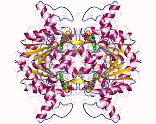
Leghemoglobin is an oxygen-carrying phytoglobin found in the nitrogen-fixing root nodules of leguminous plants. It is produced by these plants in response to the roots being colonized by nitrogen-fixing bacteria, termed rhizobia, as part of the symbiotic interaction between plant and bacterium: roots not colonized by Rhizobium do not synthesise leghemoglobin. Leghemoglobin has close chemical and structural similarities to hemoglobin, and, like hemoglobin, is red in colour. It was originally thought that the heme prosthetic group for plant leghemoglobin was provided by the bacterial symbiont within symbiotic root nodules. However, subsequent work shows that the plant host strongly expresses heme biosynthesis genes within nodules, and that activation of those genes correlates with leghemoglobin gene expression in developing nodules.

In biochemistry, an oxidoreductase is an enzyme that catalyzes the transfer of electrons from one molecule, the reductant, also called the electron donor, to another, the oxidant, also called the electron acceptor. This group of enzymes usually utilizes NADP+ or NAD+ as cofactors. Transmembrane oxidoreductases create electron transport chains in bacteria, chloroplasts and mitochondria, including respiratory complexes I, II and III. Some others can associate with biological membranes as peripheral membrane proteins or be anchored to the membranes through a single transmembrane helix.

<span class="mw-page-title-main">Nicotinamide adenine dinucleotide phosphate</span> Chemical compound

Nicotinamide adenine dinucleotide phosphate, abbreviated NADP+ or, in older notation, TPN (triphosphopyridine nucleotide), is a cofactor used in anabolic reactions, such as the Calvin cycle and lipid and nucleic acid syntheses, which require NADPH as a reducing agent ('hydrogen source'). NADPH is the reduced form of NADP+, the oxidized form. NADP+ is used by all forms of cellular life.

Microbial metabolism is the means by which a microbe obtains the energy and nutrients it needs to live and reproduce. Microbes use many different types of metabolic strategies and species can often be differentiated from each other based on metabolic characteristics. The specific metabolic properties of a microbe are the major factors in determining that microbe's ecological niche, and often allow for that microbe to be useful in industrial processes or responsible for biogeochemical cycles.

<span class="mw-page-title-main">Dihydrolipoamide dehydrogenase</span> Protein-coding gene in the species Homo sapiens

Dihydrolipoamide dehydrogenase (DLD), also known as dihydrolipoyl dehydrogenase, mitochondrial, is an enzyme that in humans is encoded by the DLD gene. DLD is a flavoprotein enzyme that oxidizes dihydrolipoamide to lipoamide.

<span class="mw-page-title-main">Glyoxylate reductase</span> Enzyme

Glyoxylate reductase, first isolated from spinach leaves, is an enzyme that catalyzes the reduction of glyoxylate to glycolate, using the cofactor NADH or NADPH.

In enzymology, a hydroxypyruvate reductase (EC 1.1.1.81) is an enzyme that catalyzes the chemical reaction

In enzymology, a mevaldate reductase (EC 1.1.1.32) is an enzyme that catalyzes the chemical reaction

In enzymology, a 4-hydroxybenzoate 3-monooxygenase [NAD(P)H] (EC 1.14.13.33) is an enzyme that catalyzes the chemical reaction

In enzymology, a 4-hydroxyphenylacetaldehyde oxime monooxygenase (EC 1.14.13.68) is an enzyme that catalyzes the chemical reaction

In enzymology, an anthraniloyl-CoA monooxygenase (EC 1.14.13.40) is an enzyme that catalyzes the chemical reaction

In enzymology, a ferredoxin–NAD+ reductase (EC 1.18.1.3) is an enzyme that catalyzes the chemical reaction:

In enzymology, a ferric-chelate reductase (EC 1.16.1.7) is an enzyme that catalyzes the chemical reaction

Flavin reductase a class of enzymes. There are a variety of flavin reductases, which bind free flavins and through hydrogen bonding, catalyze the reduction of these molecules to a reduced flavin. Riboflavin, or vitamin B, and flavin mononucleotide are two of the most well known flavins in the body and are used in a variety of processes which include metabolism of fat and ketones and the reduction of methemoglobin in erythrocytes. Flavin reductases are similar and often confused for ferric reductases because of their similar catalytic mechanism and structures.

In enzymology, a glutamate synthase (NADH) (EC 1.4.1.14) is an enzyme that catalyzes the chemical reaction

In enzymology, a hydroxylamine reductase (NADH) (EC 1.7.1.10) is an enzyme that catalyzes the chemical reaction.

<span class="mw-page-title-main">NAD(P)H dehydrogenase (quinone)</span>

In enzymology, a NAD(P)H dehydrogenase (quinone) (EC 1.6.5.2) is an enzyme that catalyzes the chemical reaction

In enzymology, a nitrite reductase [NAD(P)H] (EC 1.7.1.4) is an enzyme that catalyzes the chemical reaction

5-exo-hydroxycamphor dehydrogenase (EC 1.1.1.327, F-dehydrogenase, FdeH) is an enzyme with systematic name 5-exo-hydroxycamphor:NAD+ oxidoreductase. This enzyme catalyses the following chemical reaction

<span class="mw-page-title-main">Phytoglobin</span>

Phytoglobins are globular plant proteins classified into the globin superfamily, which contain a heme, i.e. protoporphyrin IX-Fe, prosthetic group. The earliest known phytoglobins are leghemoglobins, discovered in 1939 by Kubo after spectroscopic and chemical analysis of the red pigment of soybean root nodules. A few decades after Kubo's report the crystallization of a lupin phytoglobin by Vainshtein and collaborators revealed that the tertiary structure of this protein and that of the sperm whale myoglobin was remarkably similar, thus indicating that the phytoglobin discovered by Kubo did indeed correspond to a globin.

References

  1. Appleby C. A., The origin and functions of haemoglobin in plants, Sci. Progress, 76 (1992) 365-398.
  2. Becana M.,Klucas R. V., Oxidation and reduction of leghemoglobin in root nodules of leguminous plants, Plant Physiol., 98 (1992) 1217-1221.
  3. Lee K. K.,Klucas R. V., Reduction of ferric leghemoglobin in soybean root nodules, Plant Physiol., 74 (1984) 984-988.
  4. Lee K. K., Shearman L. L., Ericson B. K.,Klucas R. V., Ferric leghemoglobin in plant-attached leguminous nodules., Plant Physiol., 109 (1995) 261-267.
  5. Becana M.,Klucas R. V., Enzymatic and nonenzymatic mechanisms for ferric leghemoglobin reduction in legume root nodules, Proc. Natl. Acad. Sci. USA, 87 (1990) 7295-7299.
  6. Burris R. H.,Hass E., The red pigment of leguminous root nodules., J. Biol. Chem., 155 (1944) 227-229.
  7. Appleby C. A., Properties of leghaemoglobin in vivo, and its isolation as ferrous oxyleghaemoglobin, Biochim Biophys. Acta, 188 (1969) 222-229.
  8. Kretovich V. L., Melik-Sarkisyan S. S., Bashirova N. F.,Topunov A. F., Enzymatic reduction of leghemoglobin in lupin nodules, J. Appl. Biochem., 4 (1982) 209-217.
  9. 1 2 Saari L. L.,Klucas R. V., Ferric leghemoglobin reductase from soybean root nodules, Arch. Biochem. Biophys., 231 (1984) 102-113.
  10. Ji L., Wood S., Becana M.,Klucas R. V., Purification and characterization of soybean root nodule ferric leghemoglobin reductase, Plant Physiol., 96 (1991) 32-37.
  11. Ji L., Becana M., Sarath G.,Klucas R. V., Cloning and sequence analysis of a cDNA encoding ferric leghemoglobin reductase from soybean nodules, Plant Physiol., 104 (1994) 453-459.
  12. Moran J. F., Sun Z., Sarath G., Arredondo-Peter R., James E. K., Becana M.,Klucas R. V., Molecular cloning, functional characterization, and subcellular localization of soybean nodule dihydrolipoamide reductase., Plant Physiol., 128 (2002) 300-313.
  13. Luan P., Aréchaga-Ocampo E., Sarath G., Arredondo-Peter R.,Klucas R. V., Analysis of a ferric leghemoglobin reductase from cowpea (Vigna unguiculata) root nodules., Plant Sci., 154 (2000) 161-170.
  14. Gopalasubramaniam S. K., Kondapalli K. C., Millán-Pacheco C., Pastor N., Stemmler T. L., Moran J. F.,Arredondo-Peter R., Soybean dihydrolipoamide dehydrogenase (ferric leghemoglobin reductase 2) interacts with and reduces ferric non-symbiotic hemoglobin 1., ScienceJet, 2 (2013) 33.

See also