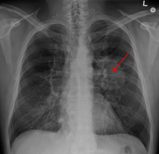
Limited-stage Small Cell Lung Carcinoma | |
---|---|
A CT scan of limited-stage small cell lung carcinoma | |
Specialty | Oncology |
Symptoms | persistent cough, chest pains, rust-coloured sputum, shortness of breath, fatigue, weight loss, wheezing, hoarseness, pneumonia, bronchitis. |
Complications | Nervous system disease |
Diagnostic method | chest x-ray, CT, biopsy |
Treatment | Surgery, chemotherapy, radiotherapy, Prophylactic cranial irradiation |
Prognosis | median overall survival time of approximately 12–16 months, with five-year survival rate of approximately 26% and the long-term survival rate of approximately 4 - 5%. |
Limited-stage small cell lung carcinoma (LS-SCLC) is a type of small cell lung cancer (SCLC) that is confined to an area which is small enough to be encompassed within a radiation portal. [1] [2] [3] [4] This generally includes cancer to one side of the lung and those might have reached the lymph nodes on the same side of the lung. 33% patients with small cell lung cancer are diagnosed with limited-stage small cell lung carcinoma when it is first found. [5] Common symptoms include but are not limited to persistent cough, chest pain, rust-coloured sputum, shortness of breath, fatigue, weight loss, wheezing, hoarseness and recurrent respiratory tract infections such as pneumonia and bronchitis. Nervous system problems, Cushing syndrome and SIADH (syndrome of inappropriate anti-diuretic hormone) can also be associated with small cell lung cancer. [6] Unlike extensive-stage small cell lung cancer, limited-stage small cell lung carcinoma is potentially curable. [4] Standard treatments consist of surgery, platinum-based combination chemotherapy, thoracic irradiation, and prophylactic cranial irradiation. Patient five-year survival rate has significantly increased from 1% with surgery to 26% after the application of combination chemotherapy. [4]
Small cell lung cancer is often treated as a systematic disease due to its tendency for early dissemination, [4] thus, instead of the traditional TNM staging system, the Veterans' Administration Lung Study Group (VALSG) introduced a simplified 2-stage system in the 1950s to divide small cell lung cancer into limited stage and extensive stage. [7] As a result of the change in policy from targeting all nodal stations by radiotherapy ports to target only obviously involved nodal stations, the definition of the limited disease varies. [4]
According to a broad medical consensus, limited-stage small cell lung carcinoma is generally considered to be encompassed within a radiation portal. [4] [5] Variability in categorising small-cell lung cancer as limited or extensive occurs with reference to the extent of pleural effusion and nodal stations involvement. [4] [8] In addition, patients with an ipsilateral pleural effusion are frequently excluded from the limited-disease category. [4] [9] In rare cases, patients with minimal pleural effusions are included in the limited-disease protocols; this includes those with demonstrated effusions only on chest CT, as well as those with blunting of the costophrenic angle on chest radiographs. [4] [10] In most cases, all patients demonstrated any positive sign of pleural effusion on any type of imaging tests are excluded. [11]
Likewise, ambiguities in the definition of limited disease also occurs in classification of the limited disease with reference to the extent of lymphadenopathy. For instance, patients with bilateral supraclavicular adenopathy are included in the limited-disease category in some cases, [12] others include only those with ipsilateral adenopathy. [10] Similarly, the presence of contralateral hilar adenopathy frequently, though not uniformly, includes patients in the limited-disease category. [13] [14]
The traditional TNM classification system is preferred over the 2-stage system when surgery is the recommended treatment option. [4]
Similar to other lung cancers, according to the American Cancer Society, the most common symptoms of limited-stage of lung cancer are: [6]
In addition, patients diagnosed with small cell lung carcinoma has an increased vulnerability to nervous system problems (i.e. Lambert-Eaton syndrome , paraneoplastic cerebellar degeneration ), Cushing syndrome and Syndrome of Inappropriate Anti-diuretic Hormone (SIADH) and can demonstrate relevant symptoms . [6]
Imaging tests are often the first diagnostic step if a patient reports symptoms that may be suggestive of lung cancer. [15] A chest x-ray is the most standard imaging test to look for any abnormality within the lung. If abnormality is present, a computed tomography (CT) scan is frequently ordered to reveal the size, shape, and position of any lung tumour and can help locate enlarged lymph nodes that might contain cancer metastasised from the lung origin. [15] Most patients with limited-stage small cell lung cancer will receive a CT scan of the chest and abdomen to search for abnormality within the lungs and lymph nodes, as well as abnormal areas in more distal organs such as adrenal glands and liver that might arise from the metastasis of lung cancer. [15]
For patients with limited-stage small cell lung cancer, a positron emission tomography (PET) scan is a useful diagnostic tool to investigate the extent of lymph node involvements, which can help determine treatment options. [15]
Though results of imaging test might be suggestive of lung cancer, the actual diagnosis is made by investigating the lung cells under the microscope via lab tests. The cells can be obtained from lung secretions (sputum cytology), fluid removed from pleural effusion ( thoracentesis ), or from a suspicious area ( needle biopsy ). [15]
The standard treatments of limited-stage small cell lung cancer are surgery, platinum-based combination chemotherapy, thoracic irradiation, and prophylactic cranial irradiation. [4] [5]
Up until the late 1960s, surgical resection remained as the mainstay treatment for both limited and extensive small cell lung cancer. [16] The principle of surgical resection in limited‐stage small cell lung carcinoma aimed to remove all viable tumors with curative intent. [17] Consideration of surgery is recommended for Stage 1 limited-stage small cell lung cancer patient with a solitary nodule, no hilar or mediastinal involvement, absence of distant metastases, and no contraindications to surgery classified by the TNM staging system. [18] Surgery is normally followed by chemotherapy. In cases where tumour were found in the lymph nodes, radiation therapy to the chest is usually advised after resection. The International Association for the Study of Lung Cancer (IASLC) Lung Cancer Staging Project demonstrated five‐year survival rates after resection as below: [8]
Pathological stage | five-year survival rate |
---|---|
I | 48% |
II | 39% |
III | 15% |
According to a broad medical consensus, the current standard treatment for fit patients is concurrent chemoradiotherapy. Concurrent chemoradiotherapy is a combination of chemotherapy and thoracic irradiation. To rapidly counter the fast-growing tumour, chemotherapy is often initiated first by physicians. Limited stage small cell lung cancer is sensitive to chemotherapy and a rapid response is often observed. Thoracic radiotherapy generally begins with the first or second cycle when chemotherapy is used according to most guidelines. [19] The time between the start and the end of chemoradiotherapy is a predicator of survival in limited stage small cell lung cancer, prolongation leads to a decrease in overall survival of 1.9% per week. [20]
Early concurrent radiochemotherapy may not be suitable for all patients. A deferred start of concurrent chemotherapy or even a sequential treatment is recommended for patients with large tumour volumes and poor fitness status. This is because early radiotherapy may increase acute and late toxicities. Depend on the size of the tumour, an increase in treatment dose may be required for large tumours, but the dosage can be reduced if initial chemotherapy shrinks the tumour to reduce late toxicities. [10]
Concurrent chemoradiotherapy also remains the mainstay treatment for patients aged 70 years or more and showed a survival benefits when compared to sequential chemoradiotherapy. [21]
Combined use of Cisplatin and Etoposide has become the first-line chemotherapy for limited-stage small cell lung cancer since 1980s. [22] Carboplatin can also be used as a substitute when patient is intolerant of cisplatin. [23] Other chemotherapy regimens including Paclitaxel and Topoisomerase I Inhibitors: Topotecan (Hycamtin) and Irinotecan (Camptosar) also reported significant response against limited-stage small-cell carcinoma during clinical trials. [24] The optimal timing of concurrent chemoradiotherapy is during the first or second cycle. Commonly used radiation schedule and dose are either 1.5 Gy twice daily to a total of 45 Gy or 1.8–2.0 Gy daily to a total dose of 60–70 Gy. [1]
High-dose chemotherapy using cyclophosphamide, cisplatin, and carmustine with hematologic stem-cell support or marrow support on patients aged 60 years or younger who had achieved complete or partial remission with conventional induction chemotherapy also showed significant increase in five-year survival rate to 41% - 53%. [25]
Once daily radiotherapy with 66 Gy and twice-daily with 70 Gy remains two standard treatments for fit patients. Both regimens may be considered depending on the patient's preference. The treatment volume covers the primary tumour and the involved lymph nodes. [19]
One of the negative impacts of thoracic radiotherapy on patients of limited-stage small cell lung carcinoma is the risk of developing brain metastasis. The risk is positively correlated with the size of tumour. [26] Prophylactic cranial irradiation is found to be beneficial in decrease central nervous system recurrence and increase disease-free survival. The risk of brain relapse in limited-stage small cell lung carcinoma is 50% to 60% without prophylactic cranial irradiation. Administration of prophylactic cranial irradiation increased 3-year survival from 15.3% to 20.7%. [10] [27] Standard radiation schedule of prophylactic cranial irradiation for limited stage small cell lung cancer patients is 25 Gy delivered at 2.5 Gy per fraction per day or 30 Gy delivered at 2 Gy per fraction per day. [1]
Unlike extensive-stage small cell lung cancer, limited-stage small cell lung cancer is potentially curable. [4] In limited small cell lung cancer, the median overall survival time is approximately 12–16 months, with five year survival rate of approximately 26% and long-term survival rate of approximately 4–5%. [19]
According to multiple studies, common prognostic factors of limited stage small cell lung cancer lies in performance status, age, life expectancy, comorbidities, extent of disease and smoking. Continued cigarette smoking negatively impacts limited-stage small cell lung cancer survival. All clinicians and care providers should strongly encourage smoking cessation at the diagnosis of limited stage small cell lung cancer. Comparing to continued smokers, patients who quit at or after diagnosis lower the risk of death by 45%. [28]
Lung cancer, also known as lung carcinoma, is a malignant tumor that begins in the lung. Lung cancer is caused by genetic damage to the DNA of cells in the airways, often caused by cigarette smoking or inhaling damaging chemicals. Damaged airway cells gain the ability to multiply unchecked, causing the growth of a tumor. Without treatment, tumors spread throughout the lung, damaging lung function. Eventually lung tumors metastasize, spreading to other parts of the body.
Radiation therapy or radiotherapy is a treatment using ionizing radiation, generally provided as part of cancer therapy to either kill or control the growth of malignant cells. It is normally delivered by a linear particle accelerator. Radiation therapy may be curative in a number of types of cancer if they are localized to one area of the body, and have not spread to other parts. It may also be used as part of adjuvant therapy, to prevent tumor recurrence after surgery to remove a primary malignant tumor. Radiation therapy is synergistic with chemotherapy, and has been used before, during, and after chemotherapy in susceptible cancers. The subspecialty of oncology concerned with radiotherapy is called radiation oncology. A physician who practices in this subspecialty is a radiation oncologist.
Brachytherapy is a form of radiation therapy where a sealed radiation source is placed inside or next to the area requiring treatment. Brachy is Greek for short. Brachytherapy is commonly used as an effective treatment for cervical, prostate, breast, esophageal and skin cancer and can also be used to treat tumours in many other body sites. Treatment results have demonstrated that the cancer-cure rates of brachytherapy are either comparable to surgery and external beam radiotherapy (EBRT) or are improved when used in combination with these techniques. Brachytherapy can be used alone or in combination with other therapies such as surgery, EBRT and chemotherapy.
Small-cell carcinoma is a type of highly malignant cancer that most commonly arises within the lung, although it can occasionally arise in other body sites, such as the cervix, prostate, and gastrointestinal tract. Compared to non-small cell carcinoma, small cell carcinoma is more aggressive, with a shorter doubling time, higher growth fraction, and earlier development of metastases.
In medicine, proton therapy, or proton radiotherapy, is a type of particle therapy that uses a beam of protons to irradiate diseased tissue, most often to treat cancer. The chief advantage of proton therapy over other types of external beam radiotherapy is that the dose of protons is deposited over a narrow range of depth; hence in minimal entry, exit, or scattered radiation dose to healthy nearby tissues.
Invasive carcinoma of no special type, invasive breast carcinoma of no special type (IBC-NST), invasive ductal carcinoma (IDC), infiltrating ductal carcinoma (IDC) or invasive ductal carcinoma, not otherwise specified (NOS) is a disease. For international audiences this article will use "invasive carcinoma NST" because it is the preferred term of the World Health Organization (WHO).
Adjuvant therapy, also known as adjunct therapy, adjuvant care, or augmentation therapy, is a therapy that is given in addition to the primary or initial therapy to maximize its effectiveness. The surgeries and complex treatment regimens used in cancer therapy have led the term to be used mainly to describe adjuvant cancer treatments. An example of such adjuvant therapy is the additional treatment usually given after surgery where all detectable disease has been removed, but where there remains a statistical risk of relapse due to the presence of undetected disease. If known disease is left behind following surgery, then further treatment is not technically adjuvant.
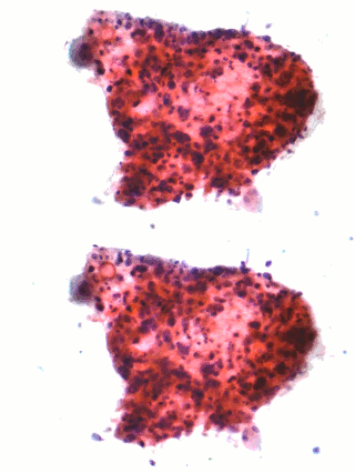
Non-small-cell lung cancer (NSCLC), or non-small-cell lung carcinoma, is any type of epithelial lung cancer other than small-cell lung cancer (SCLC). NSCLC accounts for about 85% of all lung cancers. As a class, NSCLCs are relatively insensitive to chemotherapy, compared to small-cell carcinoma. When possible, they are primarily treated by surgical resection with curative intent, although chemotherapy has been used increasingly both preoperatively and postoperatively.
Mitumomab (BEC-2) is a mouse anti-BEC-2 monoclonal antibody investigated for the treatment of small cell lung carcinoma in combination with BCG vaccination. Mitumomab attacks tumour cells, while the vaccine is thought to activate the immune system. It was developed by ImClone and Merck.
Adenocarcinoma in situ (AIS) of the lung —previously included in the category of "bronchioloalveolar carcinoma" (BAC)—is a subtype of lung adenocarcinoma. It tends to arise in the distal bronchioles or alveoli and is defined by a non-invasive growth pattern. This small solitary tumor exhibits pure alveolar distribution and lacks any invasion of the surrounding normal lung. If completely removed by surgery, the prognosis is excellent with up to 100% 5-year survival.
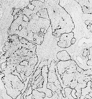
Esthesioneuroblastoma is a rare cancer of the nasal cavity. Arising from the upper nasal tract, esthesioneuroblastoma is believed to originate from sensory neuroepithelial cells, also known as neuroectodermal olfactory cells.
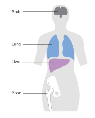
Metastatic breast cancer, also referred to as metastases, advanced breast cancer, secondary tumors, secondaries or stage IV breast cancer, is a stage of breast cancer where the breast cancer cells have spread to distant sites beyond the axillary lymph nodes. There is no cure for metastatic breast cancer; there is no stage after IV.
Human papillomavirus-positive oropharyngeal cancer, is a cancer of the throat caused by the human papillomavirus type 16 virus (HPV16). In the past, cancer of the oropharynx (throat) was associated with the use of alcohol or tobacco or both, but the majority of cases are now associated with the HPV virus, acquired by having oral contact with the genitals of a person who has a genital HPV infection. Risk factors include having a large number of sexual partners, a history of oral-genital sex or anal–oral sex, having a female partner with a history of either an abnormal Pap smear or cervical dysplasia, having chronic periodontitis, and, among men, younger age at first intercourse and a history of genital warts. HPV-positive OPC is considered a separate disease from HPV-negative oropharyngeal cancer.
A brain metastasis is a cancer that has metastasized (spread) to the brain from another location in the body and is therefore considered a secondary brain tumor. The metastasis typically shares a cancer cell type with the original site of the cancer. Metastasis is the most common cause of brain cancer, as primary tumors that originate in the brain are less common. The most common sites of primary cancer which metastasize to the brain are lung, breast, colon, kidney, and skin cancer. Brain metastases can occur months or even years after the original or primary cancer is treated. Brain metastases have a poor prognosis for cure, but modern treatments allow patients to live months and sometimes years after the diagnosis.
Combined small cell lung carcinoma is a form of multiphasic lung cancer that is diagnosed by a pathologist when a malignant tumor, arising from transformed cells originating in lung tissue, contains a component of;small cell lung carcinoma (SCLC), admixed with one components of any histological variant of non-small cell lung carcinoma (NSCLC) in any relative proportion.
Treatment of lung cancer refers to the use of medical therapies, such as surgery, radiation, chemotherapy, immunotherapy, percutaneous ablation, and palliative care, alone or in combination, in an attempt to cure or lessen the adverse impact of malignant neoplasms originating in lung tissue.
Targeted therapy of lung cancer refers to using agents specifically designed to selectively target molecular pathways responsible for, or that substantially drive, the malignant phenotype of lung cancer cells, and as a consequence of this (relative) selectivity, cause fewer toxic effects on normal cells.
Adenocarcinoma of the lung is the most common type of lung cancer, and like other forms of lung cancer, it is characterized by distinct cellular and molecular features. It is classified as one of several non-small cell lung cancers (NSCLC), to distinguish it from small cell lung cancer which has a different behavior and prognosis. Lung adenocarcinoma is further classified into several subtypes and variants. The signs and symptoms of this specific type of lung cancer are similar to other forms of lung cancer, and patients most commonly complain of persistent cough and shortness of breath.
Prophylactic cranial irradiation (PCI) is a technique used to combat the occurrence of metastasis to the brain in highly aggressive cancers that commonly metastasize to brain, most notably small-cell lung cancer. Radiation therapy is commonly used to treat known tumor occurrence in the brain, either with highly precise stereotactic radiation or therapeutic cranial irradiation. By contrast, PCI is intended as preemptive treatment in patients with no known current intracranial tumor, but with high likelihood for harboring occult microscopic disease and eventual occurrence. For small-cell lung cancer with limited and select cases of extensive disease, PCI has shown to reduce recurrence of brain metastases and improve overall survival in complete remission.
Joaquín Gómez Mira is a scientist and physician specialized in radiation oncology. Born and raised in Spain, he completed his studies and developed his career in the United States, where he moved in 1967. He is a member of the American Society of Clinical Oncology, and a Fellow and appointed councilor of the American College of Radiology. His work, achievements and lectures in the field of radiation oncology are held in high regard.