McBurney's point

Last updated
McBurney's point
McBurney's point.jpg
Location of McBurney's point (1), located two thirds the distance from the umbilicus (2) to the right anterior superior iliac spine (3).
Surface projections of the organs of the trunk.png
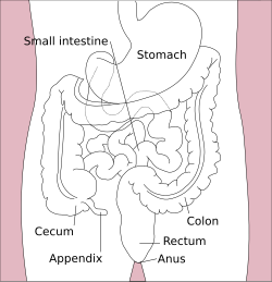
Surface projections of the organs of the trunk, with McBurney's point labeled with a red circle at bottom left at the inferior part of the cecum.
Identifiers
FMA 11362
Anatomical terminology

McBurney's point is the name given to the point over the right side of the abdomen that is one-third of the distance from the anterior superior iliac spine to the umbilicus (navel). This is near the most common location of the appendix.

Contents

Location

McBurney's point is located one third of the distance from the right anterior superior iliac spine to the umbilicus (navel). [1] [2] This point roughly corresponds to the most common location of the base of the appendix, where it is attached to the cecum. [3]

Normal location of the appendix relative to other organs of the digestive system (anterior view). Cecum and appendix are visible at bottom left. Stomach colon rectum diagram-en.svg
Normal location of the appendix relative to other organs of the digestive system (anterior view). Cecum and appendix are visible at bottom left.

Appendicitis

Deep tenderness at McBurney's point, known as McBurney's sign, is a sign of acute appendicitis. [1] [2] [4] The clinical sign of referred pain in the epigastrium when pressure is applied is also known as Aaron's sign. Specific localization of tenderness to McBurney's point indicates that inflammation is no longer limited to the lumen of the bowel (which localizes pain poorly), and is irritating the lining of the peritoneum at the place where the peritoneum comes into contact with the appendix. Tenderness at McBurney's point suggests the evolution of acute appendicitis to a later stage, and thus, the increased likelihood of rupture. Other abdominal processes can also sometimes cause tenderness at McBurney's point. [5] Thus, this sign is highly useful but neither necessary nor sufficient to make a diagnosis of acute appendicitis. [5] The anatomical position of the appendix is highly variable (for example in retrocaecal appendix, an appendix behind the caecum), which also limits the use of this sign, as many cases of appendicitis do not cause point tenderness at McBurney's point. For most open appendectomies (as opposed to laparoscopic appendectomies), the incision is made at McBurney's point. [6]

Pseudoaneurysm

A pseudoaneurysm in the aorta may be treated surgically, with an incision made between McBurney's point and the lower intercostal spaces. [7]

Peritoneal dialysis

McBurney's point may be a useful site for insertion of a peritoneal dialysis catheter. [8]

History

McBurney's point is named after American surgeon Charles McBurney (1845–1913). [1] [6] McBurney himself did not locate his point in a precise way in his original article.

The seat of greatest pain, determined by the pressure of one finger, has been very exactly between an inch and a half and two inches from the anterior spinous process of the ilium on a straight line drawn from that process to the umbilicus

Charles McBurney, "Experience with Early Operative Interference in Cases of Disease of the Vermiform Appendix"; New York Medical Journal , 1889, 50: 676–684 [pg 678].

Related Research Articles

<span class="mw-page-title-main">Appendicitis</span> Inflammation of the appendix

Appendicitis is inflammation of the appendix. Symptoms commonly include right lower abdominal pain, nausea, vomiting, and decreased appetite. However, approximately 40% of people do not have these typical symptoms. Severe complications of a ruptured appendix include widespread, painful inflammation of the inner lining of the abdominal wall and sepsis.

<span class="mw-page-title-main">Appendix (anatomy)</span> Tube attached to the intestine

The appendix is a finger-like, blind-ended tube connected to the cecum, from which it develops in the embryo. The cecum is a pouch-like structure of the large intestine, located at the junction of the small and the large intestines. The term "vermiform" comes from Latin and means "worm-shaped". The appendix was once considered a vestigial organ, but this view has changed since the early 2000s. Research suggests that the appendix may serve an important purpose as a reservoir for beneficial gut bacteria.

<span class="mw-page-title-main">Appendectomy</span> Surgical removal of the vermiform appendix

An appendectomy or appendicectomy is a surgical operation in which the vermiform appendix is removed. Appendectomy is normally performed as an urgent or emergency procedure to treat complicated acute appendicitis.

<span class="mw-page-title-main">Umbilical artery</span> Artery in the abdominal and pelvic regions

The umbilical artery is a paired artery that is found in the abdominal and pelvic regions. In the fetus, it extends into the umbilical cord.

<span class="mw-page-title-main">Rovsing's sign</span> Medical condition

Rovsing's sign, named after the Danish surgeon Niels Thorkild Rovsing (1862–1927), is a sign of appendicitis. If palpation of the left lower quadrant of a person's abdomen increases the pain felt in the right lower quadrant, the patient is said to have a positive Rovsing's sign and may have appendicitis. The phenomenon was first described by Swedish surgeon Emil Samuel Perman (1856–1945) writing in the journal Hygiea in 1904.

<span class="mw-page-title-main">Platysma muscle</span> Human neck muscle

The platysma muscle is a superficial muscle of the human neck that overlaps the sternocleidomastoid. It covers the anterior surface of the neck superficially. When it contracts, it produces a slight wrinkling of the neck, and a "bowstring" effect on either side of the neck.

<span class="mw-page-title-main">Abdominal examination</span> Physical examination of abdomen

An abdominal examination is a portion of the physical examination which a physician or nurse uses to clinically observe the abdomen of a patient for signs of disease. The abdominal examination is conventionally split into four different stages: first, inspection of the patient and the visible characteristics of their abdomen. Auscultation (listening) of the abdomen with a stethoscope. Palpation of the patient's abdomen. Finally, percussion (tapping) of the patient's abdomen and abdominal organs. Depending on the need to test for specific diseases such as ascites, special tests may be performed as a part of the physical examination. An abdominal examination may be performed because the physician suspects a disease of the organs inside the abdominal cavity (including the liver, spleen, large or small intestines), or simply as a part of a complete physical examination for other conditions. In a complete physical examination, the abdominal exam classically follows the respiratory examination and cardiovascular examination.

<span class="mw-page-title-main">Psoas sign</span> Medical sign

The psoas sign, also known as Cope's sign or Obraztsova's sign, is a medical sign that indicates irritation to the iliopsoas group of hip flexors in the abdomen, and consequently indicates that the inflamed appendix is retrocaecal in orientation.

<span class="mw-page-title-main">Common iliac artery</span> Artery in the abdomen

The common iliac artery is a large artery of the abdomen paired on each side. It originates from the aortic bifurcation at the level of the 4th lumbar vertebra. It ends in front of the sacroiliac joint, one on either side, and each bifurcates into the external and internal iliac arteries.

<span class="mw-page-title-main">Anterior superior iliac spine</span> Bony projection of the iliac bone

The anterior superior iliac spine (ASIS) is a bony projection of the iliac bone, and an important landmark of surface anatomy. It refers to the anterior extremity of the iliac crest of the pelvis. It provides attachment for the inguinal ligament, and the sartorius muscle. The tensor fasciae latae muscle attaches to the lateral aspect of the superior anterior iliac spine, and also about 5 cm away at the iliac tubercle.

<span class="mw-page-title-main">Inferior epigastric artery</span> Blood vessel

In human anatomy, the inferior epigastric artery is an artery that arises from the external iliac artery. It is accompanied by the inferior epigastric vein; inferiorly, these two inferior epigastric vessels together travel within the lateral umbilical fold The inferior epigastric artery then traverses the arcuate line of rectus sheath to enter the rectus sheath, then anastomoses with the superior epigastric artery within the rectus sheath.

Postural drainage (PD) is the drainage of lung secretions using gravity. It is used to treat a variety of conditions that cause the build-up of secretions in the lungs.

<span class="mw-page-title-main">Pericardial sinus</span>

The pericardial sinuses are impressions in the pericardial sac formed between the points where great vessels enter it.

<span class="mw-page-title-main">Retromandibular vein</span> Major face vein

The retromandibular vein is a major vein of the face. It is formed within the parotid gland by the confluence of the maxillary vein, and superficial temporal vein. It descends in the gland and splits into two branches upon emerging from the gland. Its anterior branch then joins the (anterior) facial vein forming the common facial vein, while its posterior branch joins the posterior auricular vein forming the external jugular vein.

<span class="mw-page-title-main">Duodenojejunal flexure</span> Border between the duodenum and the jejunum

The duodenojejunal flexure or duodenojejunal junction, also known as the angle of Treitz, is the border between the duodenum and the jejunum.

<span class="mw-page-title-main">Obturator sign</span> Medical condition

The obturator sign, also called Cope's obturator test, is an indicator of irritation to the obturator internus muscle.

<span class="mw-page-title-main">Glenoid labrum</span> Ligament in the shoulder blade

The glenoid labrum is a fibrocartilaginous structure rim attached around the margin of the glenoid cavity in the shoulder blade. The shoulder joint is considered a ball and socket joint. However, in bony terms the 'socket' is quite shallow and small, covering at most only a third of the 'ball'. The socket is deepened by the glenoid labrum, stabilizing the shoulder joint.

<span class="mw-page-title-main">Bare area of the liver</span> Part of the surface of the liver

The bare area of the liver is a large triangular area on the diaphragmatic surface of the liver. It is the only part of the liver with no peritoneal covering, although it is still covered by Glisson’s capsule. It is attached directly to the diaphragm by loose connective tissue. The bare area of the liver is relevant to the portacaval anastomosis, encloses the right extraperitoneal subphrenic space, and can be a site of spread of infection from the abdominal cavity to the thoracic cavity

<span class="mw-page-title-main">Rectovesical pouch</span> Part of pelvic anatomy in male mammals

The rectovesical pouch is the pocket that lies between the rectum and the bladder in males in humans and other mammals. It is lined by peritoneum.

In surgery, a surgical incision is a cut made through the skin and soft tissue to facilitate an operation or procedure. Often, multiple incisions are possible for an operation. In general, a surgical incision is made as small and unobtrusive as possible to facilitate safe and timely operating conditions.

References

  1. 1 2 3 Fasen, Geoffrey; Schirmer, Bruce; Hedrick, Traci L. (2019-01-01), Yeo, Charles J. (ed.), "Chapter 164 - Appendix", Shackelford's Surgery of the Alimentary Tract, 2 Volume Set (Eighth Edition), Philadelphia: Elsevier, pp. 1951–1958, doi:10.1016/b978-0-323-40232-3.00164-3, ISBN   978-0-323-40232-3, S2CID   239120680 , retrieved 2020-12-15
  2. 1 2 Noel, Richard J. (2018-01-01), Kliegman, Robert M.; Lye, Patricia S.; Bordini, Brett J.; Toth, Heather (eds.), "12 - Vomiting and Regurgitation", Nelson Pediatric Symptom-Based Diagnosis, Elsevier, pp. 204–234.e1, doi:10.1016/b978-0-323-39956-2.00012-1, ISBN   978-0-323-39956-2 , retrieved 2020-12-15
  3. Kyung Won Chung (2005). Gross Anatomy (Board Review) . Hagerstown, MD: Lippincott Williams & Wilkins. pp.  255. ISBN   978-0-7817-5309-8.
  4. "Definition: McBurney's sign from Online Medical Dictionary" . Retrieved 2007-12-06.
  5. 1 2 Sifri, Costi D.; Madoff, Lawrence C. (2015-01-01), Bennett, John E.; Dolin, Raphael; Blaser, Martin J. (eds.), "80 - Appendicitis", Mandell, Douglas, and Bennett's Principles and Practice of Infectious Diseases (Eighth Edition), Philadelphia: W.B. Saunders, pp. 982–985.e1, doi:10.1016/b978-1-4557-4801-3.00080-1, ISBN   978-1-4557-4801-3 , retrieved 2020-12-15
  6. 1 2 Prager, Elliot (2005-01-01), Fazio, Victor W.; Church, James M.; Delaney, Conor P. (eds.), "Acute Appendicitis", Current Therapy in Colon and Rectal Surgery (Second Edition), Philadelphia: Mosby, pp. 259–262, doi:10.1016/b978-1-55664-480-1.50051-4, ISBN   978-1-55664-480-1
  7. Byrne, John; Darling III, R. Clement (2009-01-01), Evans, Stephen R. T. (ed.), "Chapter 59 - Aortic Surgery", Surgical Pitfalls, Philadelphia: W.B. Saunders, pp. 597–612, doi:10.1016/b978-141602951-9.50075-x, ISBN   978-1-4160-2951-9 , retrieved 2020-12-15
  8. Soni, Sachin S.; Barnela, Shriganesh R.; Saboo, Sonali S.; Takalkar, Unmesh V. (2019-01-01), Ronco, Claudio; Bellomo, Rinaldo; Kellum, John A.; Ricci, Zaccaria (eds.), "Chapter 188 - Nursing and Procedure Issues in Peritoneal Dialysis", Critical Care Nephrology (Third Edition), Philadelphia: Elsevier, pp. 1134–1136.e1, doi:10.1016/b978-0-323-44942-7.00188-6, ISBN   978-0-323-44942-7, S2CID   87745135 , retrieved 2020-12-15

Bibliography