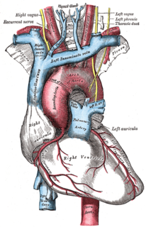
Related Research Articles
Venous thrombosis is thrombosis in a vein, caused by a thrombus. A common form of venous thrombosis is deep vein thrombosis (DVT), when a blood clot forms in the deep veins. If a thrombus breaks off (embolizes) and flows towards the lungs, it can become a pulmonary embolism (PE), a blood clot in the lungs. The conditions of DVT only, DVT with PE, and PE only are captured by the term venous thromboembolism (VTE).
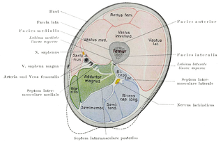
In human anatomy, the thigh is the area between the hip (pelvis) and the knee. Anatomically, it is part of the lower limb.

The femoral artery is a large artery in the thigh and the main arterial supply to the thigh and leg. The femoral artery gives off the deep femoral artery or profunda femoris artery and descends along the anteromedial part of the thigh in the femoral triangle. It enters and passes through the adductor canal, and becomes the popliteal artery as it passes through the adductor hiatus in the adductor magnus near the junction of the middle and distal thirds of the thigh.

The great saphenous vein is a large, subcutaneous, superficial vein of the leg. It is the longest vein in the body, running along the length of the lower limb, returning blood from the foot, leg and thigh to the deep femoral vein at the femoral triangle.
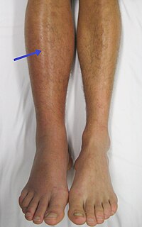
Deep vein thrombosis (DVT) is the formation of a blood clot in a deep vein, most commonly in the legs or pelvis. A minority of DVTs occur in the arms. Symptoms can include pain, swelling, redness, and enlarged veins in the affected area, but some DVTs have no symptoms. The most common life-threatening concern with DVT is the potential for a clot to embolize, travel as an embolus through the right side of the heart, and become lodged in a pulmonary artery that supplies blood to the lungs. This is called a pulmonary embolism (PE). DVT and PE comprise the cardiovascular disease of venous thromboembolism (VTE). About two-thirds of VTE manifests as DVT only, with one-third manifesting as PE with or without DVT. The most frequent long-term DVT complication is post-thrombotic syndrome, which can cause pain, swelling, a sensation of heaviness, itching, and in severe cases, ulcers. Recurrent VTE occurs in about 30% of those in the ten years following an initial VTE.

Esophageal varices are extremely dilated sub-mucosal veins in the lower third of the esophagus. They are most often a consequence of portal hypertension, commonly due to cirrhosis. People with esophageal varices have a strong tendency to develop severe bleeding which left untreated can be fatal. Esophageal varices are typically diagnosed through an esophagogastroduodenoscopy.

Portal hypertension is abnormally increased portal venous pressure – blood pressure in the portal vein and its branches, that drain from most of the intestine to the liver. Portal hypertension is defined as a hepatic venous pressure gradient greater than 5 mmHg. Cirrhosis is the most common cause of portal hypertension; other, less frequent causes are therefore grouped as non-cirrhotic portal hypertension. When it becomes severe enough to cause symptoms or complications, treatment may be given to decrease portal hypertension itself or to manage its complications.

In the human body, the femoral vein is a blood vessel that accompanies the femoral artery in the femoral sheath. It begins at the adductor hiatus and is a continuation of the popliteal vein. It ends at the inferior margin of the inguinal ligament, where it becomes the external iliac vein. The femoral vein bears valves which are mostly bicuspid and whose number is variable between individuals and often between left and right leg.

Cavernous sinus thrombosis (CST) is the formation of a blood clot within the cavernous sinus, a cavity at the base of the brain which drains deoxygenated blood from the brain back to the heart. This is a rare disorder and can be of two types–septic cavernous thrombosis and aseptic cavernous thrombosis. Most commonly the form is of septic cavernous sinus thrombosis. The cause is usually from a spreading infection in the nose, sinuses, ears, or teeth. Staphylococcus aureus and Streptococcus are often the associated bacteria.
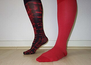
Compression stockings are a specialized hosiery designed to help prevent the occurrence of, and guard against further progression of, venous disorders such as edema, phlebitis and thrombosis. Compression stockings are elastic compression garments worn around the leg, compressing the limb. This reduces the diameter of distended veins and increases venous blood flow velocity and valve effectiveness. Compression therapy helps decrease venous pressure, prevents venous stasis and impairments of venous walls, and relieves heavy and aching legs.
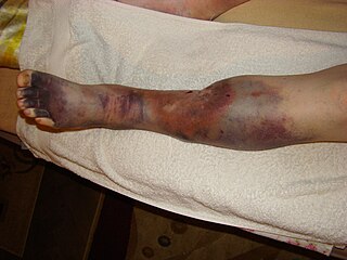
Post-thrombotic syndrome (PTS), also called postphlebitic syndrome and venous stress disorder is a medical condition that may occur as a long-term complication of deep vein thrombosis (DVT).
Charles Theodore Dotter was a pioneering US vascular radiologist who is credited with developing interventional radiology. Dotter, with his trainee Dr Melvin P. Judkins, described angioplasty in 1964.
Venous cutdown is an emergency procedure in which the vein is exposed surgically and then a cannula is inserted into the vein under direct vision. It is used to get vascular access in trauma and hypovolemic shock patients when peripheral cannulation is difficult or impossible. The saphenous vein is most commonly used. This procedure has fallen out of favor with the development of safer techniques for central venous catheterization such as the Seldinger technique, the modified Seldinger technique, intraosseous infusion, as well as the use of ultrasound guidance for placement of central venous catheters without using the cutdown technique.

Cerebral venous sinus thrombosis (CVST), cerebral venous and sinus thrombosis or cerebral venous thrombosis (CVT), is the presence of a blood clot in the dural venous sinuses, the cerebral veins, or both. Symptoms may include severe headache, visual symptoms, any of the symptoms of stroke such as weakness of the face and limbs on one side of the body, and seizures.
Congenital stenosis of vena cava is a congenital anomaly in which the superior vena cava or inferior vena cava has an aberrant interruption or coarctation.
Bancroft's sign, also known as Moses' sign, is a clinical sign found in patients with deep vein thrombosis of the lower leg involving the posterior tibial veins. The sign is positive if pain is elicited when the calf muscle is compressed forwards against the tibia, but not when the calf muscle is compressed from side to side. Like other clinical signs for deep vein thrombosis, such as Homans sign and Lowenberg's sign, this sign is neither sensitive nor specific for the presence of thrombosis.
The Pratt Test is a simple test to check for deep vein thrombosis in the leg. It involves having the patient lie supine with the leg bent at the knee, grasping the calf with both hands and pressing on the popliteal vein in the proximal calf. If the patient feels pain, it is a sign that a deep vein thrombosis exists.

Ultrasonography of suspected or previously confirmed chronic venous insufficiency of leg veins is a risk-free, non-invasive procedure. It gives information about the anatomy, physiology and pathology of mainly superficial veins. As with heart ultrasound (echocardiography) studies, venous ultrasonography requires an understanding of hemodynamics in order to give useful examination reports. In chronic venous insufficiency, sonographic examination is of most benefit; in confirming varicose disease, making an assessment of the hemodynamics, and charting the progression of the disease and its response to treatment. It has become the reference standard for examining the condition and hemodynamics of the lower limb veins. Particular veins of the deep venous system (DVS), and the superficial venous system (SVS) are looked at. The great saphenous vein (GSV), and the small saphenous vein (SSV) are superficial veins which drain into respectively, the common femoral vein and the popliteal vein. These veins are deep veins. Perforator veins drain superficial veins into the deep veins. Three anatomic compartments are described, (N1) containing the deep veins, (N2) containing the perforator veins, and (N3) containing the superficial veins, known as the saphenous compartment. This compartmentalisation makes it easier for the examiner to systematize and map. The GSV can be located in the saphenous compartment where together with the Giacomini vein and the accessory saphenous vein (ASV) an image resembling an eye, known as the 'eye sign' can be seen. The ASV which is often responsible for varicose veins, can be located at the 'alignment sign', where it is seen to align with the femoral vessels.
Venous access is any method used to access the bloodstream through the veins, either to administer intravenous therapy, parenteral nutrition, to obtain blood for analysis, or to provide an access point for blood-based treatments such as dialysis or apheresis. Access is most commonly achieved via the Seldinger technique, and guidance tools such as ultrasound and fluoroscopy can also be used to assist with visualizing access placement.
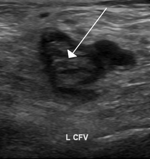
Ultrasonography in suspected deep vein thrombosis focuses primarily on the femoral vein and the popliteal vein, because thrombi in these veins are associated with the greatest risk of harmful pulmonary embolism.
References
| | This medical sign article is a stub. You can help Wikipedia by expanding it. |