
Thrombosis is the formation of a blood clot inside a blood vessel, obstructing the flow of blood through the circulatory system. When a blood vessel is injured, the body uses platelets (thrombocytes) and fibrin to form a blood clot to prevent blood loss. Even when a blood vessel is not injured, blood clots may form in the body under certain conditions. A clot, or a piece of the clot, that breaks free and begins to travel around the body is known as an embolus.
Venous thrombosis is thrombosis in a vein, caused by a thrombus. A common form of venous thrombosis is deep vein thrombosis (DVT), when a blood clot forms in the deep veins. If a thrombus breaks off (embolizes) and flows towards the lungs, it can become a pulmonary embolism (PE), a blood clot in the lungs. The conditions of DVT only, DVT with PE, and PE only are captured by the term venous thromboembolism (VTE).
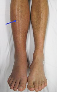
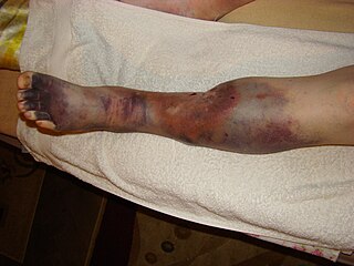
Deep vein thrombosis (DVT) is the formation of a blood clot in a deep vein, most commonly in the legs or pelvis. A minority of DVTs occur in the arms. Symptoms can include pain, swelling, redness, and enlarged veins in the affected area, but some DVTs have no symptoms. The most common life-threatening concern with DVT is the potential for a clot to embolize, travel as an embolus through the right side of the heart, and become lodged in a pulmonary artery that supplies blood to the lungs. This is called a pulmonary embolism (PE). DVT and PE comprise the cardiovascular disease of venous thromboembolism (VTE). About two-thirds of VTE manifests as DVT only, with one-third manifesting as PE with or without DVT. The most frequent long-term DVT complication is post-thrombotic syndrome, which can cause pain, swelling, a sensation of heaviness, itching, and in severe cases, ulcers. Recurrent VTE occurs in about 30% of those in the ten years following an initial VTE.
A Baker's cyst, also known as a popliteal cyst, is a type of fluid collection behind the knee. Often there are no symptoms. If symptoms do occur these may include swelling and pain behind the knee, or knee stiffness. If the cyst breaks open, pain may significantly increase with swelling of the calf. Rarely complications such as deep vein thrombosis, peripheral neuropathy, ischemia, or compartment syndrome may occur.

Heparin-induced thrombocytopenia (HIT) is the development of thrombocytopenia, due to the administration of various forms of heparin, an anticoagulant. HIT predisposes to thrombosis because platelets release microparticles that activate thrombin, thereby leading to thrombosis. When thrombosis is identified the condition is called heparin-induced thrombocytopenia and thrombosis (HITT). HIT is caused by the formation of abnormal antibodies that activate platelets. If someone receiving heparin develops new or worsening thrombosis, or if the platelet count falls, HIT can be confirmed with specific blood tests.
Thrombophilia is an abnormality of blood coagulation that increases the risk of thrombosis. Such abnormalities can be identified in 50% of people who have an episode of thrombosis that was not provoked by other causes. A significant proportion of the population has a detectable thrombophilic abnormality, but most of these develop thrombosis only in the presence of an additional risk factor.
In medicine, Homans' sign is considered by some physicians to be a sign of deep vein thrombosis (DVT). It was defined by John Homans in 1941 as discomfort behind the knee upon forced dorsiflexion of the foot. After many examples of false-positive Homans' signs were reported, Homans redefined it in 1944, stating that "discomfort need have no part in the reaction", and that increased resistance, involuntary flexure of the knee or pain in the calf upon forced dorsiflexion should be considered positive responses.

May–Thurner syndrome (MTS), also known as the iliac vein compression syndrome, is a condition in which compression of the common venous outflow tract of the left lower extremity may cause discomfort, swelling, pain or clots in the iliofemoral veins.
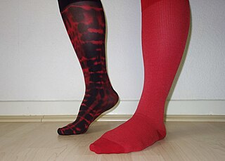
Compression stockings are a specialized hosiery designed to help prevent the occurrence of, and guard against further progression of, venous disorders such as edema, phlebitis and thrombosis. Compression stockings are elastic compression garments worn around the leg, compressing the limb. This reduces the diameter of distended veins and increases venous blood flow velocity and valve effectiveness. Compression therapy helps decrease venous pressure, prevents venous stasis and impairments of venous walls, and relieves heavy and aching legs.
Post-thrombotic syndrome (PTS), also called postphlebitic syndrome and venous stress disorder is a medical condition that may occur as a long-term complication of deep vein thrombosis (DVT).
The term venous translucence has been used in phlebology since 1996 by surgeon Pedro Fernandes Neto during ambulatory clinical exams in Brazil. His results were published in the annals of the national and international congresses of angiology. Venous translucence is the process of reflective image visualization of veins by light, which reaches up to the superficial venous system. It is a non-invasive method. Since it is a simple, low-cost technique it can be repeated as needed, which is useful in disease-process monitoring. It is a new diagnostic procedure, still undergoing investigation; more analysis is necessary to hone its technical aspects. Venous translucence is based on optical physics. It is caused by the refraction, absorption and reflection of light. The color which is not absorbed is reflected, and is the one that is seen. Therefore, venous translumination is based on the incidence of luminosity on the vein, where part of the light is absorbed and another reflected.

Cerebral venous sinus thrombosis (CVST), cerebral venous and sinus thrombosis or cerebral venous thrombosis (CVT), is the presence of a blood clot in the dural venous sinuses, the cerebral veins, or both. Symptoms may include severe headache, visual symptoms, any of the symptoms of stroke such as weakness of the face and limbs on one side of the body, and seizures.

Chronic venous insufficiency (CVI) is a medical condition in which blood pools in the veins, straining the walls of the vein. The most common cause of CVI is superficial venous reflux which is a treatable condition. As functional venous valves are required to provide for efficient blood return from the lower extremities, this condition typically affects the legs. If the impaired vein function causes significant symptoms, such as swelling and ulcer formation, it is referred to as chronic venous disease. It is sometimes called chronic peripheral venous insufficiency and should not be confused with post-thrombotic syndrome in which the deep veins have been damaged by previous deep vein thrombosis.
Pratt's sign is an indication of femoral deep vein thrombosis. It is seen as the presence of dilated pretibial veins in the affected leg, which remain dilated on raising the leg.

Venostasis, or venous stasis, is a condition of slow blood flow in the veins, usually of the legs.
Pseudothrombophlebitis is a clinical condition where there are signs and symptoms of phlebitis in the absence of a thrombophlebitis lesion. Symptoms include pain, swelling, erythema and tenderness evolving over hours or days. It is often associated with the rupture or dissection of a popliteal cyst otherwise known as a Baker's cyst, although it can be associated with other disorders such as the arthritides. It may also occur as an orthopaedic surgical complication, secondary to trauma or as a presentation of septic arthritis. It is crucial to differentiate this condition from deep vein thrombosis as the treatment for DVT can cause adverse effects in patients with pseudothrombophlebitis.
Bancroft's sign, also known as Moses' sign, is a clinical sign found in patients with deep vein thrombosis of the lower leg involving the posterior tibial veins. The sign is positive if pain is elicited when the calf muscle is compressed forwards against the tibia, but not when the calf muscle is compressed from side to side. Like other clinical signs for deep vein thrombosis, such as Homans sign and Lowenberg's sign, this sign is neither sensitive nor specific for the presence of thrombosis.
Lowenberg's sign is a clinical sign found in patients with deep vein thrombosis of the lower leg. The sign is positive when pain is elicited rapidly when a blood pressure cuff is placed around the calf and inflated to 80mmHg. Like other signs of deep vein thrombosis, such as Homans sign and Bancroft's sign, this sign is neither sensitive nor specific for the presence of thrombosis.
Peabody's sign is a clinical sign which may be found in patients with deep vein thrombosis (DVT). The sign is positive when calf muscle spasm occurs on raising the affected leg with the foot extended. The sign is neither sensitive nor specific for the presence of DVT.
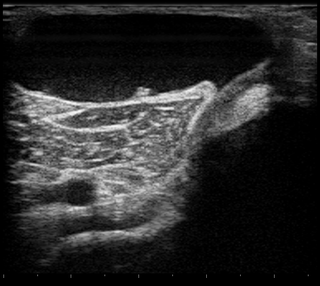
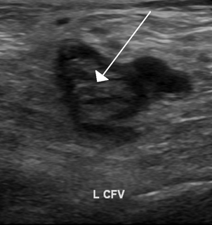
Ultrasonography in suspected deep vein thrombosis focuses primarily on the femoral vein and the popliteal vein, because thrombi in these veins are associated with the greatest risk of harmful pulmonary embolism.