Prosector's wart

Last updated
Prosector's wart
Photographic atlas of the diseases of the skin a series of ninety-six plates, comprising nearly two hundred illustrations, with descriptive text, and a treatise on cutaneous therapeutics (1905) (14597583520).jpg
Classification and external resources
Specialty infectious disease
ICD-10 A18.4 (ILDS A18.440)
ICD-9-CM 017.0

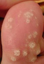
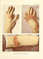
Tuberculosis verrucosa cutis (also known as "lupus verrucosus", [1] "prosector's wart", [1] and "warty tuberculosis" [1] ) is a rash of small, red papular nodules in the skin that may appear 2–4 weeks after inoculation by Mycobacterium tuberculosis in a previously infected and immunocompetent individual.

Rash

A rash is a change of the human skin which affects its color, appearance, or texture.

Skin soft outer covering organ of vertebrates

Skin is the soft outer tissue covering of vertebrates with three main functions: protection, regulation, and sensation.

<i>Mycobacterium tuberculosis</i> Species of bacterium

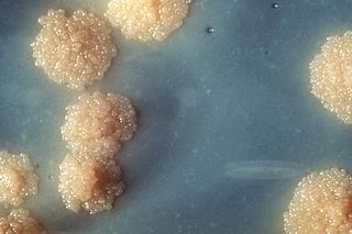
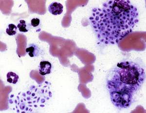
Mycobacterium tuberculosis is a species of pathogenic bacteria in the family Mycobacteriaceae and the causative agent of tuberculosis. First discovered in 1882 by Robert Koch, M. tuberculosis has an unusual, waxy coating on its cell surface primarily due to the presence of mycolic acid. This coating makes the cells impervious to Gram staining, and as a result, M. tuberculosis can appear either Gram-negative or Gram-positive. Acid-fast stains such as Ziehl-Neelsen, or fluorescent stains such as auramine are used instead to identify M. tuberculosis with a microscope. The physiology of M. tuberculosis is highly aerobic and requires high levels of oxygen. Primarily a pathogen of the mammalian respiratory system, it infects the lungs. The most frequently used diagnostic methods for tuberculosis are the tuberculin skin test, acid-fast stain, culture, and polymerase chain reaction.

Contents

It is so called because it was a common occupational disease of prosectors, the preparers of dissections and autopsies. Reinfection by tuberculosis via the skin, therefore, can result from accidental exposure to human tuberculous tissue in physicians, pathologists and laboratory workers; or to tissues of other infected animals, in veterinarians, butchers, etc. Other names given to this form of skin tuberculosis are anatomist's wart and verruca necrogenica (literally, generated by corpses).

An occupational disease is any chronic ailment that occurs as a result of work or occupational activity. It is an aspect of occupational safety and health. An occupational disease is typically identified when it is shown that it is more prevalent in a given body of workers than in the general population, or in other worker populations. The first such disease to be recognised, squamous-cell carcinoma of the scrotum, was identified in chimney sweep boys by Sir Percival Pott in 1775. Occupational hazards that are of a traumatic nature are not considered to be occupational diseases.

Prosector person with the task of preparing a dissection for demonstration

A prosector is a person with the special task of preparing a dissection for demonstration, usually in medical schools or hospitals. Many important anatomists began their careers as prosectors working for lecturers and demonstrators in anatomy and pathology.

Dissection cutting procedure used in anatomy

Dissection is the dismembering of the body of a deceased animal or plant to study its anatomical structure. Autopsy is used in pathology and forensic medicine to determine the cause of death in humans. Less extensive dissection of plants and smaller animals preserved in a formaldehyde solution is typically carried out or demonstrated in biology and natural science classes in middle school and high school, while extensive dissections of cadavers of adults and children, both fresh and preserved are carried out by medical students in medical schools as a part of the teaching in subjects such as anatomy, pathology and forensic medicine. Consequently, dissection is typically conducted in a morgue or in an anatomy lab.

TVC is one of the many forms of cutaneous tuberculosis, such as the tuberculous chancre (which results from the inoculation in people without immunity), and the reactivation cutaneous tuberculosis (the most common form, which appears in previously infected patients). Other forms of cutaneous tuberculosis are: lupus vulgaris, scrofuloderma, lichen scrofulosorum, erythema induratum and the papulonecrotic tuberculid.

Tuberculosis Infectious disease caused by the bacterium Mycobacterium tuberculosis

Tuberculosis (TB) is an infectious disease usually caused by Mycobacterium tuberculosis (MTB) bacteria. Tuberculosis generally affects the lungs, but can also affect other parts of the body. Most infections do not have symptoms, in which case it is known as latent tuberculosis. About 10% of latent infections progress to active disease which, if left untreated, kills about half of those affected. The classic symptoms of active TB are a chronic cough with blood-containing sputum, fever, night sweats, and weight loss. It was historically called "consumption" due to the weight loss. Infection of other organs can cause a wide range of symptoms.

Lupus vulgaris Human disease

Lupus vulgaris are painful cutaneous tuberculosis skin lesions with nodular appearance, most often on the face around the nose, eyelids, lips, cheeks, ears and neck. It is the most common Mycobacterium tuberculosis skin infection. The lesions may ultimately develop into disfiguring skin ulcers if left untreated.

Scrofuloderma

Scrofuloderma is a skin condition caused by tuberculous involvement of the skin by direct extension, usually from underlying tuberculous lymphadenitis.

It was described by René Laennec in 1826. [2]

Presentation

Because the TVC's entry point usually is the site of a trauma, wound or puncture in the skin (during an autopsy, for example), the most frequent site for the wart are the hands. But it can occur anywhere in the skin, such as in the sole of the feet, in the anus, and, in the case of children from developing countries, in the buttocks and knees. This is because children from countries of high incidence of tuberculosis can contract the lesion after contact with tuberculous sputum, by walking barefoot, sitting or playing on the ground.

Wound injury where the skin is torn

A wound is a type of injury which happens relatively quickly in which skin is torn, cut, or punctured, or where blunt force trauma causes a contusion. In pathology, it specifically refers to a sharp injury which damages the dermis of the skin.

Hand extremity at the end of an arm or forelimb

A hand is a prehensile, multi-fingered appendage located at the end of the forearm or forelimb of primates such as humans, chimpanzees, monkeys, and lemurs. A few other vertebrates such as the koala are often described as having "hands" instead of paws on their front limbs. The raccoon is usually described as having "hands" though opposable thumbs are lacking.

Anus Opening at the opposite end of an animals digestive tract from its mouth

The anus is an opening at the opposite end of an animal's digestive tract from the mouth. Its function is to control the expulsion of feces, unwanted semi-solid matter produced during digestion, which, depending on the type of animal, may include: matter which the animal cannot digest, such as bones; food material after all the nutrients have been extracted, for example cellulose or lignin; ingested matter which would be toxic if it remained in the digestive tract; and dead or excess gut bacteria and other endosymbionts.

When recent, the skin lesion has the outside appearance of a wart or verruca, thus it can be confused with other kinds of warts. It evolves to an annular red-brown plaque with time, with central healing and gradual expansion in the periphery. In this phase, it can be confused with fungal infections such as blastomycosis and chromoblastomycosis.

Wart Small, rough growth resembling a cauliflower or a solid blister

Warts are typically small, rough, and hard growths that are similar in color to the rest of the skin. They typically do not result in other symptoms, except when on the bottom of the feet where they may be painful. While they usually occur on the hands and feet they can also affect other locations. One or many warts may appear. They are not cancerous.

Blastomycosis primary systemic mycosis that results in a systemic fungal infection, has material basis in Blastomyces dermatitidis, transmitted by airborne spores and has symptom skin lesions, has symptom lung lesions and has symptom pleural thickening

Blastomycosis is a fungal infection of humans and other animals, notably dogs and occasionally cats, caused by the organism Blastomyces dermatitidis. Endemic to portions of North America, blastomycosis causes clinical symptoms similar to histoplasmosis. The disease occurs in several endemic areas, the most important of which is in eastern North America, particularly in the western and northern periphery of the Great Lakes Basin, extending eastward along the south shore of the St. Lawrence River Valley and southward in the territory spanned by the central Appalachian Mountains in the east, to the Mississippi River Valley in the west. Sporadic cases have been reported in continental Africa, the Arabian Peninsula and the Indian subcontinent.

Chromoblastomycosis long-term fungal infection of the skin and subcutaneous tissue

Chromoblastomycosis is a long-term fungal infection of the skin and subcutaneous tissue. The infection occurs most commonly in tropical or subtropical climates, often in rural areas. It can be caused by many different types of fungi which become implanted under the skin, often by thorns or splinters. Chromoblastomycosis spreads very slowly; it is rarely fatal and usually has a good prognosis, but it can be very difficult to cure. The several treatment options include medication and surgery.

Cause

Diagnosis

The diagnosis is confirmed by a skin biopsy and a positive culture for acid-fast bacilli. A PPD test may also result positive.

Diagnosis is the identification of the nature and cause of a certain phenomenon. Diagnosis is used in many different disciplines, with variations in the use of logic, analytics, and experience, to determine "cause and effect". In systems engineering and computer science, it is typically used to determine the causes of symptoms, mitigations, and solutions.

Biopsy medical test involving sampling of cells or tissues for examination

A biopsy is a medical test commonly performed by a surgeon, interventional radiologist, or an interventional cardiologist involving extraction of sample cells or tissues for examination to determine the presence or extent of a disease. The tissue is generally examined under a microscope by a pathologist, and can also be analyzed chemically. When an entire lump or suspicious area is removed, the procedure is called an excisional biopsy. An incisional biopsy or core biopsy samples a portion of the abnormal tissue without attempting to remove the entire lesion or tumor. When a sample of tissue or fluid is removed with a needle in such a way that cells are removed without preserving the histological architecture of the tissue cells, the procedure is called a needle aspiration biopsy. Biopsies are most commonly performed for insight into possible cancerous and inflammatory conditions.

Treatment

Therapy for cutaneous tuberculosis is the same as for systemic tuberculosis, and usually consists of a 4-drug regimen, i.e., isoniazid, rifampin, pyrazinamide, and ethambutol or streptomycin.

See also

Notes

  1. 1 2 3 Rapini, Ronald P.; Bolognia, Jean L.; Jorizzo, Joseph L. (2007). Dermatology: 2-Volume Set. St. Louis: Mosby. pp. Chapter 74. ISBN   1-4160-2999-0.
  2. Tigoulet F, Fournier V, Caumes E (January 2003). "[Clinical forms of the cutaneous tuberculosis]". Bull Soc Pathol Exot (in French). 96 (5): 362–7. PMID   15015840.

Related Research Articles

Plantar wart viral infectious disease that results in benign epithelial tumors located in skin of the sole or toes of the foot, has material basis in human papillomavirus (types 1, 2, 4 or 63), has symptom lesions that appear on the sole of the foot

A plantar wart is a wart occurring on the bottom of the foot or toes. Their color is typically similar to that of the skin. Small black dots often occur on the surface. One or more may occur in an area. They may result in pain with pressure such that walking is difficult.

Panniculitis Inflammation of the subcutaneous adipose tissue

Panniculitis is a group of diseases whose hallmark is inflammation of subcutaneous adipose tissue. Symptoms include tender skin nodules, and systemic signs such as weight loss and fatigue.

Nocardiosis is an infectious disease affecting either the lungs or the whole body. It is due to infection by bacterium of the genus Nocardia, most commonly Nocardia asteroides or Nocardia brasiliensis.

Tinea capitis Human disease

Tinea capitis is a cutaneous fungal infection (dermatophytosis) of the scalp. The disease is primarily caused by dermatophytes in the Trichophyton and Microsporum genera that invade the hair shaft. The clinical presentation is typically single or multiple patches of hair loss, sometimes with a 'black dot' pattern, that may be accompanied by inflammation, scaling, pustules, and itching. Uncommon in adults, tinea capitis is predominantly seen in pre-pubertal children, more often boys than girls.

Miliary tuberculosis extrapulmonary tuberculosis that results in formation of tiny lesions in all the organs.

Miliary tuberculosis is a form of tuberculosis that is characterized by a wide dissemination into the human body and by the tiny size of the lesions (1–5 mm). Its name comes from a distinctive pattern seen on a chest radiograph of many tiny spots distributed throughout the lung fields with the appearance similar to millet seeds—thus the term "miliary" tuberculosis. Miliary TB may infect any number of organs, including the lungs, liver, and spleen. Miliary tuberculosis is present in about 2% of all reported cases of tuberculosis and accounts for up to 20% of all extra-pulmonary tuberculosis cases.

Keratoderma is a hornlike skin condition.

Sporotrichosis primary systemic mycosis that results in a systemic fungal infection, has material basis in Sporothrix schenckii in animals and humans and results in formation of red papule at the site of inoculation

Sporotrichosis is a disease caused by the infection of the fungus Sporothrix schenckii. This fungal disease usually affects the skin, although other rare forms can affect the lungs, joints, bones, and even the brain. Because roses can spread the disease, it is one of a few diseases referred to as rose-thorn or rose-gardeners' disease.

Angiokeratoma benign cutaneous lesion of capillaries, resulting in small marks of red to blue color and characterized by hyperkeratosis

Angiokeratoma is a benign cutaneous lesion of capillaries, resulting in small marks of red to blue color and characterized by hyperkeratosis. Angiokeratoma corporis diffusum refers to Fabry's disease, but this is usually considered a distinct condition.

Koebner phenomenon skin lesion on lines of trauma

The Koebner phenomenon or Köbner phenomenon, also called the Koebner response or the isomorphic response, attributed to Heinrich Köbner, is the appearance of skin lesions on lines of trauma. The Koebner phenomenon may result from either a linear exposure or irritation. Conditions demonstrating linear lesions after a linear exposure to a causative agent include: molluscum contagiosum, warts and toxicodendron dermatitis. Warts and molluscum contagiosum lesions can be spread in linear patterns by self-scratching ("auto-inoculation"). Toxicodendron dermatitis lesions are often linear from brushing up against the plant. Causes of the Koebner phenomenon that are secondary to scratching rather than an infective or chemical cause include vitiligo, psoriasis, lichen planus, lichen nitidus, pityriasis rubra pilaris, and keratosis follicularis.

Leukemia cutis is the infiltration of neoplastic leukocytes or their precursors into the skin resulting in clinically identifiable cutaneous lesions. This condition may be contrasted with leukemids, which are skin lesions that occur with leukemia, but which are not related to leukemic cell infiltration. Leukemia cutis can occur in most forms of leukemia, including chronic myeloid leukemia, acute lymphoblastic leukemia, chronic lymphocytic leukemia, acute myeloid leukemia, and prolymphocytic leukemia.

Cutis verticis gyrata is a medical condition usually associated with thickening of the scalp. People show visible folds, ridges or creases on the surface of the top of the scalp. The number of folds can vary from two to roughly ten and are typically soft and spongy. These folds cannot be corrected with pressure. The condition typically affects the central and rear regions of the scalp, but sometimes can involve the entire scalp.

Geotrichosis opportunistic mycosis that has material basis in Galactomyces geotrichum, results in systemic infection in immunocompromised people

Geotrichosis is a mycosis caused by Geotrichum candidum.

Primary inoculation tuberculosis is a skin condition that develops at the site of inoculation of tubercle bacilli into a tuberculosis-free individual.

Tuberculosis cutis orificialis is a form of cutaneous tuberculosis that occurs at the mucocutaneous borders of the nose, mouth, anus, urinary meatus, and vagina, and on the mucous membrane of the mouth or tongue.

Majocchi's granuloma, which is a localized form of fungal folliculitis, is a skin condition characterized by a deep, pustular plaques and is a form of tinea corporis. Lesions often have a pink and scaly central component with pustules or folliculocentric papules at the periphery. The name Majocchi's comes from the Professor Domenico Majocchi who first discovered the disorder in 1883. Domenico Majocchi was a professor of dermatology at the University of Parma and later the University of Bologna. The most common dermatophyte is called Trichophyton rubrum. This disease commonly affects both immunocompetent and immunocompromised hosts. However, immunocompromised individuals have a higher risk.

Tuberculous gumma is a skin condition characterized histologically by massive necrosis. Restated, this is a skin condition that results from hematogenous dissemination of mycobacteria from a primary focus, resulting in firm, nontender erythematous nodules that soften, ulcerate, and form sinuses.

References

Goldman, G.; Bolognia, J.L. Pinpointing cutaneous signs of tuberculosis: is it a common wart, or tuberculosis verrucosa cutis? Journal of Critical Illness, Dec. 2002.