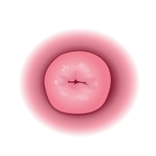
The cervix or cervix uteri is a dynamic fibromuscular sexual organ of the female reproductive system that connects the vagina with the uterine cavity. The human female cervix has been documented anatomically since at least the time of Hippocrates, over 2,000 years ago. The cervix is approximately 4 cm long with a diameter of approximately 3 cm and tends to be described as a cylindrical shape, although the front and back walls of the cervix are contiguous. The size of the cervix changes throughout a woman's life cycle. For example, during the fertile years of a woman's reproductive cycle, females tend to have a larger cervix in comparison to postmenopausal females; likewise, females who have produced offspring have a larger sized cervix than females who have not produced offspring.

The endometrium is the inner epithelial layer, along with its mucous membrane, of the mammalian uterus. It has a basal layer and a functional layer: the basal layer contains stem cells which regenerate the functional layer. The functional layer thickens and then is shed during menstruation in humans and some other mammals, including other apes, Old World monkeys, some species of bat, the elephant shrew and the Cairo spiny mouse. In most other mammals, the endometrium is reabsorbed in the estrous cycle. During pregnancy, the glands and blood vessels in the endometrium further increase in size and number. Vascular spaces fuse and become interconnected, forming the placenta, which supplies oxygen and nutrition to the embryo and fetus. The speculated presence of an endometrial microbiota has been argued against.
Horse breeding is reproduction in horses, and particularly the human-directed process of selective breeding of animals, particularly purebred horses of a given breed. Planned matings can be used to produce specifically desired characteristics in domesticated horses. Furthermore, modern breeding management and technologies can increase the rate of conception, a healthy pregnancy, and successful foaling.

The uterus or womb is the organ in the reproductive system of most female mammals, including humans, that accommodates the embryonic and fetal development of one or more fertilized eggs until birth. The uterus is a hormone-responsive sex organ that contains glands in its lining that secrete uterine milk for embryonic nourishment.
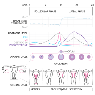
The menstrual cycle is a series of natural changes in hormone production and the structures of the uterus and ovaries of the female reproductive system that makes pregnancy possible. The ovarian cycle controls the production and release of eggs and the cyclic release of estrogen and progesterone. The uterine cycle governs the preparation and maintenance of the lining of the uterus (womb) to receive an embryo. These cycles are concurrent and coordinated, normally last between 21 and 35 days, with a median length of 28 days. Menarche usually occurs around the age of 12 years; menstrual cycles continue for about 30–45 years.

The Ocicat is an all-domestic breed of cat which resembles a wild cat but has no recent wild DNA in its gene pool. It is named for its resemblance to the ocelot. The breed was established from the Siamese and Abyssinian and later on American Shorthair would be added.

Uterine cancer, also known as womb cancer, includes two types of cancer that develop from the tissues of the uterus. Endometrial cancer forms from the lining of the uterus, and uterine sarcoma forms from the muscles or support tissue of the uterus. Endometrial cancer accounts for approximately 90% of all uterine cancers in the United States. Symptoms of endometrial cancer include changes in vaginal bleeding or pain in the pelvis. Symptoms of uterine sarcoma include unusual vaginal bleeding or a mass in the vagina.
Neutering, from the Latin neuter, is the removal of a non-human animal's reproductive organ, either all of it or a considerably large part. The male-specific term is castration, while spaying is usually reserved for female animals. Colloquially, both terms are often referred to as fixing. In male horses, castrating is referred to as gelding. An animal that has not been neutered is sometimes referred to as entire or intact. Often the term neuter[ing] is used to specifically mean castration, e.g. in phrases like "spay and neuter".

The human female reproductive system is made up of the internal and external sex organs that function in the reproduction of new offspring. The reproductive system is immature at birth and develops at puberty to be able to release matured ova from the ovaries, facilitate their fertilization, and create a protective environment for the developing fetus during pregnancy. The female reproductive tract is made of several connected internal sex organs—the vagina, uterus, and fallopian tubes—and is prone to infections. The vagina allows for sexual intercourse, and is connected to the uterus at the cervix. The uterus accommodates the embryo by developing the uterine lining.

Uterine fibroids, also known as uterine leiomyomas, fibromyoma or fibroids, are benign smooth muscle tumors of the uterus, part of the female reproductive system. Most people with fibroids have no symptoms while others may have painful or heavy periods. If large enough, they may push on the bladder, causing a frequent need to urinate. They may also cause pain during penetrative sex or lower back pain. Someone can have one uterine fibroid or many. It is uncommon but possible that fibroids may make it difficult to become pregnant.
In mammalian species, pseudopregnancy is a physical state whereby all the signs and symptoms of pregnancy are exhibited, with the exception of the presence of a fetus, creating a false pregnancy. The corpus luteum is responsible for the development of maternal behavior and lactation, which are mediated by the continued production of progesterone by the corpus luteum through some or all of pregnancy. In most species, the corpus luteum is degraded in the absence of a pregnancy. However, in some species, the corpus luteum may persist in the absence of pregnancy and cause "pseudopregnancy", in which the female will exhibit clinical signs of pregnancy.
The estrous cycle is a set of recurring physiological changes induced by reproductive hormones in females of mammalian subclass Theria. Estrous cycles start after sexual maturity in females and are interrupted by anestrous phases, otherwise known as "rest" phases, or by pregnancies. Typically, estrous cycles repeat until death. These cycles are widely variable in duration and frequency depending on the species. Some animals may display bloody vaginal discharge, often mistaken for menstruation. Many mammals used in commercial agriculture, such as cattle and sheep, may have their estrous cycles artificially controlled with hormonal medications for optimum productivity. The male equivalent, seen primarily in ruminants, is called rut.

Endometritis is inflammation of the inner lining of the uterus (endometrium). Symptoms may include fever, lower abdominal pain, and abnormal vaginal bleeding or discharge. It is the most common cause of infection after childbirth. It is also part of spectrum of diseases that make up pelvic inflammatory disease.
Endometrial ablation is a surgical procedure that is used to remove (ablate) or destroy the endometrial lining of the uterus. The goal of the procedure is to decrease the amount of blood loss during menstruation (periods). Endometrial ablation is most often employed in people with excessive menstrual bleeding following unsuccessful medical therapy. It is less effective than hysterectomy, but with a lower risk of adverse events.

Veterinary surgery is surgery performed on non-human animals by veterinarians, whereby the procedures fall into three broad categories: orthopaedics, soft tissue surgery, and neurosurgery. Advanced surgical procedures such as joint replacement, fracture repair, stabilization of cranial cruciate ligament deficiency, oncologic (cancer) surgery, herniated disc treatment, complicated gastrointestinal or urogenital procedures, kidney transplant, skin grafts, complicated wound management, and minimally invasive procedures are performed by veterinary surgeons. Most general practice veterinarians perform routine surgeries such as neuters and minor mass excisions; some also perform additional procedures.
Canine reproduction is the process of sexual reproduction in domestic dogs, wolves, coyotes and other canine species.

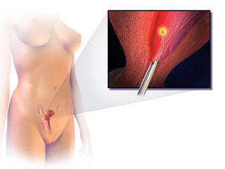
The endometrial biopsy is a medical procedure that involves taking a tissue sample of the lining of the uterus. The tissue subsequently undergoes a histologic evaluation which aids the physician in forming a diagnosis.

Prostaglandin F2α, pharmaceutically termed dinoprost, is a naturally occurring prostaglandin used in medicine to induce labor and as an abortifacient. Prostaglandins are lipids throughout the entire body that have a hormone-like function. In pregnancy, PGF2α is medically used to sustain contracture and provoke myometrial ischemia to accelerate labor and prevent significant blood loss in labor. Additionally, PGF2α has been linked to being naturally involved in the process of labor. It has been seen that there are higher levels of PGF2α in maternal fluid during labor when compared to at term. This signifies that there is likely a biological use and significance to the production and secretion of PGF2α in labor. Prostaglandin is also used to treat uterine infections in domestic animals.

Placental expulsion occurs when the placenta comes out of the birth canal after childbirth. The time between the expulsion of the baby and the expulsion of the placenta is called the third stage of labor.


