
The tongue is a muscular organ in the mouth of a typical tetrapod. It manipulates food for chewing and swallowing as part of the digestive process, and is the primary organ of taste. The tongue's upper surface (dorsum) is covered by taste buds housed in numerous lingual papillae. It is sensitive and kept moist by saliva and is richly supplied with nerves and blood vessels. The tongue also serves as a natural means of cleaning the teeth. A major function of the tongue is the enabling of speech in humans and vocalization in other animals.

The humerus is a long bone in the arm that runs from the shoulder to the elbow. It connects the scapula and the two bones of the lower arm, the radius and ulna, and consists of three sections. The humeral upper extremity consists of a rounded head, a narrow neck, and two short processes. The body is cylindrical in its upper portion, and more prismatic below. The lower extremity consists of 2 epicondyles, 2 processes, and 3 fossae. As well as its true anatomical neck, the constriction below the greater and lesser tubercles of the humerus is referred to as its surgical neck due to its tendency to fracture, thus often becoming the focus of surgeons.

In neuroanatomy, the central sulcus is a sulcus, or groove, in the cerebral cortex in the brains of vertebrates. It is sometimes confused with the longitudinal fissure.
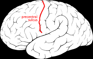
The precentral sulcus is a part of the human brain that lies parallel to, and in front of, the central sulcus. A sulcus is one of the prominent grooves on the surface of the human brain.
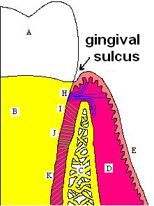
In biological morphology and anatomy, a sulcus is a furrow or fissure. It may be a groove, natural division, deep furrow, elongated cleft, or tear in the surface of a limb or an organ, most notably on the surface of the brain, but also in the lungs, certain muscles, as well as in bones, and elsewhere. Many sulci are the product of a surface fold or junction, such as in the gums, where they fold around the neck of the tooth.
The radial groove is a broad but shallow oblique depression for the radial nerve and deep brachial artery. It is located on the center of the lateral border of the humerus bone. It is situated alongside the posterior margin of the deltoid tuberosity, ending at its inferior margin.

The bicipital groove is a deep groove on the humerus that separates the greater tubercle from the lesser tubercle. It allows for the long tendon of the biceps brachii muscle to pass.
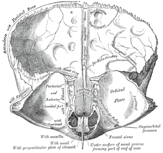
The sagittal sulcus is a midline groove that runs across the internal surfaces of part of the squamous part of the frontal bone, the parietal bones, and part of the occipital bones. The sagittal sulcus accommodates the superior sagittal sinus. The falx cerebri attaches to the edge of the sagittal sulcus on either side.

In neuroanatomy, a sulcus is a depression or groove in the cerebral cortex. It surrounds a gyrus, creating the characteristic folded appearance of the brain in humans and other mammals. The larger sulci are usually called fissures.

The coronary sulcus is a groove on the surface of the heart at the base of right auricle that separates the atria from the ventricles. The structure contains the trunks of the nutrient vessels of the heart, and is deficient in front, where it is crossed by the root of the pulmonary trunk. On the posterior surface of the heart, the coronary sulcus contains the coronary sinus. The right coronary artery, circumflex branch of left coronary artery, and small cardiac vein all travel along parts of the coronary sulcus.

The hypothalamic sulcus is a groove in the lateral wall of the third ventricle, marking the boundary between the thalamus and hypothalamus. The upper and lower portions of the lateral wall of the third ventricle correspond to the alar lamina and basal lamina, respectively, of the lateral wall of the fore-brain vesicle and are separated from each other by a furrow, the hypothalamic sulcus, which extends from the interventricular foramen to the cerebral aqueduct.

The sulcus limitans is a groove on either side of the midline in the rhomboid fossa. It separates the cranial nerve motor nuclei, and the sensory nuclei. It is parallel to the median sulcus.
The terminal sulcus is a groove on the outer surface of the right atrium of the heart marking the transition between the sinus venarum cavarum and the rest of the right atrium. The terminal sulcus corresponds to the position of the terminal crest on the inner surface of the right atium. The terminal sulcus indicate the postition of the sinoatrial node.
The anterior interventricular sulcus is one of two grooves separating the ventricles of the heart. It is situated on the sternocostal surface of the heart, close to the left margin of the heart. It extends between the coronary sulcus, and the apex of the heart; upon reaching the diaphragmatic surface of the heart, it ends at the notch of cardiac apex. It contains the anterior interventricular branch of the left coronary artery, and great cardiac vein.

The posterior interventricular sulcus or posterior longitudinal sulcus is one of the two grooves separating the ventricles of the heart. It is located on the diaphragmatic surface of the heart near the right margin. It extends between the coronary sulcus, and the apex of the heart. It contains the posterior interventricular artery, and middle cardiac vein.

The chiasmatic groove is a transverse groove upon the superior aspect of the body of sphenoid bone within the middle cranial fossa. It is bounded anteriorly by the sphenoidal limbus, and posteriorly by the tuberculum sellae. The opening of each optic canal is placed at either lateral end of the chiasmatic sulcus. The optic chiasm is situated superior and quite posterior to the chiasmatic groove.

The posterior median sulcus of medulla oblongata is a narrow groove; and exists only in the closed part of the medulla oblongata; it becomes gradually shallower from below upward, and finally ends about the middle of the medulla oblongata, where the central canal expands into the cavity of the fourth ventricle.

The intercondylar fossa of femur is a deep notch between the rear surfaces of the medial and lateral epicondyle of the femur, two protrusions on the distal end of the femur that joins the knee. On the front of the femur, the condyles are but much less prominent and are separated from one another by a smooth shallow articular depression called the patellar surface because it articulates with the posterior surface of the patella (kneecap).
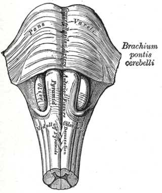
The basilar sulcus is a groove in the pons, part of the brainstem.