Related Research Articles

The human shoulder is made up of three bones: the clavicle (collarbone), the scapula, and the humerus as well as associated muscles, ligaments and tendons. The articulations between the bones of the shoulder make up the shoulder joints. The shoulder joint, also known as the glenohumeral joint, is the major joint of the shoulder, but can more broadly include the acromioclavicular joint. In human anatomy, the shoulder joint comprises the part of the body where the humerus attaches to the scapula, and the head sits in the glenoid cavity. The shoulder is the group of structures in the region of the joint.
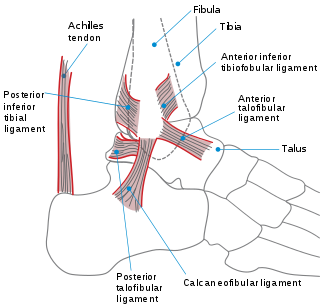
The ankle, or the talocrural region, is the region where the foot and the leg meet. The ankle includes three joints: the ankle joint proper or talocrural joint, the subtalar joint, and the inferior tibiofibular joint. The movements produced at this joint are dorsiflexion and plantarflexion of the foot. In common usage, the term ankle refers exclusively to the ankle region. In medical terminology, "ankle" can refer broadly to the region or specifically to the talocrural joint.

Ankylosing spondylitis (AS) is a type of arthritis in which there is a long-term inflammation of the joints of the spine. Typically the joints where the spine joins the pelvis are also affected. Occasionally other joints such as the shoulders or hips are involved. Eye and bowel problems may also occur. Back pain is a characteristic symptom of AS, and it often comes and goes. Stiffness of the affected joints generally worsens over time.

The anterior cruciate ligament (ACL) is one of a pair of cruciate ligaments in the human knee. The two ligaments are also called "cruciform" ligaments, as they are arranged in a crossed formation. In the quadruped stifle joint, based on its anatomical position, it is also referred to as the cranial cruciate ligament. The term cruciate translates to cross. This name is fitting because the ACL crosses the posterior cruciate ligament to form an “X”. It is composed of strong, fibrous material and assists in controlling excessive motion. This is done by limiting mobility of the joint. The anterior cruciate ligament is one of the four main ligaments of the knee, providing 85% of the restraining force to anterior tibial displacement at 30 and 90° of knee flexion. The ACL is the most injured ligament of the four located in the knee.

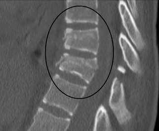
Lumbar spinal stenosis (LSS) is a medical condition in which the spinal canal narrows and compresses the nerves and blood vessels at the level of the lumbar vertebrae. Spinal stenosis may also affect the cervical or thoracic region, in which case it is known as cervical spinal stenosis or thoracic spinal stenosis. Lumbar spinal stenosis can cause pain in the low back or buttocks, abnormal sensations, and the absence of sensation (numbness) in the legs, thighs, feet, or buttocks, or loss of bladder and bowel control.
Osteophytes are exostoses that form along joint margins. They should not be confused with enthesophytes, which are bony projections that form at the attachment of a tendon or ligament. Osteophytes are not always distinguished from exostoses in any definite way, although in many cases there are a number of differences. Osteophytes are typically intra-articular.

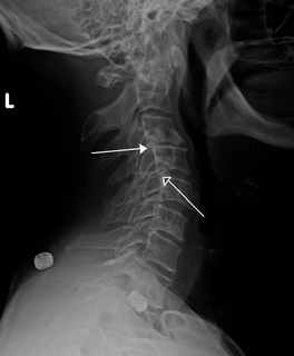
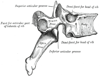
In tetrapods, cervical vertebrae are the vertebrae of the neck, immediately below the skull. Truncal vertebrae lie caudal of cervical vertebrae. In sauropsid species, the cervical vertebrae bear cervical ribs. In lizards and saurischian dinosaurs, the cervical ribs are large; in birds, they are small and completely fused to the vertebrae. The vertebral transverse processes of mammals are homologous to the cervical ribs of other amniotes. Most mammals have seven cervical vertebrae, with the only three known exceptions being the manatee with six, the two-toed sloth with five or six, and the three-toed sloth with nine.

Degenerative disc disease (DDD) is a medical condition in which there are anatomic changes and a loss of function of varying degrees of one or more intervertebral discs of the spine of sufficient magnitude as to cause symptoms. The root cause is thought to be loss of soluble proteins within the fluid contained in the disc with resultant reduction of the oncotic pressure, which in turn causes loss of fluid volume. Normal downward forces cause the affected disc to lose height, and the distance between vertebrae is reduced. The anulus fibrosus, the rigid outer shell of a disc, also weakens. This loss of height causes laxity of the longitudinal ligaments, which may allow anterior, posterior, or lateral shifting of the vertebral bodies, causing facet joint malalignment and arthritis; scoliosis; cervical hyperlordosis; thoracic hyperkyphosis; lumbar hyperlordosis; narrowing of the space available for the spinal tract within the vertebra ; or narrowing of the space through which a spinal nerve exits with resultant inflammation and impingement of a spinal nerve, causing a radiculopathy.
A retrolisthesis is a posterior displacement of one vertebral body with respect to the subjacent vertebra to a degree less than a luxation (dislocation). Retrolistheses are most easily diagnosed on lateral x-ray views of the spine. Views where care has been taken to expose for a true lateral view without any rotation offer the best diagnostic quality.

Spondylolisthesis is the displacement of one spinal vertebra compared to another. While some medical dictionaries define spondylolisthesis specifically as the forward or anterior displacement of a vertebra over the vertebra inferior to it, it is often defined in medical textbooks as displacement in any direction. Spondylolisthesis is graded based upon the degree of slippage of one vertebral body relative to the subsequent adjacent vertebral body. Spondylolisthesis is classified as one of the six major etiologies: degenerative, traumatic, dysplastic, isthmic, pathologic, or post-surgical. Spondylolisthesis most commonly occurs in the lumbar spine, primarily at the L5-S1 level with the L5 vertebral body anteriorly translating over the S1 vertebral body.

An ankle fracture is a break of one or more of the bones that make up the ankle joint. Symptoms may include pain, swelling, bruising, and an inability to walk on the injured leg. Complications may include an associated high ankle sprain, compartment syndrome, stiffness, malunion, and post-traumatic arthritis.
The unhappy triad, also known as a blown knee among other names, is an injury to the anterior cruciate ligament, medial collateral ligament, and meniscus. Analysis during the 1990s indicated that this 'classic' O'Donoghue triad is actually an unusual clinical entity among athletes with knee injuries. Some authors mistakenly believe that in this type of injury, "combined anterior cruciate and medial collateral ligament disruptions that were incurred during athletic endeavors" always present with concomitant medial meniscus injury. However, the 1990 analysis showed that lateral meniscus tears are more common than medial meniscus tears in conjunction with sprains of the ACL.

Pseudoachondroplasia is an inherited disorder of bone growth. It is a genetic autosomal dominant disorder. It is generally not discovered until 2–3 years of age, since growth is normal at first. Pseudoachondroplasia is usually first detected by a drop of linear growth in contrast to peers, a waddling gait or arising lower limb deformities.
A Chance fracture is a type of vertebral fracture that results from excessive flexion of the spine. Symptoms may include abdominal bruising, or less commonly paralysis of the legs. In around half of cases there is an associated abdominal injury such as a splenic rupture, small bowel injury, pancreatic injury, or mesenteric tear. Injury to the bowel may not be apparent in the first day.

Diffuse idiopathic skeletal hyperostosis (DISH) is a condition characterized by abnormal calcification/bone formation (hyperostosis) of the soft tissues surrounding the joints of the spine, and also of the peripheral or appendicular skeleton. In the spine, there is bone formation along the anterior longitudinal ligament and sometimes the posterior longitudinal ligament, which may lead to partial or complete fusion of adjacent vertebrae. The facet and sacroiliac joints tend to be uninvolved. The thoracic spine is the most common level involved. In the peripheral skeleton, DISH manifests as a calcific enthesopathy, with pathologic bone formation at sites where ligaments and tendons attach to bone.
Baastrup's sign is an orthopedic and radiographic disorder that often occurs in elderly humans. It is characterized by enlargement of the posterior spinous processes of the lumbar spine, with normal intervertebral disc height and neuroforamina. The reason it is referred to as kissing spine is because the posterior spinous processes 'kiss' and touch one another as the individual goes into lumbar extension, for example when flat on their stomach. The condition has been seen in humans, canines, particularly with boxer breeds, and certain breeds of horses. This disorder is named after Christian Ingerslev Baastrup.

Sacroiliac joint dysfunction generally refers to pain in the sacroiliac joint region that is caused by abnormal motion in the sacroiliac joint, either too much motion or too little motion. It typically results in inflammation of the sacroiliac joint, and can be debilitating.
Srb's anomaly is the clinical condition describing synostosis, or fusion, between the first and second ribs. It may be either a partial or complete fusion between the two ribs to create an entirely indistinguishable new rib.

In radiology, the Terry Thomas sign is a scapholunate ligament dissociation on an anteroposterior view of the wrist. Most commonly a result of a fall on the outstretched hand (FOOSH), the scapholunate ligament ruptures resulting in separation of the lunate and scaphoid bones. This burst causes the scaphoid bone to dorsally rotate. A gap of more than 3mm is pathognomonic for scapholunate dissociation.
In radiology, a Romanus lesion is the erosion of the anterior and posterior vertebral endplates in patients with an inflammatory spondyloarthropathy – such as ankylosing spondylitis or an enteropathic arthropathy. The anterior erosion in particular causes a loss of anterior vertebral body concavity, causing the vertebra to display a squared contour or even a barrel-shape. Healing of the erosion results in a sclerotic increase in density causing what is known as a shiny corner sign, which can later result in syndesmophyte formation. It is most easily diagnosed using MRI, compared to conventional radiography.
References
- ↑ Yochum, Terry R. (2005). Yochum and Rowe's Essentials of Skeletal Radiology . Lippincott/Williams & Wilkins. pp. 955, 1039–1042. ISBN 9780781739467 . Retrieved 16 October 2018.
Essentials of Skeletal Radiology Ed3 Vol.1.
- ↑ Forrester, D. M.; Brown, John Cavendish (1987). The Radiology of Joint Disease. Saunders. p. 453. ISBN 9780721638270 . Retrieved 16 October 2018.