Related Research Articles

The blood vessels are the components of the circulatory system that transport blood throughout the human body. These vessels transport blood cells, nutrients, and oxygen to the tissues of the body. They also take waste and carbon dioxide away from the tissues. Blood vessels are needed to sustain life, because all of the body's tissues rely on their functionality.
The circulatory system, also called the cardiovascular system or the vascular system, is an organ system that permits blood to circulate and transport nutrients, oxygen, carbon dioxide, hormones, and blood cells to and from the cells in the body to provide nourishment and help in fighting diseases, stabilize temperature and pH, and maintain homeostasis.

Edema, also spelled oedema, and also known as fluid retention, dropsy, hydropsy and swelling, is the build-up of fluid in the body's tissue. Most commonly, the legs or arms are affected. Symptoms may include skin which feels tight, the area may feel heavy, and affected joints may be hard to move. Other symptoms depend on the underlying cause.
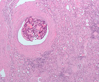
An embolism is the lodging of an embolus, a blockage-causing piece of material, inside a blood vessel. The embolus may be a blood clot (thrombus), a fat globule, a bubble of air or other gas, amniotic fluid, or foreign material. An embolism can cause partial or total blockage of blood flow in the affected vessel. Such a blockage may affect a part of the body distant from the origin of the embolus. An embolism in which the embolus is a piece of thrombus is called a thromboembolism.

The portal vein or hepatic portal vein (HPV) is a blood vessel that carries blood from the gastrointestinal tract, gallbladder, pancreas and spleen to the liver. This blood contains nutrients and toxins extracted from digested contents. Approximately 75% of total liver blood flow is through the portal vein, with the remainder coming from the hepatic artery proper. The blood leaves the liver to the heart in the hepatic veins.

The renal veins are veins that drain the kidney. They connect the kidney to the inferior vena cava. They carry the blood filtered by the kidney.

In human anatomy, the superior mesenteric artery (SMA) arises from the anterior surface of the abdominal aorta, just inferior to the origin of the celiac trunk, and supplies blood to the intestine from the lower part of the duodenum through two-thirds of the transverse colon, as well as the pancreas.

An arteriovenous fistula is an abnormal connection or passageway between an artery and a vein. It may be congenital, surgically created for hemodialysis treatments, or acquired due to pathologic process, such as trauma or erosion of an arterial aneurysm.

The hepatic veins are the veins that drain de-oxygenated blood from the liver into the inferior vena cava. There are usually three upper hepatic veins draining from the left, middle, and right parts of the liver. These are larger than the group of lower hepatic veins that can number from six to twenty. All of the hepatic veins drain into the inferior vena cava.

The cavernous sinus within the human head is one of the dural venous sinuses creating a cavity called the lateral sellar compartment bordered by the temporal bone of the skull and the sphenoid bone, lateral to the sella turcica.

In human anatomy, inferior epigastric vein refers to the vein that drains into the external iliac vein and anastomoses from the superior epigastric vein. Along its course, it is accompanied by a similarly named artery, the inferior epigastric artery.
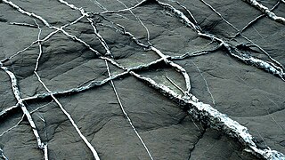
In geology, a vein is a distinct sheetlike body of crystallized minerals within a rock. Veins form when mineral constituents carried by an aqueous solution within the rock mass are deposited through precipitation. The hydraulic flow involved is usually due to hydrothermal circulation.
The Suprarenal veins are two in number:

The pterygoid canal is a passage in the sphenoid bone of the skull leading from just anterior to the foramen lacerum in the middle cranial fossa to the pterygopalatine fossa.
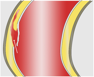
A dissection is a tear within the wall of a blood vessel, which allows blood to separate the wall layers. Usually, a dissection is an arterial wall dissection, but rarely it may be a vein wall dissection (VWD).

The intercostal arteries are a group of arteries that supply the area between the ribs ("costae"), called the intercostal space. The highest intercostal artery is an artery in the human body that usually gives rise to the first and second posterior intercostal arteries, which supply blood to their corresponding intercostal space. It usually arises from the costocervical trunk, which is a branch of the subclavian artery. Some anatomists may contend that there is no supreme intercostal artery, only a supreme intercostal vein.

In human anatomy, the dorsal veins of the penis comprise the superficial dorsal vein of the penis and the deep dorsal vein of the penis.
In the course of the round ligament of liver, small veins (paraumbilical) are found which establish an anastomosis between the veins of the anterior abdominal wall and the hepatic portal, hypogastric, and iliac veins.
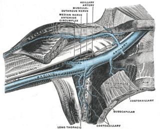
The lateral thoracic vein is a tributary of the axillary vein. It runs with the lateral thoracic artery and drains the Serratus anterior muscle and the Pectoralis major muscle.
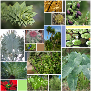
A leaf is the principal lateral appendage of the vascular plant stem, usually borne above ground and specialized for photosynthesis. The leaves, stem, flower and fruit together form the shoot system. Leaves are collectively referred to as foliage, as in "autumn foliage". In most leaves, the primary photosynthetic tissue, the palisade mesophyll, is located on the upper side of the blade or lamina of the leaf but in some species, including the mature foliage of Eucalyptus, palisade mesophyll is present on both sides and the leaves are said to be isobilateral. Most leaves are flattened and have distinct upper and lower surfaces that differ in color, hairiness, the number of stomata, the amount and structure of epicuticular wax and other features. Leaves are mostly green in color due to the presence of a compound called chlorophyll that is essential for photosynthesis as it absorbs light energy from the sun. A leaf with lighter-colored or white patches or edges is called a variegated leaf.
References
- ↑ Standring, Susan (2016). Gray's Anatomy (41 ed.). Elsevier. pp. 1179–1187.
| | This cardiovascular system article is a stub. You can help Wikipedia by expanding it. |