Phosphatidylethanolamine

Last updated
Biosynthesis of various phospholipids (including phosphatidylethanolamine) in bacteria Biosynthesis of phosphatidylglycerol, phosphatidylserine, and phosphatidylethanolamine.svg
Biosynthesis of various phospholipids (including phosphatidylethanolamine) in bacteria

Phosphatidylethanolamine (PE) is a class of phospholipids found in biological membranes. [1] They are synthesized by the addition of cytidine diphosphate-ethanolamine to diglycerides, releasing cytidine monophosphate. S-Adenosyl methionine can subsequently methylate the amine of phosphatidylethanolamines to yield phosphatidylcholines.

Contents

Function

The major membrane lipids: phosphatidylcholine (PtdCho); phosphatidylethanolamine (PtdEtn); phosphatidylinositol (PtdIns); phosphatidylserine (PtdSer). Membrane Lipids.svg
The major membrane lipids: phosphatidylcholine (PtdCho); phosphatidylethanolamine (PtdEtn); phosphatidylinositol (PtdIns); phosphatidylserine (PtdSer).

In cells

Phosphatidylethanolamines are found in all living cells, composing 25% of all phospholipids. In human physiology, they are found particularly in nervous tissue such as the white matter of brain, nerves, neural tissue, and in spinal cord, where they make up 45% of all phospholipids. [2]

Phosphatidylethanolamines play a role in membrane fusion and in disassembly of the contractile ring during cytokinesis in cell division. [3] Additionally, it is thought that phosphatidylethanolamine regulates membrane curvature. Phosphatidylethanolamine is an important precursor, substrate, or donor in several biological pathways. [2]

As a polar head group, phosphatidylethanolamine creates a more viscous lipid membrane compared to phosphatidylcholine. For example, the melting temperature of di-oleoyl-phosphatidylethanolamine is -16 °C while the melting temperature of di-oleoyl-phosphatidylcholine is -20 °C. If the lipids had two palmitoyl chains, phosphatidylethanolamine would melt at 63 °C while phosphatidylcholine would melt already at 41 °C. [4] Lower melting temperatures correspond, in a simplistic view, to more fluid membranes.

In humans

In humans, metabolism of phosphatidylethanolamine is thought to be important in the heart. When blood flow to the heart is restricted, the asymmetrical distribution of phosphatidylethanolamine between membrane leaflets is disrupted, and as a result the membrane is disrupted. Additionally, phosphatidylethanolamine plays a role in the secretion of lipoproteins in the liver. This is because vesicles for secretion of very low-density lipoproteins coming off of the Golgi apparatus have a significantly higher phosphatidylethanolamine concentration when compared to other vesicles containing very low-density lipoproteins. [5] Phosphatidylethanolamine has also shown to be able to propagate infectious prions without the assistance of any proteins or nucleic acids, which is a unique characteristic of it. [6] Phosphatidylethanolamine is also thought to play a role in blood clotting, as it works with phosphatidylserine to increase the rate of thrombin formation by promoting binding to factor V and factor X, two proteins which catalyze the formation of thrombin from prothrombin. [7] The synthesis of endocannabinoid anandamide is performed from the phosphatidylethanolamine by the successive action of two enzymes, N-acetyltransferase and phospholipase-D. [8]

In bacteria

Where phosphatidylcholine is the principal phospholipid in animals, phosphatidylethanolamine is the principal one in bacteria. One of the primary roles for phosphatidylethanolamine in bacterial membranes is to spread out the negative charge caused by anionic membrane phospholipids. In the bacterium E. coli, phosphatidylethanolamine play a role in supporting lactose permeases active transport of lactose into the cell, and may play a role in other transport systems as well. Phosphatidylethanolamine plays a role in the assembly of lactose permease and other membrane proteins. It acts as a 'chaperone' to help the membrane proteins correctly fold their tertiary structures so that they can function properly. When phosphatidylethanolamine is not present, the transport proteins have incorrect tertiary structures and do not function correctly. [9]

Phosphatidylethanolamine also enables bacterial multidrug transporters to function properly and allows the formation of intermediates that are needed for the transporters to properly open and close. [10]

Structure

Ethanolamine Ethanolamine.svg
Ethanolamine

As a lecithin, phosphatidylethanolamine consists of a combination of glycerol esterified with two fatty acids and phosphoric acid. Whereas the phosphate group is combined with choline in phosphatidylcholine, it is combined with ethanolamine in phosphatidylethanolamine. The two fatty acids may be identical or different, and are usually found in positions 1,2 (less commonly in positions 1,3).

Synthesis

The phosphatidylserine decarboxylation pathway and the cytidine diphosphate-ethanolamine pathways are used to synthesize phosphatidylethanolamine. Phosphatidylserine decarboxylase is the enzyme that is used to decarboxylate phosphatidylserine in the first pathway. The phosphatidylserine decarboxylation pathway is the main source of synthesis for phosphatidylethanolamine in the membranes of the mitochondria. Phosphatidylethanolamine produced in the mitochondrial membrane is also transported throughout the cell to other membranes for use. In a process that mirrors phosphatidylcholine synthesis, phosphatidylethanolamine is also made via the cytidine diphosphate-ethanolamine pathway, using ethanolamine as the substrate. Through several steps taking place in both the cytosol and endoplasmic reticulum, the synthesis pathway yields the end product of phosphatidylethanolamine. [11] Phosphatidylethanolamine is also found abundantly in soy or egg lecithin and is produced commercially using chromatographic separation.

Regulation

Synthesis of phosphatidylethanolamine through the phosphatidylserine decarboxylation pathway occurs rapidly in the inner mitochondrial membrane. However, phosphatidylserine is made in the endoplasmic reticulum. Because of this, the transport of phosphatidylserine from the endoplasmic reticulum to the mitochondrial membrane and then to the inner mitochondrial membrane limits the rate of synthesis via this pathway. The mechanism for this transport is currently unknown but may play a role in the regulation of the rate of synthesis in this pathway. [12]

Presence in food, health issues

Phosphatidylethanolamines in food break down to form phosphatidylethanolamine-linked Amadori products as a part of the Maillard reaction. [13] These products accelerate membrane lipid peroxidation, causing oxidative stress to cells that come in contact with them. [14] Oxidative stress is known to cause food deterioration and several diseases. Significant levels of Amadori-phosphatidylethanolamine products have been found in a wide variety of foods such as chocolate, soybean milk, infant formula, and other processed foods. The levels of Amadori-phosphatidylethanolamine products are higher in foods with high lipid and sugar concentrations that have high temperatures in processing. [13] Additional studies have found that Amadori-phosphatidylethanolamine may play a role in vascular disease, [15] act as the mechanism by which diabetes can increase the incidence of cancer, [16] and potentially play a role in other diseases as well. Amadori-phosphatidylethanolamine has a higher plasma concentration in diabetes patients than healthy people, indicating it may play a role in the development of the disease or be a product of the disease. [17]

See also

Related Research Articles

<span class="mw-page-title-main">Lipid</span> Substance of biological origin that is soluble in nonpolar solvents

Lipids are a broad group of organic compounds which include fats, waxes, sterols, fat-soluble vitamins, monoglycerides, diglycerides, phospholipids, and others. The functions of lipids include storing energy, signaling, and acting as structural components of cell membranes. Lipids have applications in the cosmetic and food industries, and in nanotechnology.

<span class="mw-page-title-main">Phospholipid</span> Class of lipids

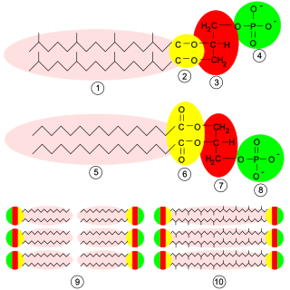
Phospholipids are a class of lipids whose molecule has a hydrophilic "head" containing a phosphate group and two hydrophobic "tails" derived from fatty acids, joined by an alcohol residue. Marine phospholipids typically have omega-3 fatty acids EPA and DHA integrated as part of the phospholipid molecule. The phosphate group can be modified with simple organic molecules such as choline, ethanolamine or serine.

<span class="mw-page-title-main">Choline</span> Chemical compound and essential nutrient

Choline ( KOH-leen) is an essential nutrient for humans and many other animals, which was formerly classified as a B vitamin (vitamin B4). It is a structural part of phospholipids and a methyl donor in metabolic one-carbon chemistry. The compound is related to trimethylglycine in the latter respect. It is a cation with the chemical formula [(CH3)3NCH2CH2OH]+. Choline forms various salts, for example choline chloride and choline bitartrate.

<span class="mw-page-title-main">Phosphatidylcholine</span> Class of phospholipids

Phosphatidylcholines (PC) are a class of phospholipids that incorporate choline as a headgroup. They are a major component of biological membranes and can be easily obtained from a variety of readily available sources, such as egg yolk or soybeans, from which they are mechanically or chemically extracted using hexane. They are also a member of the lecithin group of yellow-brownish fatty substances occurring in animal and plant tissues. Dipalmitoylphosphatidylcholine (lecithin) is a major component of the pulmonary surfactant, and is often used in the lecithin–sphingomyelin ratio to calculate fetal lung maturity. While phosphatidylcholines are found in all plant and animal cells, they are absent in the membranes of most bacteria, including Escherichia coli. Purified phosphatidylcholine is produced commercially.

<span class="mw-page-title-main">Sphingomyelin</span> Class of chemical compounds

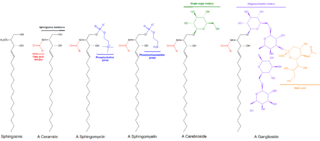
Sphingomyelin is a type of sphingolipid found in animal cell membranes, especially in the membranous myelin sheath that surrounds some nerve cell axons. It usually consists of phosphocholine and ceramide, or a phosphoethanolamine head group; therefore, sphingomyelins can also be classified as sphingophospholipids. In humans, SPH represents ~85% of all sphingolipids, and typically make up 10–20 mol % of plasma membrane lipids.

<span class="mw-page-title-main">Glycerophospholipid</span> Class of lipids

Glycerophospholipids or phosphoglycerides are glycerol-based phospholipids. They are the main component of biological membranes in eukaryotic cells. They are a type of lipid, of which its composition affects membrane structure and properties. Two major classes are known: those for bacteria and eukaryotes and a separate family for archaea.

<span class="mw-page-title-main">Inner mitochondrial membrane</span>

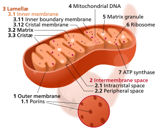
The inner mitochondrial membrane (IMM) is the mitochondrial membrane which separates the mitochondrial matrix from the intermembrane space.

<span class="mw-page-title-main">Phosphatidylserine</span> Chemical compound

Phosphatidylserine is a phospholipid and is a component of the cell membrane. It plays a key role in cell cycle signaling, specifically in relation to apoptosis. It is a key pathway for viruses to enter cells via apoptotic mimicry. Its exposure on the outer surface of a membrane marks the cell for destruction via apoptosis.

<span class="mw-page-title-main">Phospholipid scramblase</span> Protein

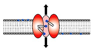
Scramblase is a protein responsible for the translocation of phospholipids between the two monolayers of a lipid bilayer of a cell membrane. In humans, phospholipid scramblases (PLSCRs) constitute a family of five homologous proteins that are named as hPLSCR1–hPLSCR5. Scramblases are members of the general family of transmembrane lipid transporters known as flippases. Scramblases are distinct from flippases and floppases. Scramblases, flippases, and floppases are three different types of enzymatic groups of phospholipid transportation enzymes. The inner-leaflet, facing the inside of the cell, contains negatively charged amino-phospholipids and phosphatidylethanolamine. The outer-leaflet, facing the outside environment, contains phosphatidylcholine and sphingomyelin. Scramblase is an enzyme, present in the cell membrane, that can transport (scramble) the negatively charged phospholipids from the inner-leaflet to the outer-leaflet, and vice versa.

Phospholipase D (EC 3.1.4.4, lipophosphodiesterase II, lecithinase D, choline phosphatase, PLD; systematic name phosphatidylcholine phosphatidohydrolase) is an enzyme of the phospholipase superfamily that catalyses the following reaction

<span class="mw-page-title-main">Citicoline</span> Chemical compound

Citicoline (INN), also known as cytidine diphosphate-choline (CDP-Choline) or cytidine 5'-diphosphocholine is an intermediate in the generation of phosphatidylcholine from choline, a common biochemical process in cell membranes. Citicoline is naturally occurring in the cells of human and animal tissue, in particular the organs.

<span class="mw-page-title-main">Phosphatidylethanolamine N-methyltransferase</span> Protein-coding gene in the species Homo sapiens

Phosphatidylethanolamine N-methyltransferase is a transferase enzyme which converts phosphatidylethanolamine (PE) to phosphatidylcholine (PC) in the liver. In humans it is encoded by the PEMT gene within the Smith–Magenis syndrome region on chromosome 17.

Choline kinase is an enzyme which catalyzes the first reaction in the choline pathway for phosphatidylcholine (PC) biosynthesis. This reaction involves the transfer of a phosphate group from adenosine triphosphate (ATP) to choline in order to form phosphocholine.

Membrane contact sites (MCS) are close appositions between two organelles. Ultrastructural studies typically reveal an intermembrane distance in the order of the size of a single protein, as small as 10 nm or wider, with no clear upper limit. These zones of apposition are highly conserved in evolution. These sites are thought to be important to facilitate signalling, and they promote the passage of small molecules, including ions, lipids and reactive oxygen species. MCS are important in the function of the endoplasmic reticulum (ER), since this is the major site of lipid synthesis within cells. The ER makes close contact with many organelles, including mitochondria, Golgi, endosomes, lysosomes, peroxisomes, chloroplasts and the plasma membrane. Both mitochondria and sorting endosomes undergo major rearrangements leading to fission where they contact the ER. Sites of close apposition can also form between most of these organelles most pairwise combinations. First mentions of these contact sites can be found in papers published in the late 1950s mainly visualized using electron microscopy (EM) techniques. Copeland and Dalton described them as “highly specialized tubular form of endoplasmic reticulum in association with the mitochondria and apparently in turn, with the vascular border of the cell”.

<span class="mw-page-title-main">Choline/ethanolamine kinase family</span>

In molecular biology, the choline/ethanolamine kinase family includes choline kinase(EC 2.7.1.32) and ethanolamine kinase.

<span class="mw-page-title-main">1-Lysophosphatidylcholine</span>

1-Lysophosphatidylcholines are a class of phospholipids that are intermediates in the metabolism of lipids. They result from the hydrolysis of an acyl group from the sn-1 position of phosphatidylcholine. They are also called 2-acyl-sn-glycero-3-phosphocholines. The synthesis of phosphatidylcholines with specific fatty acids occurs through the synthesis of 1-lysoPC. The formation of various other lipids generates 1-lysoPC as a by-product.

In enzymology, a ceramide phosphoethanolamine synthase is an enzyme that catalyzes the chemical reaction

<span class="mw-page-title-main">CDP-choline pathway</span>

The CDP-choline pathway, first identified by Eugene P. Kennedy in 1956, is the predominant mechanism by which mammalian cells synthesize phosphatidylcholine (PC) for incorporation into membranes or lipid-derived signalling molecules. The CDP-choline pathway represents one half of what is known as the Kennedy pathway. The other half is the CDP-ethanolamine pathway which is responsible for the biosynthesis of the phospholipid phosphatidylethanolamine (PE).

<span class="mw-page-title-main">Mitochondria associated membranes</span> Cellular structure

Mitochondria-associated membranes (MAMs) represent regions of the endoplasmic reticulum (ER) which are reversibly tethered to mitochondria. These membranes are involved in import of certain lipids from the ER to mitochondria and in regulation of calcium homeostasis, mitochondrial function, autophagy and apoptosis. They also play a role in development of neurodegenerative diseases and glucose homeostasis.

<span class="mw-page-title-main">Jean Vance</span> British-Canadian biochemist

Jean Vance is a British-Canadian biochemist. She is known for her pioneering work on subcellular organelles and for her discovery of a connection between the endoplasmic reticulum and the mitochondrial membrane. She is a Professor of Medicine at the University of Alberta, Canada and a Fellow of the Royal Society of Canada.

References

  1. Wellner, Niels; Diep, Thi Ai; Janfelt, Christian; Hansen, Harald Severin (2012). "N-acylation of phosphatidylethanolamine and its biological functions in mammals". Biochimica et Biophysica Acta (BBA) - Molecular and Cell Biology of Lipids. 1831 (3): 652–62. doi:10.1016/j.bbalip.2012.08.019. PMID   23000428.
  2. 1 2 Vance, Jean E.; Tasseva, Guergana (2012). "Formation and function of phosphatidylserine and phosphatidylethanolamine in mammalian cells". Biochimica et Biophysica Acta (BBA) - Molecular and Cell Biology of Lipids. 1831 (3): 543–54. doi:10.1016/j.bbalip.2012.08.016. PMID   22960354.
  3. Emoto, K.; Kobayashi, T; Yamaji, A; Aizawa, H; Yahara, I; Inoue, K; Umeda, M (1996). "Redistribution of phosphatidylethanolamine at the cleavage furrow of dividing cells during cytokinesis". Proceedings of the National Academy of Sciences. 93 (23): 12867–72. Bibcode:1996PNAS...9312867E. doi: 10.1073/pnas.93.23.12867 . JSTOR   40713. PMC   24012 . PMID   8917511.
  4. See references in Wan et al. Biochemistry 47 2008[ verification needed ]
  5. Vance, J. E. (2008). "Thematic Review Series: Glycerolipids. Phosphatidylserine and phosphatidylethanolamine in mammalian cells: Two metabolically related aminophospholipids". The Journal of Lipid Research. 49 (7): 1377–87. doi: 10.1194/jlr.R700020-JLR200 . PMID   18204094.
  6. Deleault, N. R.; Piro, J. R.; Walsh, D. J.; Wang, F.; Ma, J.; Geoghegan, J. C.; Supattapone, S. (2012). "Isolation of phosphatidylethanolamine as a solitary cofactor for prion formation in the absence of nucleic acids". Proceedings of the National Academy of Sciences. 109 (22): 8546–51. Bibcode:2012PNAS..109.8546D. doi: 10.1073/pnas.1204498109 . PMC   3365173 . PMID   22586108.
  7. Majumder, R.; Liang, X.; Quinn-Allen, M. A.; Kane, W. H.; Lentz, B. R. (2011). "Modulation of Prothrombinase Assembly and Activity by Phosphatidylethanolamine". Journal of Biological Chemistry. 286 (41): 35535–42. doi: 10.1074/jbc.M111.260141 . PMC   3195639 . PMID   21859710.
  8. Isidro, F. (2014). "Cannabinoids for treatment of Alzheimer's disease: moving toward the clinic". Frontiers in Pharmacology. 5: 37. doi: 10.3389/fphar.2014.00037 . PMC   3942876 . PMID   24634659.
  9. Christie, W.W. (April 16, 2012). "Phosphatidylethanolamine and Related Lipids". The AOCS Lipid Library. Archived from the original on August 21, 2012. Retrieved September 3, 2012.
  10. Gbaguidi, B.; Hakizimana, P.; Vandenbussche, G.; Ruysschaert, J.-M. (2007). "Conformational changes in a bacterial multidrug transporter are phosphatidylethanolamine-dependent" (PDF). Cellular and Molecular Life Sciences. 64 (12): 1571–82. doi:10.1007/s00018-007-7031-0. PMID   17530171. S2CID   2078590.
  11. Kelly, Karen (July 28, 2011). "Phospholipid Biosynthesis". The AOCS Lipid Library. Retrieved September 3, 2012.
  12. Kuge, Osamu; Nishijima, Masahiro (1 April 2003). "Biosynthetic Regulation and Intracellular Transport of phosphatidylserine in Mammalian Cells". The Journal of Biochemistry. 133 (4): 397–403. doi:10.1093/jb/mvg052. PMID   12761285. Archived from the original on 30 January 2021. Retrieved 30 January 2021.
  13. 1 2 Oak, Jeong-Ho; Nakagawa, Kiyotaka; Miyazawa, Teruo (2002). "UV analysis of Amadori-glycated phosphatidylethanolamine in foods and biological samples". The Journal of Lipid Research. 43 (3): 523–9. doi: 10.1016/S0022-2275(20)30158-9 . PMID   11893788.
  14. Oak, Jeong-Ho; Nakagawa, Kiyotaka; Miyazawa, Teruo (2000). "Synthetically prepared Amadori-glycated phosphatidylethanolamine can trigger lipid peroxidation via free radical reactions". FEBS Letters. 481 (1): 26–30. Bibcode:2000FEBSL.481...26O. doi: 10.1016/S0014-5793(00)01966-9 . PMID   10984609. S2CID   23265125.
  15. Oak, Jeong-Ho; Nakagawa, Kiyotaka; Oikawa, Shinichi; Miyazawa, Teruo (2003). "Amadori-glycated phosphatidylethanolamine induces angiogenic differentiations in cultured human umbilical vein endothelial cells". FEBS Letters. 555 (2): 419–23. Bibcode:2003FEBSL.555..419O. doi:10.1016/S0014-5793(03)01237-7. PMID   14644453. S2CID   33974755.
  16. Eitsuka, Takahiro; Nakagawa, Kiyotaka; Ono, Yuichi; Tatewaki, Naoto; Nishida, Hiroshi; Kurata, Tadao; Shoji, Naoki; Miyazawa, Teruo (2012). "Amadori-glycated phosphatidylethanolamine up-regulates telomerase activity in PANC-1 human pancreatic carcinoma cells". FEBS Letters. 586 (16): 2542–7. Bibcode:2012FEBSL.586.2542E. doi: 10.1016/j.febslet.2012.06.027 . PMID   22750441. S2CID   5452160.
  17. Ariizumi, Ken; Koike, T; Ohara, S; Inomata, Y; Abe, Y; Iijima, K; Imatani, A; Oka, T; Shimosegawa, T (2008). "Incidence of reflux esophagitis and H pylori infection in diabetic patients". World Journal of Gastroenterology. 14 (20): 3212–7. doi: 10.3748/wjg.14.3212 . PMC   2712855 . PMID   18506928.