Zoonotic origins of COVID-19

Last updated

SARS-CoV-2, the causative agent of COVID-19, was first introduced to humans through zoonosis (transmission of a pathogen to a human from an animal), and a zoonotic spillover event is the origin of COVID-19 that is considered most plausible by the scientific community. [lower-alpha 1] Human coronaviruses including SARS-CoV-2 are zoonotic diseases that are often acquired through spillover infection from animals. [2]

Contents

Background

Previous emergence of SARS-CoV-1 and MERS-CoV showed that Betacoronaviruses represent a risk for emergence of diseases threatening to humans. [3] [4] Increased awareness due to the 2002–2004 SARS outbreak motivated research into the potential for other coronavirus outbreaks and the animal reservoirs which could lead to them. [5] Bats, in particular, are known to harbor persistent populations of coronaviruses, and under conditions of persistent infection, coronaviruses tend to accumulate mutations that allow their receptor binding domains to interact with cross-species orthologs of the target receptors. [6] Based on serological and molecular studies, Chinese horseshoe bats were identified as the most likely reservoir for SARS-CoV-1. [7] Bats were also a likely reservoir for the related betacoronavirus MERS-CoV, though the evidence for this is less conclusive than the role of camels as a reservoir for MERS. [8] By 2010, in vitro experiments had confirmed that modifications to the spike protein receptor binding domain could enable human infection by several SARS-related coronaviruses. [9] Virologists Rachel Graham and Ralph S. Baric at that time wrote, "that the question of emergence of another pathogenic human coronavirus from bat reservoirs might be more appropriately expressed as 'when' than as 'if'." [10] [ better source needed ]

Origin in bats

The likely origin of SARS-CoV-2 from bats aligns with the origins of other coronaviruses in its genus Betacoronavirus, subgenus Sarbecovirus. [11] [12] [13] [14] Several bat species have special cellular mechanisms to resist proinflammatory cytokines associated with Betacoronavirus virulence, [15] for example, spike proteins in SARS-related coronaviruses coevolve with bat ACE2 receptors in an evolutionary arms race. [16]

Phylogenetic tree of SARS-CoV-2 and closely related betacoronaviruses (left) and their geographic context (right) Phylogenetic analysis of SARS-CoV-2 and representative sarbecoviruses and geographical context.webp
Phylogenetic tree of SARS-CoV-2 and closely related betacoronaviruses (left) and their geographic context (right)

Bats, along with their viruses, have large overlapping geographic ranges in Southeast Asia, [17] and there is a particularly great concentration and diversity of bat-related coronaviruses in Southern and Southwest China. [15] The most similar known viruses to SARS-CoV-2 include bat coronaviruses RpYN06 with 94.5% identity, [15] and RmYN02 with 93% identity [18] RaTG13 was not the direct progenitor of SARS-CoV-2. [19] Temmam et al. found no serological evidence for exposure to BANAL-52 among bat handlers and guano collectors in the area of Laos where it was sampled. [20] Lytras et al. wrote that "SARS-CoV-2 can be unambiguously traced to horseshoe bats". [21] They estimated SARS-CoV-2's most recent common ancestors with RmYN02 and RaTG13 to have diverged 40 and 50 years ago, respectively. [22]

Novel features of SARS-CoV-2

The receptor binding domain of the SARS-CoV-2 spike protein has an insertion of amino acids between its S1 and S2 subunits. [23] Among Sarbecoviruses, only SARS-CoV-2 and RmYN02 have such an insertion, suggesting differences in reservoir species, intermediate hosts, or evolutionary pathway. [18] The receptor binding motif is the portion of SARS-CoV-2 that is most diverged from RaTG13. [24] The SARS-CoV-2 receptor binding domain is more similar to those of pangolin coronaviruses. [18] Viruses including BANAL-52 isolated from bats in Laos showed high similarity to the SARS-CoV-2 receptor binding domain in amino acid residues but less than 76.4% nucleotide identity across the spike protein. [2] The observed binding of N-acetylneuraminic acid by the NTD [25] of the spike protein and loss of that binding through mutation of the corresponding sugar binding pocket in emergent variants of concern has suggested a potential role for transient sugar-binding in the zoonosis of SARS-CoV-2, consistent with prior evolutionary proposals. [26]

Within genus Betacoronavirus, furin cleavage sites are common in subgenera Merbecovirus and Embecovirus . [27] Furin cleavage sites have independently evolved six times in different clades of Betacoronavirus. [27] Furin cleavage sites also evolved in Alphacoronaviruses and Gammacoronaviruses independently of Betacoronavirus. [27]

Phylogenetic tree of Betacoronaviruses. Those with furin cleavage sites are indicated in red text. 1-s2.0-S1873506120304165-ga1 lrg.jpg
Phylogenetic tree of Betacoronaviruses. Those with furin cleavage sites are indicated in red text.

Furin cleavage contributes strongly to the transmissibility and pathogenicity of SARS-CoV-2 in humans. [28] SARS-CoV-2 variants lacking the furin cleavage site are transmissible between humans, but much less effectively. [29] The furin cleavage site is sometimes described as "polybasic" on account of its particular motif of basic amino acids. [28] SARS-CoV-2 shares amino acid identity with a furin cleavage site of human ENaC α subunit. [30] [31] [lower-alpha 2] Human ENaC is identical only to that of a few great apes and Pipistrellus kuhli. [32]

SARS-CoV-2 is also distinct among human coronaviruses for having a single intact ORF8 gene rather than "a" and "b" subunits. [1]

Processes of host adaptation

SARS-CoV-2 has an expanded host range compared to SARS-CoV-1 and MERS-CoV. [33] [34] SARS-CoV-2 (along with SARS-CoV-1 and MERS-CoV) are generalist viruses, not specifically adapted to humans, meaning they have potential to spill over to many species and establish new natural reservoirs after adaptive evolutionary changes. [35]

Within a single host, a variety of single-nucleotide variations arise through random mutations and genetic drift giving rise to viral quasispecies. [36] SARS-CoV-2 mutates at a slower rate than is typical of RNA viruses. [36] The main host-derived driver of mutation is editing by APOBEC proteins. [36] Negative selection by host immune processes causes convergent evolution towards immune escape. [36] Persistence of infection is correlated with quasispecies diversity, but the direction of causality for this is unknown. [36]

Actual host susceptibility can differ significantly from in silico predictions. [35] [37] The process of host adaptation has been studied in humanized mice as well as by generating mouse-adapted viral strains through serial passage. [38] Wild-type mice are only weakly susceptible to the original SARS-CoV-2 strain. [38]

Recombination

Compared to other single-stranded RNA viruses, coronaviruses have increased tendency to undergo genetic recombination, which allows them to exchange genetic material with close relatives co-infecting the same organism. [39] The origin of SARS-CoV-1 is believed to involve multiple recombination events. [40] Recombination between strains of SARS-CoV-1 is common. [41] Inferred phylogenies of SARS2-related coronaviruses might be explained by recombination. [42] Recombination between various SARS-CoV-2 variants of concern has been reported. [43]

Illustrations of replicative and nonreplicative RNA recombination Viruses-11-00859-g004.png
Illustrations of replicative and nonreplicative RNA recombination

Lytras et al. identified the SARS-CoV-2 spike open reading frame as a recombination hotspot. [44] They speculate that the SARS-CoV-2 genome may involve repeated recombination events overprinting regions that were already themselves products of recombination. [45] Temmam et al. wrote that due to the limited diffusion of bat viruses in mammals, co-infection required for recombination in mammals was unlikely. [46] Therefore, they considered it more likely that the furin cleavage site arose in a bat reservoir prior to spillover.

Selection

After cross-species transmission of a virus, rapid evolution and positive selection are expected. [47] Several studies found only weak signs of adaptive evolution early in the COVID-19 pandemic. [lower-alpha 3] Kang et al. wrote that SARS-CoV-2 had exhibited relatively little genetic variation by 2021. [47] Tai et al. wrote that population expansion rather than positive selection explained the mutation frequency spectrum during the early pandemic. [49] Cagliani et al. wrote that the SARS-CoV-2 genome overall shows evidence of "strong to moderate" purifying selection. [50] Accessory open reading frames, especially ORF8, showed weak to neutral selection. The general lack of positive selection during the early outbreak of SARS-CoV-2 contrasted with the evolutionary course of SARS-CoV-1. [51]

SARS-CoV-2 spike homotrimer with one protein subunit highlighted. The ACE2 binding domain is magenta. 6VSB spike protein SARS-CoV-2 monomer in homotrimer.png
SARS‑CoV‑2 spike homotrimer with one protein subunit highlighted. The ACE2 binding domain is magenta.

Strong evidence of positive selection was found however in the spike protein S1 subunit, which contains the receptor binding domain. [24] [52]

Genome instability in the spike protein is typical of coronaviruses in general, favoring the production of numerous spike protein variants. [43] The receptor binding domain is a significant factor in host tropism, or the variety of species a virus can infect. [53] [54] Coronavirus adaptation to a new host often requires mutations in the receptor binding domain. [55] Kang et al. identified a single nucleotide polymorphism relative to RaTG13 in the spike protein, consistent among all of more than 180,000 SARS-CoV-2 samples, affecting glycosylation of the receptor binding domain. [56] Using a reverse genetics system to generate an ancestral-like mutant, they confirmed that the putative ancestral form of this SNP was much less transmissible in human cells. [57]

Host proteases

The majority of sarbecoviruses are not dependent on ACE2 for cell entry. [58] Guo et al. divide the sarbecoviruses into four clades, the first being ACE2-using respiratory viruses including SARS-CoV-1, SARS-CoV-2, and WIV1. Relative to clade 1, clades 3 and 4 have a one residue deletion in the receptor binding domain and diminished ability to use ACE2. Clade 2 has two deletions and does not interact with ACE2. Clades 2 through 4 are more difficult to isolate or propagate in cell culture, and consequently have been less studied. ACE2-independent infection by sarbecoviruses depends on high levels of trypsin, a digestive protein, in the host environment. Trypsin may compensate for other missing or compatible host proteases. The ubiquity of furin compared to tripsin would allow the gain of a furin cleavage site to expand viral tissue tropism. [59] Guo et al. identified clade 1 and 2 sarbecoviruses in Rhinolophus fecal samples, suggesting that both types naturally replicate in the bat digestive tract. [58] Bats with SARS-like infections in the wild showed no viral replication in respiratory droplets. [60] A fecal-oral transmission is an alternative route for some respiratory viruses. [58] No higher incidence of Sarbecoviruses has been reported in workers who come into direct contact with guano. [13] Temmam et al. found that BANAL-236, a SARS-CoV-2-related virus isolated from bats in Laos, acts as an enteric virus in macaques. [61]

Timing of spillover

In contrast to SARS-CoV-1, the initial outbreak of SARS-CoV-2 was reported in only one city. [62] Chinese epidemiological surveillance did not report any other pneumonia outbreaks in autumn 2019. [63] Graham and Baric wrote that in the case of SARS-CoV-1, virus populations circulated and adapted to civets and humans over the course of two years before the recognized outbreak. [60]

Reverse zoonosis

Transmission of SARS-CoV-2 from humans to animals, known as reverse zoonosis or anthroponosis, is possible. [64] Reverse zoonotic or experimental infection with SARS-CoV-2 has been reported in 31 animal species. [65] It is not believed that wildlife plays a significant role in the ongoing circulation of SARS-CoV-2 among humans. [66]

Mink in a Polish farm c. 2013 DSC 0377 (8413679878).jpg
Mink in a Polish farm c.2013

SARS-CoV-2 variants adapted to mink were found co-circulating among humans and farmed mink. [2] Mink are the only animal in Europe or North America to experience widespread SARS-CoV-2 outbreaks. Transmission from human to mink has occurred multiple times, in most cases not resulting in a sustained mink outbreak. [51] Strong evidence was seen of positive selection in mink after spillover, concentrated in the receptor binding motif. [67]

Transmission back to humans has been documented for mink, hamsters, and cats. [66] Transmission to humans from deer is suspected. [66] Transmission of SARS-CoV-2 among domestic cats has been confirmed. [68]

Li et al. wrote that SARS-CoV-2 may be less able than SARS-CoV-1 to pass from humans to animals. [54]

Hypothesized intermediate hosts

In the outbreak of SARS-CoV-1, palm civets, raccoon dogs, ferret badgers, red foxes, domestic cats, and rice field rats were possible vectors. [7] Graham and Baric wrote that human and civet infections likely stemmed from an unknown common progenitor. [69] Patrick Berche wrote that the emergences of SARS-CoV-1 and MERS-CoV appeared to be sequential processes involving intermediate hosts, co-infections, and recombination. [70] In contrast with the rapid identification of animal hosts for SARS-CoV-1 and MERS-CoV, no direct animal source for SARS-CoV-2 has been found. [71] Holmes et al. wrote that the lack of intermediate host is likely because the right animal has not been tested so far. [19] Frutos et al. proposed that rather than a discrete spillover event, SARS-CoV-2 arose in accordance with a circulation model, involving repeated horizontal transfer among humans, bats, and other mammals without establishing significant reservoirs in any of them until the pandemic. [72]

Sunda pangolin Pangolin borneo.jpg
Sunda pangolin

Pangolins have been considered a possible reservoir of SARS-CoV-2. [73] Pangolins are sometimes sold in wet markets in China, where they are considered a culinary delicacy and a component of traditional medicine. [13] The highest sequence similarity to the SARS-CoV-2 spike receptor binding domain was found in a coronavirus infecting Sunda pangolins in Guangdong province. [33] Pangolins are frequently smuggled to China. [45] Lytras et al. wrote that, consistent with a lack of reported infections of pangolins in Malaysia, they were likely infected after being trafficked into China. [45]

The SARS-CoV-2 receptor binding domain shares more synonymous substitutions with a pangolin-CoV than RaTG13. [74] The binding potential of SARS-CoV-2 to pangolin ACE2 however is very low. [37] The receptor binding domain of SARS-CoV-2 has been hypothesized to originate from recombination of the relevant portion of pangolin-CoV with an RaTG13-like virus. [75]

Deer mice are highly susceptible to SARS-CoV-2, making them potential reservoir or intermediate hosts. [76] Raccoon dogs could be capable of transmitting SARS-CoV-2 to other animals under farm-like conditions. [68] Transmission between raccoon dogs was shown under laboratory conditions. [65] White-tailed deer are a potential vector for SARS-CoV-2. [68]

Wet market hypothesis

Spillover has been proposed to have occurred at one or more wet markets in China. [77] Wild and semi-wild game animals are commonly traded and consumed in China, and this practice has expanded in recent decades. The close contact among animals in unsanitary conditions creates the potential for diseases to thrive. In the 2002 outbreak in Guangdong, most living animals in the markets showed serological evidence of exposure to SARS-CoV-1. [70]

Exterior of Huanan seafood market undergoing sterilization in March 2020 Huanan Seafood Wholesale Market.png
Exterior of Huanan seafood market undergoing sterilization in March 2020

Many early cases were associated with the Huanan seafood market in Wuhan. [21] The first documented COVID-19 infection was in a worker at a seafood stall in the Huanan market. [78] Sequences from that patient have not been published; however, SARS-CoV-2 belonging to lineage B/L were detected in environmental samples from the patient's stall. [78] No bats or pangolins were reported to be at the market between May 2017 and November 2019. Raccoon dogs, which are hypothesized to be succeptible to SARS-CoV-2, were at the market. [79] A survey by He et al. in 2021 identified 102 mammal-affecting viruses in Chinese game animals, 65 of which were identified then for the first time. [80] They did not find any SARS-like viruses, but did find evidence for the transmission of a MERS-like virus from bats to hedgehogs. [80]

Between January 1 and March 30, epidemiologists from China CDC collected 457 samples from animal matter including carcasses and feces and collected 923 environmental samples. All animal samples tested negative for SARS-CoV-2. [81] SARS-CoV-2 was found in 73 environmental samples. Live virus was isolated from three samples, two of which came from stalls belonging to known patients. [81] No significant association was found between environmental virus titer and the type of product sold at particular stalls. [82] SARS-CoV-2 was identified in all four sewer wells in the market. Overground drainage from the surrounding area concentrated under the market.

One environmental sample contained lineage A/S SARS-CoV-2, while all others belonged to lineage B/L. [83] The sample containing lineage A/S also contained evidence of humans and livestock, but no wild animals. [84] Overall, wildlife including racoon dogs were detected at very low levels and mostly associated with negative samples. [85] Liu et al. wrote that there was not enough information to determine the origin of the virus. [85] In particular, they wrote that the evidence does not prove the presence of an infected raccoon dog or the occurrence of multiple zoonotic spillovers at the market as proposed by Pekar et al.

Origins of variants

The World Health Organization defines variants of concern as a variant with evidence of increase transmissibility, severity, or immune escape. [86] All variants of concern so far independently evolved from the original strain, rather than from each other. [66]

The evolution of the Omicron variant appeared to be more rapid than other variants. [55] Theories for the origin of the omicron variant include long-term circulation among humans outside areas where genetic surveillance was performed, mutation in an immunocompromised individual, or adaptation in an animal species after reverse zoonosis. [87] [88] The Omicron variant is proposed to have originated in mice. [41] [88]

Notes

  1.  : [1] ... "the proximal phylogenetic origin of SARS-CoV-2 and its mode of introduction into human circulation remain unclear. There is no evidence for SARS-CoV-2 having been 'engineered' in a lab. Conversely, the escape of a strain from a lab or an accidental contamination during field work cannot formally be ruled out at this stage. However, a zoonotic spillover event in nature is considered as the most plausible scenario in the scientific community"
  2. Harrison and Sachs' analysis of the furin cleavage site have been disputed in a letter to the editor Garry, Robert F. (September 29, 2022). "SARS-CoV-2 furin cleavage site was not engineered". PNAS. 119 (40): e2211107119. Bibcode:2022PNAS..11911107G. doi: 10.1073/pnas.2211107119 . PMC   9546612 . PMID   36173950., with a reply from the authors Harrison, Neil L.; Sachs, Jeffery D. (November 2, 2022a). "Reply to Garry: The origin of SARS-CoV-2 remains unresolved". PNAS. 119 (45): e2215826119. Bibcode:2022PNAS..11915826H. doi:10.1073/pnas.2215826119. PMC   9659384 . PMID   36322733.
  3.  : [48] "Weak signs of adaptive evolution during the early phase of the epidemic of SARS-CoV-2 in humans have been observed in many studies (Chaw et al. 2020; Tang et al. 2020; MacLean et al. 2021; Martin et al. 2021)."

Related Research Articles

<span class="mw-page-title-main">Coronavirus</span> Subfamily of viruses in the family Coronaviridae

Coronaviruses are a group of related RNA viruses that cause diseases in mammals and birds. In humans and birds, they cause respiratory tract infections that can range from mild to lethal. Mild illnesses in humans include some cases of the common cold, while more lethal varieties can cause SARS, MERS and COVID-19. In cows and pigs they cause diarrhea, while in mice they cause hepatitis and encephalomyelitis.

<span class="mw-page-title-main">SARS-related coronavirus</span> Species of coronavirus causing SARS and COVID-19

Severe-acute-respiratory-syndrome–related coronavirus is a species of virus consisting of many known strains. Two strains of the virus have caused outbreaks of severe respiratory diseases in humans: severe acute respiratory syndrome coronavirus 1, the cause of the 2002–2004 outbreak of severe acute respiratory syndrome (SARS), and severe acute respiratory syndrome coronavirus 2 (SARS-CoV-2), the cause of the pandemic of COVID-19. There are hundreds of other strains of SARSr-CoV, which are only known to infect non-human mammal species: bats are a major reservoir of many strains of SARSr-CoV; several strains have been identified in Himalayan palm civets, which were likely ancestors of SARS-CoV-1.

<span class="mw-page-title-main">Angiotensin-converting enzyme 2</span> Exopeptidase enzyme that acts on angiotensin I and II

Angiotensin-converting enzyme 2 (ACE2) is an enzyme that can be found either attached to the membrane of cells (mACE2) in the intestines, kidney, testis, gallbladder, and heart or in a soluble form (sACE2). Both membrane bound and soluble ACE2 are integral parts of the renin–angiotensin–aldosterone system (RAAS) that exists to keep the body's blood pressure in check. mACE2 is cleaved by the enzyme ADAM17 in a process regulated by substrate presentation. ADAM17 cleavage releases the extracellular domain creating soluble ACE2 (sACE2). ACE2 enzyme activity opposes the classical arm of the RAAS by lowering blood pressure through catalyzing the hydrolysis of angiotensin II into angiotensin (1–7). Angiotensin (1-7) in turns binds to MasR receptors creating localized vasodilation and hence decreasing blood pressure. This decrease in blood pressure makes the entire process a promising drug target for treating cardiovascular diseases.

<span class="mw-page-title-main">SARS-CoV-1</span> Virus that causes SARS

Severe acute respiratory syndrome coronavirus 1 (SARS-CoV-1), previously known as severe acute respiratory syndrome coronavirus (SARS-CoV), is a strain of coronavirus that causes severe acute respiratory syndrome (SARS), the respiratory illness responsible for the 2002–2004 SARS outbreak. It is an enveloped, positive-sense, single-stranded RNA virus that infects the epithelial cells within the lungs. The virus enters the host cell by binding to angiotensin-converting enzyme 2. It infects humans, bats, and palm civets. The SARS-CoV-1 outbreak was largely brought under control by simple public health measures. Testing people with symptoms, isolating and quarantining suspected cases, and restricting travel all had an effect. SARS-CoV-1 was most transmissible when patients were sick, so its spread could be effectively suppressed by isolating patients with symptoms.

<i>Human coronavirus NL63</i> Species of virus

Human coronavirus NL63 (HCoV-NL63) is a species of coronavirus, specifically a Setracovirus from among the Alphacoronavirus genus. It was identified in late 2004 in patients in the Netherlands by Lia van der Hoek and Krzysztof Pyrc using a novel virus discovery method VIDISCA. Later on the discovery was confirmed by the researchers from the Rotterdam, the Netherlands The virus is an enveloped, positive-sense, single-stranded RNA virus which enters its host cell by binding to ACE2. Infection with the virus has been confirmed worldwide, and has an association with many common symptoms and diseases. Associated diseases include mild to moderate upper respiratory tract infections, severe lower respiratory tract infection, croup and bronchiolitis.

<span class="mw-page-title-main">Furin</span> Enzyme found in humans

Furin is a protease, a proteolytic enzyme activated by substrate presentation that in humans and other animals is encoded by the FURIN gene. Some proteins are inactive when they are first synthesized, and must have sections removed in order to become active. Furin cleaves these sections and activates the proteins. It was named furin because it was in the upstream region of an oncogene known as FES. The gene was known as FUR and therefore the protein was named furin. Furin is also known as PACE. A member of family S8, furin is a subtilisin-like peptidase.

<i>Murine coronavirus</i> Species of virus

Murine coronavirus (M-CoV) is a virus in the genus Betacoronavirus that infects mice. Belonging to the subgenus Embecovirus, murine coronavirus strains are enterotropic or polytropic. Enterotropic strains include mouse hepatitis virus (MHV) strains D, Y, RI, and DVIM, whereas polytropic strains, such as JHM and A59, primarily cause hepatitis, enteritis, and encephalitis. Murine coronavirus is an important pathogen in the laboratory mouse and the laboratory rat. It is the most studied coronavirus in animals other than humans, and has been used as an animal disease model for many virological and clinical studies.

<span class="mw-page-title-main">TMPRSS2</span> Protein-coding gene in the species Homo sapiens

Transmembrane protease, serine 2 is an enzyme that in humans is encoded by the TMPRSS2 gene. It belongs to the TMPRSS family of proteins, whose members are transmembrane proteins which have a serine protease activity. The TMPRSS2 protein is found in high concentration in the cell membranes of epithelial cells of the lung and of the prostate, but also in the heart, liver and gastrointestinal tract.

Novel coronavirus (nCoV) is a provisional name given to coronaviruses of medical significance before a permanent name is decided upon. Although coronaviruses are endemic in humans and infections normally mild, such as the common cold, cross-species transmission has produced some unusually virulent strains which can cause viral pneumonia and in serious cases even acute respiratory distress syndrome and death.

<span class="mw-page-title-main">3C-like protease</span> Class of enzymes

The 3C-like protease (3CLpro) or main protease (Mpro), formally known as C30 endopeptidase or 3-chymotrypsin-like protease, is the main protease found in coronaviruses. It cleaves the coronavirus polyprotein at eleven conserved sites. It is a cysteine protease and a member of the PA clan of proteases. It has a cysteine-histidine catalytic dyad at its active site and cleaves a Gln–(Ser/Ala/Gly) peptide bond.

<i>Betacoronavirus</i> Genus of viruses

Betacoronavirus is one of four genera of coronaviruses. Member viruses are enveloped, positive-strand RNA viruses that infect mammals, including humans. The natural reservoir for betacoronaviruses are bats and rodents. Rodents are the reservoir for the subgenus Embecovirus, while bats are the reservoir for the other subgenera.

<i>Human coronavirus 229E</i> Species of virus

Human coronavirus 229E (HCoV-229E) is a species of coronavirus which infects humans and bats. It is an enveloped, positive-sense, single-stranded RNA virus which enters its host cell by binding to the APN receptor. Along with Human coronavirus OC43, it is one of the viruses responsible for the common cold. HCoV-229E is a member of the genus Alphacoronavirus and subgenus Duvinacovirus.

<span class="mw-page-title-main">Positive-strand RNA virus</span> Class of viruses in the Baltimore classification

Positive-strand RNA viruses are a group of related viruses that have positive-sense, single-stranded genomes made of ribonucleic acid. The positive-sense genome can act as messenger RNA (mRNA) and can be directly translated into viral proteins by the host cell's ribosomes. Positive-strand RNA viruses encode an RNA-dependent RNA polymerase (RdRp) which is used during replication of the genome to synthesize a negative-sense antigenome that is then used as a template to create a new positive-sense viral genome.

<span class="mw-page-title-main">SARS-CoV-2</span> Virus that causes COVID-19

Severe acute respiratory syndrome coronavirus 2 (SARS‑CoV‑2) is a strain of coronavirus that causes COVID-19, the respiratory illness responsible for the COVID-19 pandemic. The virus previously had the provisional name 2019 novel coronavirus (2019-nCoV), and has also been called human coronavirus 2019. First identified in the city of Wuhan, Hubei, China, the World Health Organization designated the outbreak a public health emergency of international concern from January 30, 2020, to May 5, 2023. SARS‑CoV‑2 is a positive-sense single-stranded RNA virus that is contagious in humans.

SHC014-CoV is a SARS-like coronavirus (SL-COV) which infects horseshoe bats. It was discovered in Kunming in Yunnan Province, China. It was discovered along with SL-CoV Rs3367, which was the first bat SARS-like coronavirus shown to directly infect a human cell line. The line of Rs3367 that infected human cells was named Bat SARS-like coronavirus WIV1.

Bat coronavirus RaTG13 is a SARS-like betacoronavirus identified in the droppings of the horseshoe bat Rhinolophus affinis. It was discovered in 2013 in bat droppings from a mining cave near the town of Tongguan in Mojiang county in Yunnan, China. In February 2020, it was identified as the closest known relative of SARS-CoV-2, the virus that causes COVID-19, sharing 96.1% nucleotide identity. However, in 2022, scientists found three closer matches in bats found 530 km south, in Feuang, Laos, designated as BANAL-52, BANAL-103 and BANAL-236.

<span class="mw-page-title-main">COVID-19 lab leak theory</span> Proposed theory on the origins of COVID-19

The COVID-19 lab leak theory, or lab leak hypothesis, is the idea that SARS-CoV-2, the virus that caused the COVID-19 pandemic, came from a laboratory. This claim is highly controversial; most scientists believe the virus spilled into human populations through natural zoonosis, similar to the SARS-CoV-1 and MERS-CoV outbreaks, and consistent with other pandemics in human history. Available evidence suggests that the SARS-CoV-2 virus was originally harbored by bats, and spread to humans from infected wild animals, functioning as an intermediate host, at the Huanan Seafood Market in Wuhan, Hubei, China, in December 2019. Several candidate animal species have been identified as potential intermediate hosts. There is no evidence SARS-CoV-2 existed in any laboratory prior to the pandemic, or that any suspicious biosecurity incidents happened in any laboratory.

Civet SARS-CoV is a coronavirus associated with severe acute respiratory syndrome coronavirus (SARS-CoV), which infected humans and caused SARS events from 2002 to 2003. It infected the masked palm civet. The severe acute respiratory syndrome coronavirus (SARS-CoV) is highly similar, with a genome sequence similarity of about 99.8%. Because several patients infected at the early stage of the epidemic had contact with fruit-eating Japanese raccoon dog in the market, tanuki may be a direct source of human SARS coronavirus. At the end of 2003, four more people in Guangzhou, China, were infected with the disease. Sequence analysis found that the similarity with the tanuki virus reached 99.9%, and the SARS coronavirus was also caused by cases of tanuki transmission.

<span class="mw-page-title-main">Coronavirus spike protein</span> Glycoprotein spike on a viral capsid or viral envelope

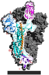
Spike (S) glycoprotein is the largest of the four major structural proteins found in coronaviruses. The spike protein assembles into trimers that form large structures, called spikes or peplomers, that project from the surface of the virion. The distinctive appearance of these spikes when visualized using negative stain transmission electron microscopy, "recalling the solar corona", gives the virus family its main name.

<span class="mw-page-title-main">Universal coronavirus vaccine</span> Vaccine that prevents infection from all strains of coronaviruses

A universal coronavirus vaccine, also known as a pan-coronavirus vaccine, is a theoretical coronavirus vaccine that would be effective against all coronavirus strains. A universal vaccine would provide protection against coronavirus strains that have caused disease in humans, such as SARS-CoV-2, while also providing protection against future coronavirus strains. Such a vaccine has been proposed to prevent or mitigate future coronavirus epidemics and pandemics.

References

Citations

  1. 1 2 Balloux et al. 2022, p. 4.
  2. 1 2 3 Kane, Wong & Gao 2023, p. 15.
  3. Kane, Wong & Gao 2023, p. 23.
  4. Kane, Wong & Gao 2023, pp. 20, 21, 23.
  5. Graham & Baric 2010, p. 3135.
  6. Graham & Baric 2010, pp. 3139–3140.
  7. 1 2 Kane, Wong & Gao 2023, p. 12.
  8. Kane, Wong & Gao 2023, p. 13.
  9. Graham & Baric 2010.
  10. Graham & Baric 2010, pp. 3142.
  11. Kane, Wong & Gao 2023, pp. 2–3.
  12. Cagliani et al. 2020, p. 1.
  13. 1 2 3 Frutos et al. 2020, p. 1.
  14. Kane, Wong & Gao 2023, pp. 12, 23.
  15. 1 2 3 Kane, Wong & Gao 2023, p. 17.
  16. Guo et al. 2020.
  17. Lytras et al. 2022.
  18. 1 2 3 Kane, Wong & Gao 2023, p. 3.
  19. 1 2 Holmes et al. 2021, p. 4850.
  20. Temmam et al. 2023, p. 8,10.
  21. 1 2 Lytras et al. 2022, p. 1.
  22. Lytras et al. 2022, p. 4.
  23. Kane, Wong & Gao 2023, pp. 3, 14.
  24. 1 2 Cagliani et al. 2020, p. 5.
  25. Buchanan, Charles J.; et al. (2022). "Pathogen-sugar interactions revealed by universal saturation transfer analysis". Science. 377 (6604). doi:10.1126/science.abm3125. hdl:1983/355cbd8f-c424-4cc0-adb2-881c04ab3bf0. PMID   35737812.
  26. Rossmann, M. G. (1989). "The Canyon Hypothesis". Journal of Biological Chemistry. 264 (25): 14587–14590. doi: 10.1016/S0021-9258(18)63732-9 .
  27. 1 2 3 Wu & Zhao 2021.
  28. 1 2 Hossain et al. 2021, p. 1816.
  29. Hossain et al. 2021, p. 1817.
  30. Anand et al. 2020.
  31. Harrison & Sachs 2022.
  32. Harrison & Sachs 2022, p. 4.
  33. 1 2 Kane, Wong & Gao 2023, p. 14.
  34. Li et al. 2023.
  35. 1 2 Li et al. 2023, p. 557.
  36. 1 2 3 4 5 Biancolella et al. 2023, p. 3.
  37. 1 2 Frutos et al. 2020, p. 3.
  38. 1 2 Kane, Wong & Gao 2023, p. 8.
  39. Kane, Wong & Gao 2023, p. 19.
  40. Graham & Baric 2010, p. 3139.
  41. 1 2 Kane, Wong & Gao 2023, p. 20.
  42. Qian et al. 2022, p. 8.
  43. 1 2 Kane, Wong & Gao 2023, p. 4.
  44. Lytras et al. 2022, p. 3.
  45. 1 2 3 Lytras et al. 2022, p. 6.
  46. Temmam et al. 2023, p. 10.
  47. 1 2 Kang et al. 2021, p. 4393.
  48. Tai et al. 2022, p. 4.
  49. Tai et al. 2022, p. 8.
  50. Cagliani et al. 2020, p. 3.
  51. 1 2 Tai et al. 2022, p. 2.
  52. Qian et al. 2022.
  53. Kane, Wong & Gao 2023, p. 16.
  54. 1 2 Li et al. 2023, p. 554.
  55. 1 2 Kane, Wong & Gao 2023, p. 21.
  56. Kang et al. 2021, p. 4396.
  57. Kang et al. 2021, p. 4397.
  58. 1 2 3 Guo et al. 2022.
  59. Jaimes, Millet & Whittaker 2020, p. 2.
  60. 1 2 Graham & Baric 2010, p. 3141.
  61. Temmam et al. 2023, p. 5.
  62. Berche 2023, p. 1,5.
  63. Berche 2023, p. 5.
  64. Qian et al. 2022, p. 16.
  65. 1 2 Reggiani, Rugna & Bonilauri 2022, p. 4.
  66. 1 2 3 4 Reggiani, Rugna & Bonilauri 2022, p. 2.
  67. Tai et al. 2022, p. 2-3.
  68. 1 2 3 Kane, Wong & Gao 2023, p. 11.
  69. Graham & Baric 2010, pp. 3141, 3142.
  70. 1 2 Berche 2023, p. 1.
  71. Deng, Xing & He 2022.
  72. Frutos et al. 2021.
  73. Frutos et al. 2020.
  74. Cagliani et al. 2020, p. 6.
  75. Kane, Wong & Gao 2023, pp. 19–20.
  76. Kane, Wong & Gao 2023, p. 10.
  77. Lytras et al. 2022, p. 2.
  78. 1 2 Pekar et al. 2022, p. 4.
  79. Liu et al. 2023, p. 3.
  80. 1 2 He et al. 2022.
  81. 1 2 Liu et al. 2023, p. 7.
  82. Liu et al. 2023, p. 6.
  83. Liu et al. 2023, pp. 8–9.
  84. Liu et al. 2023, p. 10.
  85. 1 2 Liu et al. 2023, p. 12.
  86. Qian et al. 2022, p. 13.
  87. Qian et al. 2022, p. 14.
  88. 1 2 Reggiani, Rugna & Bonilauri 2022, p. 3.

Bibliography