Arginine:glycine amidinotransferase

Last updated
Glycine amidinotransferase
Amidinotransferase structure diagram.JPG
Stereo view of AGAT in standard orientation with the handles of basket at the top of the model [1]
Identifiers
EC no. 2.1.4.1
CAS no. 9027-35-4
Databases
IntEnz IntEnz view
BRENDA BRENDA entry
ExPASy NiceZyme view
KEGG KEGG entry
MetaCyc metabolic pathway
PRIAM profile
PDB structures RCSB PDB PDBe PDBsum
Gene Ontology AmiGO / QuickGO
Search
PMC articles
PubMed articles
NCBI proteins
Role of AGAT during creatine synthesis Amidinotransferase reaction mechanism in creatine metabolism.JPG
Role of AGAT during creatine synthesis
Transamidination reaction mechanism of AGAT Reaction mechanism amidino group transfer in AGAT.JPG
Transamidination reaction mechanism of AGAT

L-Arginine:glycine amidinotransferase (AGAT; EC 2.1.4.1) is the enzyme that catalyses the transfer of an amidino group from L-arginine to glycine. The products are L-ornithine and glycocyamine, also known as guanidinoacetate, the immediate precursor of creatine. Creatine and its phosphorylated form play a central role in the energy metabolism of muscle and nerve tissues. Creatine is in highest concentrations in the skeletal muscle, heart, spermatozoa and photoreceptor cells. Creatine helps buffer the rapid changes in ADP/ATP ratio in muscle and nerve cells during active periods. Creatine is also synthesized in other tissues, such as pancreas, kidneys, and liver, where amidinotransferase is located in the cytoplasm, including the intermembrane space of the mitochondria, of the cells that make up those tissues. [2]

Contents

Function

L-Arginine:glycine amidinotransferase catalyses the first, which is also the committed step in the formation of creatine. The second step of the process, producing the actual creatine molecule, occurs solely in the cytosol, where the second enzyme, S-adenosylmethionine:guanidinoacetate methyltransferase (GAMT), is found. The creatine is then transported through the bloodstream and taken up through sodium-dependent creatine transporters by cells that require creatine. [1]

Structure

The crystal structure of AGAT was determined by Humm, Fritsche, Steinbacher, and Huber of the Max Planck Institute of Biochemistry in Martinsried, Germany in 1997. X-ray examinations of the structure reveal a novel symmetry with fivefold pseudosymmetry of beta beta alphabeta modules. The overall structure of the molecule resembles a basket with handles. The active site lies at the bottom of a long, narrow channel and includes a Cys-His-Asp catalytic triad. The intermediate structure involves the amidino group temporarily covalently bonding to the Cys residue on the catalytic triad, while the His residue takes part in general acid/base catalysis, meaning it acts as a proton donator/receiver itself. [2]

Reaction

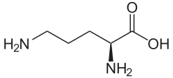
The actual reaction catalyzed by AGAT is the synthesis of guanidinoacetate from arginine and glycine, with ornithine as a byproduct. The guanidinoacetate produced is then combined with S-Adenosyl-L-methionine, a reaction catalyzed by GAMT, to produce creatine and S-Adenosyl-L-homocysteine. The mechanism by which the AGAT catalyzes this committed step follows a ping-pong mechanism, and involves the transferring of an amidino group to the Cys407 residue on the protein from L-arginine, which leaves as L-ornithine. The His303 residue then extracts a proton from glycine, which then picks up the amidino group from Cys407 in exchange for a proton to become guanidinoacetate and renew the catalyst. [2]

Regulation of expression and activity

The formation of guanidinoacetate is normally the rate-limiting step of creatine biosynthesis. [3] Consequently, the AGAT reaction is the most likely control step in the pathway, a hypothesis that is supported by a great deal of experimental work. Most important in this respect is the feedback repression of AGAT by creatine, the end-product of the pathway. Cyclocreatine, N-acetimidoylsarcosine, and N-ethylguanidinoacetate display repressor activity like creatine as well. L-Arginine and guanidinoacetate have only "apparent" repressor activity. They exert no effect on AGAT expression by themselves but are readily converted to creatine, which then acts as the true repressor. [4] It has been suggested that AGAT activity in tissues is regulated in a number of ways including induction by growth hormone and thyroxine, [5] inhibition of the enzyme by ornithine, [6] and repression of its synthesis by creatine. [7] [8]

Sex hormones may regulate the activity of AGAT. [9] Treatment of male rats with testosterone propionate increases AGAT activity. In contrast, estrogen treatment decreases AGAT activity and induces weight loss. It is currently unclear whether the changes in the level of AGAT transcript results from altered mRNA stability or enhanced transcriptional rate. If estrogen-mediated alteration results from transcriptional regulation, the site of estrogen action is yet to be determined. [10]

GATM expression within the mouse placenta has been shown to be imprinted meaning only the maternal copy of GATM is expressed . Due to this it is thought that GATM acts as a growth suppressor within the placenta.

Clinical significance

Deficiency

In 2000, The American Journal of Human Genetics reported two female siblings, aged 4 and 6 years, with intellectual disability and severe creatine deficiency in the brain. [11] Arginine:glycine amidinotransferase (AGAT) catalyzes the first step of creatine synthesis, resulting in the formation of guanidinoacetate, which is a substrate for creatine formation. In two female siblings with intellectual disability who had brain creatine deficiency that was reversible by means of oral creatine supplementation and had low urinary guanidinoacetate concentrations, Arginine:glycine amidinotransferase deficiency was identified as a new genetic defect in creatine metabolism. It is one of three cerebral creatine deficiencies.

Patients with brain creatine deficiency present nonspecific neurologic symptoms, including intellectual disability, language disorders, epilepsy, autistic-like behavior, neurologic deterioration, and movement disorders. A deficiency in AGAT results in a creatine deficiency in the body. The treatment for this is creatine supplements since the body cannot make the creatine on its own. The positive results of creatine treatment (in AGAT deficiencies) and the observation that fetal and early postnatal development are normal in these patients support the hypothesis that earlier diagnosis and treatment can substantially improve the final prognosis of these diseases. Brain 1H-MRS examination is a reliable and minimally invasive technique to assess brain creatine disorders. Because of its limited availability and high cost, the 1H-MRS technique cannot be proposed for all children whose clinical condition suggests the diagnosis of brain creatine depletion. [12]

AGAT deficiency is, along with GAMT and creatine transporter defect, one of three inborn errors of the creatine biosynthesis/transport pathway. The prevalence of these defects is unknown, however they have been observed to occur in high frequency in intellectually disabled children. The actual genetic mutation associated with AGAT involves a tryptophan codon being converted to a stop codon at residue 149. [11]

Heart failure

Microarray analysis from one report shows a significant decrease in myocardial arginine:glycine amidinotransferase (AGAT) gene expression during the late-stage heart failure. This suggests that the reduced AGAT may correlate with loss of heart function. Increase of AGAT expression in the myocardium after heart failure due to increase in creatine synthesis was associated with favorable outcome. [13]

Related Research Articles

<span class="mw-page-title-main">Arginine</span> Amino acid

Arginine is the amino acid with the formula (H2N)(HN)CN(H)(CH2)3CH(NH2)CO2H. The molecule features a guanidino group appended to a standard amino acid framework. At physiological pH, the carboxylic acid is deprotonated (−CO2) and both the amino and guanidino groups are protonated, resulting in a cation. Only the l-arginine (symbol Arg or R) enantiomer is found naturally. Arg residues are common components of proteins. It is encoded by the codons CGU, CGC, CGA, CGG, AGA, and AGG. The guanidine group in arginine is the precursor for the biosynthesis of nitric oxide. Like all amino acids, it is a white, water-soluble solid.

<span class="mw-page-title-main">Ornithine</span> Chemical compound

Ornithine is a non-proteinogenic amino acid that plays a role in the urea cycle. Ornithine is abnormally accumulated in the body in ornithine transcarbamylase deficiency. The radical is ornithyl.

<span class="mw-page-title-main">Ornithine transcarbamylase</span> Mammalian protein found in Homo sapiens

Ornithine transcarbamylase (OTC) is an enzyme that catalyzes the reaction between carbamoyl phosphate (CP) and ornithine (Orn) to form citrulline (Cit) and phosphate (Pi). There are two classes of OTC: anabolic and catabolic. This article focuses on anabolic OTC. Anabolic OTC facilitates the sixth step in the biosynthesis of the amino acid arginine in prokaryotes. In contrast, mammalian OTC plays an essential role in the urea cycle, the purpose of which is to capture toxic ammonia and transform it into urea, a less toxic nitrogen source, for excretion.

<span class="mw-page-title-main">Creatine</span> Chemical compound

Creatine is an organic compound with the nominal formula (H2N)(HN)CN(CH3)CH2CO2H. It exists in various tautomers in solutions. Creatine is found in vertebrates where it facilitates recycling of adenosine triphosphate (ATP), primarily in muscle and brain tissue. Recycling is achieved by converting adenosine diphosphate (ADP) back to ATP via donation of phosphate groups. Creatine also acts as a buffer.

<span class="mw-page-title-main">Phosphocreatine</span> Chemical compound

Phosphocreatine, also known as creatine phosphate (CP) or PCr (Pcr), is a phosphorylated form of creatine that serves as a rapidly mobilizable reserve of high-energy phosphates in skeletal muscle, myocardium and the brain to recycle adenosine triphosphate, the energy currency of the cell.

<span class="mw-page-title-main">Arginase</span> Manganese-containing enzyme

Arginase (EC 3.5.3.1, arginine amidinase, canavanase, L-arginase, arginine transamidinase) is a manganese-containing enzyme. The reaction catalyzed by this enzyme is:

<span class="mw-page-title-main">Aminolevulinic acid synthase</span> Class of enzymes

Aminolevulinic acid synthase (ALA synthase, ALAS, or delta-aminolevulinic acid synthase) is an enzyme (EC 2.3.1.37) that catalyzes the synthesis of δ-aminolevulinic acid (ALA) the first common precursor in the biosynthesis of all tetrapyrroles such as hemes, cobalamins and chlorophylls. The reaction is as follows:

In molecular biology, biosynthesis is a multi-step, enzyme-catalyzed process where substrates are converted into more complex products in living organisms. In biosynthesis, simple compounds are modified, converted into other compounds, or joined to form macromolecules. This process often consists of metabolic pathways. Some of these biosynthetic pathways are located within a single cellular organelle, while others involve enzymes that are located within multiple cellular organelles. Examples of these biosynthetic pathways include the production of lipid membrane components and nucleotides. Biosynthesis is usually synonymous with anabolism.

<span class="mw-page-title-main">Argininosuccinate synthase</span> Enzyme

Argininosuccinate synthase or synthetase is an enzyme that catalyzes the synthesis of argininosuccinate from citrulline and aspartate. In humans, argininosuccinate synthase is encoded by the ASS gene located on chromosome 9.

<i>N</i>-Acetylglutamate synthase Class of enzymes

N-Acetylglutamate synthase (NAGS) is an enzyme that catalyses the production of N-acetylglutamate (NAG) from glutamate and acetyl-CoA.

<span class="mw-page-title-main">Amino acid synthesis</span> The set of biochemical processes by which amino acids are produced

Amino acid synthesis is the set of biochemical processes by which the amino acids are produced. The substrates for these processes are various compounds in the organism's diet or growth media. Not all organisms are able to synthesize all amino acids. For example, humans can synthesize 11 of the 20 standard amino acids. These 11 are called the non-essential amino acids).

<span class="mw-page-title-main">Guanidinoacetate methyltransferase deficiency</span> Medical condition

Guanidinoacetate methyltransferase deficiency is an autosomal recessive cerebral creatine deficiency that primarily affects the nervous system and muscles. It is the first described disorder of creatine metabolism, and results from deficient activity of guanidinoacetate methyltransferase, an enzyme involved in the synthesis of creatine. Clinically, affected individuals often present with hypotonia, seizures and developmental delay. Diagnosis can be suspected on clinical findings, and confirmed by specific biochemical tests, brain magnetic resonance spectroscopy, or genetic testing. Biallelic pathogenic variants in GAMT are the underlying cause of the disorder. After GAMT deficiency is diagnosed, it can be treated by dietary adjustments, including supplementation with creatine. Treatment is highly effective if started early in life. If treatment is started late, it cannot reverse brain damage which has already taken place.

Cerebral creatine deficiencies are a small group of inherited disorders that result from defects in creatine biosynthesis and utilization. Commonly affected tissues include the brain and muscles. There are three distinct CCDs. The most common is creatine transporter defect (CTD), an X-linked disorder caused by pathogenic variants in SLC6A8. The main symptoms of CTD are intellectual disability and developmental delay, and these are caused by a lack of creatine in the brain, due to the defective transporter. There are also two enzymatic defects of creatine biosynthesis, arginine:glycine amidinotransferase deficiency, caused by variants in GATM and guanidinoacetate methyltransferase deficiency, caused by variants in GAMT. The single enzyme defects are both inherited in an autosomal recessive manner.

<span class="mw-page-title-main">Guanidinoacetate N-methyltransferase</span> Mammalian protein found in Homo sapiens

Guanidinoacetate N-methyltransferase is an enzyme that catalyzes the chemical reaction and is encoded by gene GAMT located on chromosome 19p13.3.

<span class="mw-page-title-main">GATM (gene)</span> Protein-coding gene in the species Homo sapiens

Glycine amidinotransferase, mitochondrial is an enzyme that in humans is encoded by the GATM gene.

<span class="mw-page-title-main">Glycocyamine</span> Chemical compound

Glycocyamine is a metabolite of glycine in which the amino group has been converted into a guanidine by guanylation. In vertebrate organism it is then transformed into creatine by methylation.

A ureohydrolase is a type of hydrolase enzyme. The ureohydrolase superfamily includes arginase, agmatinase, formiminoglutamase and proclavaminate amidinohydrolase. These enzymes share a 3-layer alpha-beta-alpha structure, and play important roles in arginine/agmatine metabolism, the urea cycle, histidine degradation, and other pathways.

<span class="mw-page-title-main">Homoarginine</span> Chemical compound

Homoarginine is an nonproteinogenic alpha-amino acid. It is structurally equivalent to a one-methylene group-higher homolog of arginine and to the guanidino derivative of lysine. L-Homoarginine is the naturally-occurring enantiomer. Physiologically, homoarginine increases nitric oxide (NO) supply and betters endothelial functions in the body, with a particular correlation and effect towards cardiovascular outcome and mortality. At physiological pH, homoarginine is cationic: the guanidino group is protonated.

<span class="mw-page-title-main">Creatine transporter defect</span> Medical condition

Creatine transporter deficiency (CTD) is an inborn error of creatine metabolism in which creatine is not properly transported to the brain and muscles due to defective creatine transporters. CTD is an X-linked disorder caused by mutation in SLC6A8. SLC6A8 is located at Xq28. Hemizygous males with CTD express speech and behavior abnormalities, intellectual disabilities, development delay, seizures, and autistic behavior. Heterozygous females with CTD generally express fewer, less severe symptoms. CTD is one of three different types of cerebral creatine deficiency (CCD). The other two types of CCD are guanidinoacetate methyltransferase (GAMT) deficiency and L-arginine:glycine amidinotransferase (AGAT) deficiency. Clinical presentation of CTD is similar to that of GAMT and AGAT deficiency. CTD was first identified in 2001 with the presence of a hemizygous nonsense change in SLC6A8 in a male patient.

<span class="mw-page-title-main">Arginine:glycine amidinotransferase deficiency</span> Medical condition

Arginine:glycine amidinotransferase deficiency or AGAT deficiency is an autosomal recessive cerebral creatine deficiency caused by a deficiency of the enzyme arginine:glycine amidinotransferase. This enzyme deficiency results in decreased creatine synthesis, and is caused by biallelic pathogenic variants in GATM. Individuals with AGAT deficiency are intellectually disabled and have muscle weakness. The symptoms of AGAT deficiency are caused by the lack of creatine in specific tissues, most notably muscle and brain. Oral creatine supplementation can be used to treat AGAT deficiency, with early intervention providing the best results. All creatine deficiencies are rare, and there have been fewer than 20 individuals reported in medical literature with AGAT deficiency. This disorder was first described in 2000.

References

  1. 1 2 3 4 Humm A, Fritsche E, Steinbacher S, Huber R (June 1997). "Crystal structure and mechanism of human L-arginine:glycine amidinotransferase: a mitochondrial enzyme involved in creatine biosynthesis". EMBO J. 16 (12): 3373–85. doi:10.1093/emboj/16.12.3373. PMC   1169963 . PMID   9218780.
  2. 1 2 3 Humm A, Fritsche E, Mann K, Göhl M, Huber R (March 1997). "Recombinant expression and isolation of human L-arginine:glycine amidinotransferase and identification of its active-site cysteine residue". Biochem. J. 322 (3): 771–6. doi:10.1042/bj3220771. PMC   1218254 . PMID   9148748.
  3. Walker JB (2006). "Creatine: Biosynthesis, Regulation, and Function". Advances in Enzymology and Related Areas of Molecular Biology. pp. 177–242. doi:10.1002/9780470122952.ch4. ISBN   9780470122952. PMID   386719.{{cite book}}: |journal= ignored (help)
  4. Wyss M, Kaddurah-Daouk R (July 2000). "Creatine and creatinine metabolism". Physiol. Rev. 80 (3): 1107–213. doi:10.1152/physrev.2000.80.3.1107. PMID   10893433.
  5. McGuire DM, Tormanen CD, Segal IS, Van Pilsum JF (February 1980). "The effect of growth hormone and thyroxine on the amount of L-arginine:glycine amidinotransferase in kidneys of hypophysectomized rats. Purification and some properties of rat kidney transamidinase". J. Biol. Chem. 255 (3): 1152–9. doi: 10.1016/S0021-9258(19)86155-0 . PMID   6766137.
  6. Sipilä I (1980). "Inhibition of arginine-glycine amidinotransferase by ornithine. A possible mechanism for the muscular and chorioretinal atrophies in gyrate atrophy of the choroid and retina with hyperornithinemia". Biochim. Biophys. Acta. 613 (1): 79–84. doi:10.1016/0005-2744(80)90194-1. PMID   7378422.
  7. McGuire DM, Gross MD, Van Pilsum JF, Towle HC (October 1984). "Repression of rat kidney L-arginine:glycine amidinotransferase synthesis by creatine at a pretranslational level". J. Biol. Chem. 259 (19): 12034–8. doi: 10.1016/S0021-9258(20)71316-5 . PMID   6384218.
  8. Guthmiller P, Van Pilsum JF, Boen JR, McGuire DM (July 1994). "Cloning and sequencing of rat kidney L-arginine:glycine amidinotransferase. Studies on the mechanism of regulation by growth hormone and creatine". J. Biol. Chem. 269 (26): 17556–60. doi: 10.1016/S0021-9258(17)32477-8 . PMID   8021264.
  9. Kriskó I, Walker JB (December 1966). "Influence of sex hormones on amidinotransferase levels. Metabolic control of creatine biosynthesis". Acta Endocrinol. 53 (4): 655–62. doi:10.1530/acta.0.0530655. PMID   5953691.
  10. Zhu Y, Evans MI (May 2001). "Estrogen modulates the expression of L-arginine:glycine amidinotransferase in chick liver". Mol. Cell. Biochem. 221 (1–2): 139–45. doi:10.1023/A:1010946414017. PMID   11506177. S2CID   23212603.
  11. 1 2 Item CB, Stöckler-Ipsiroglu S, Stromberger C, Mühl A, Alessandrì MG, Bianchi MC, Tosetti M, Fornai F, Cioni G (November 2001). "Arginine:glycine amidinotransferase deficiency: the third inborn error of creatine metabolism in humans". Am. J. Hum. Genet. 69 (5): 1127–33. doi:10.1086/323765. PMC   1274356 . PMID   11555793.
  12. Carducci C, Birarelli M, Leuzzi V, Carducci C, Battini R, Cioni G, Antonozzi I (October 2002). "Guanidinoacetate and creatine plus creatinine assessment in physiologic fluids: an effective diagnostic tool for the biochemical diagnosis of arginine:glycine amidinotransferase and guanidinoacetate methyltransferase deficiencies". Clin. Chem. 48 (10): 1772–8. doi: 10.1093/clinchem/48.10.1772 . PMID   12324495.
  13. Cullen ME, Yuen AH, Felkin LE, Smolenski RT, Hall JL, Grindle S, Miller LW, Birks EJ, Yacoub MH, Barton PJ (July 2006). "Myocardial expression of the arginine:glycine amidinotransferase gene is elevated in heart failure and normalized after recovery: potential implications for local creatine synthesis". Circulation. 114 (1 Suppl): I16–20. doi: 10.1161/CIRCULATIONAHA.105.000448 . PMID   16820567.