The articular bone is part of the lower jaw of most vertebrates, including most jawed fish, amphibians, birds and various kinds of reptiles, as well as ancestral mammals.
The articular bone is part of the lower jaw of most vertebrates, including most jawed fish, amphibians, birds and various kinds of reptiles, as well as ancestral mammals.
In most vertebrates, the articular bone is connected to two other lower jaw bones, the suprangular and the angular. [1] Developmentally, it originates from the embryonic mandibular cartilage. The most caudal portion of the mandibular cartilage ossifies to form the articular bone, while the remainder of the mandibular cartilage either remains cartilaginous or disappears. [1]
In snakes, the articular, surangular, and prearticular bones have fused to form the compound bone. The mandible is suspended from the quadrate bone and articulates at this compound bone. [2]
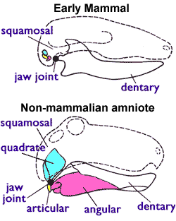
In most tetrapods, the articular bone forms the lower portion of the jaw joint. The upper jaw articulates at the quadrate bone. [3]
In mammals, the articular bone evolves to form the malleus , one of the mammalian ossicles of the middle ear. This is an apomorphy of the mammalian clade, [4] and is used to determine the fossil transition to mammals. [5] It is analogous to, but not homologous to the articular process of the lower jaw.
After the loss of the quadrate-articular joint, the squamosal and dentary bones form the new jaw joint in mammals. [6]

The middle ear is the portion of the ear medial to the eardrum, and distal to the oval window of the cochlea.

The rib cage or thoracic cage is an endoskeletal enclosure in the thorax of most vertebrates that comprises the ribs, vertebral column and sternum, which protect the vital organs of the thoracic cavity, such as the heart, lungs and great vessels and support the shoulder girdle to form the core part of the axial skeleton.
The ossicles are three bones in either middle ear that are among the smallest bones in the human body. They serve to transmit sound vibrations sent from the ear drum to the fluid-filled labyrinth (cochlea). The absence of the auditory ossicles would constitute a moderate-to-severe hearing loss. The term "ossicle" literally means "tiny bone". Though the term may refer to any small bone throughout the body, it typically refers to the malleus, incus, and stapes of the middle ear.

Synapsida is one of the two major clades of vertebrate animals in the group Amniota, the other being the Sauropsida. The synapsids were the dominant land animals in the late Paleozoic and early Mesozoic, but the only group that survived into the Cenozoic are mammals. Unlike other amniotes, synapsids have a single temporal fenestra, an opening low in the skull roof behind each eye orbit, leaving a bony arch beneath each; this accounts for their name. The distinctive temporal fenestra developed about 318 million years ago during the Late Carboniferous period, when synapsids and sauropsids diverged, but was subsequently merged with the orbit in early mammals.

The jaws are a pair of opposable articulated structures at the entrance of the mouth, typically used for grasping and manipulating food. The term jaws is also broadly applied to the whole of the structures constituting the vault of the mouth and serving to open and close it and is part of the body plan of humans and most animals.

The quadrate bone is a skull bone in most tetrapods, including amphibians, sauropsids, and early synapsids.
The quadratojugal is a skull bone present in many vertebrates, including some living reptiles and amphibians.

The pharyngeal arches, also known as visceral arches, are transient structures seen in the embryonic development of humans and other vertebrates, that are recognisable precursors for many structures. In fish, the arches support the gills and are known as the branchial arches, or gill arches.

In humans, the cartilaginous bar of the mandibular arch is formed by what are known as Meckel's cartilages also known as Meckelian cartilages; above this the incus and malleus are developed. Meckel's cartilage arises from the first pharyngeal arch.

The squamosal is a skull bone found in most reptiles, amphibians, and birds. In fishes, it is also called the pterotic bone.

The evolution of mammalian auditory ossicles was an evolutionary process that resulted in the formation of the bones of the mammalian middle ear. These bones, or ossicles, are a defining characteristic of all mammals. The event is well-documented and important as a demonstration of transitional forms and exaptation, the re-purposing of existing structures during evolution.
The splanchnocranium is the portion of the cranium that is derived from pharyngeal arches. Splanchno indicates to the gut because the face forms around the mouth, which is an end of the gut. The splanchnocranium consists of cartilage and endochondral bone. In mammals, the splanchnocranium comprises the three ear ossicles, as well as the alisphenoid, the styloid process, the hyoid apparatus, and the thyroid cartilage.

NK3 homeobox 2 also known as NKX3-2 is a human gene. It is a homolog of bagpipe (bap) in Drosophila and therefore also known as Bapx1. The protein encoded by this gene is a homeodomain containing transcription factor.

The sternum or breastbone is a long flat bone located in the central part of the chest. It connects to the ribs via cartilage and forms the front of the rib cage, thus helping to protect the heart, lungs, and major blood vessels from injury. Shaped roughly like a necktie, it is one of the largest and longest flat bones of the body. Its three regions are the manubrium, the body, and the xiphoid process. The word sternum originates from Ancient Greek στέρνον (stérnon) 'chest'.
In jawed vertebrates, the mandible, lower jaw, or jawbone is a bone that makes up the lower – and typically more mobile – component of the mouth.

Most bony fishes have two sets of jaws made mainly of bone. The primary oral jaws open and close the mouth, and a second set of pharyngeal jaws are positioned at the back of the throat. The oral jaws are used to capture and manipulate prey by biting and crushing. The pharyngeal jaws, so-called because they are positioned within the pharynx, are used to further process the food and move it from the mouth to the stomach.
This glossary explains technical terms commonly employed in the description of dinosaur body fossils. Besides dinosaur-specific terms, it covers terms with wider usage, when these are of central importance in the study of dinosaurs or when their discussion in the context of dinosaurs is beneficial. The glossary does not cover ichnological and bone histological terms, nor does it cover measurements.

Each vertebra is an irregular bone with a complex structure composed of bone and some hyaline cartilage, that make up the vertebral column or spine, of vertebrates. The proportions of the vertebrae differ according to their spinal segment and the particular species.
The postdentary trough is a skeletal feature seen in Mesozoic mammals. It is found on the inside of the lower jaw (dentary), at the back behind the molar teeth. It is the hollow in which the postdentary bones and Meckel's cartilage sit. These bones form the middle ear in later mammal groups ; they include the incus (quadrate), malleus (articular), ectotympanic (angular) and prearticular.

The malleus, or hammer, is a hammer-shaped small bone or ossicle of the middle ear. It connects with the incus, and is attached to the inner surface of the eardrum. The word is Latin for 'hammer' or 'mallet'. It transmits the sound vibrations from the eardrum to the incus (anvil).
| | This vertebrate anatomy–related article is a stub. You can help Wikipedia by expanding it. |