Related Research Articles

Thrombosis is the formation of a blood clot inside a blood vessel, obstructing the flow of blood through the circulatory system. When a blood vessel is injured, the body uses platelets (thrombocytes) and fibrin to form a blood clot to prevent blood loss. Even when a blood vessel is not injured, blood clots may form in the body under certain conditions. A clot, or a piece of the clot, that breaks free and begins to travel around the body is known as an embolus.

The great saphenous vein is a large, subcutaneous, superficial vein of the leg. It is the longest vein in the body, running along the length of the lower limb, returning blood from the foot, leg and thigh to the deep femoral vein at the femoral triangle.

The 'inferior vena cava is a large vein that carries the deoxygenated blood from the lower and middle body into the right atrium of the heart. It is formed by the joining of the right and the left common iliac veins, usually at the level of the fifth lumbar vertebra.
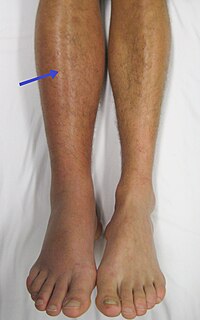
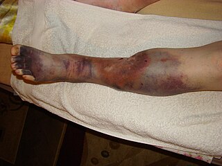
Deep vein thrombosis (DVT) is the formation of a blood clot in a deep vein, most commonly in the legs or pelvis. A minority of DVTs occur in the arms. Symptoms can include pain, swelling, redness, and enlarged veins in the affected area, but some DVTs have no symptoms. The most common life-threatening concern with DVT is the potential for a clot to embolize, travel as an embolus through the right side of the heart, and become lodged in a pulmonary artery that supplies blood to the lungs. This is called a pulmonary embolism (PE). DVT and PE comprise the cardiovascular disease of venous thromboembolism (VTE). About two-thirds of VTE manifests as DVT only, with one-third manifesting as PE with or without DVT. The most frequent long-term DVT complication is post-thrombotic syndrome, which can cause pain, swelling, a sensation of heaviness, itching, and in severe cases, ulcers. Recurrent VTE occurs in about 30% of those in the ten years following an initial VTE.

In the human body, the femoral vein is a blood vessel that accompanies the femoral artery in the femoral sheath. It begins at the adductor hiatus and is a continuation of the popliteal vein. It ends at the inferior margin of the inguinal ligament, where it becomes the external iliac vein. The femoral vein bears valves which are mostly bicuspid and whose number is variable between individuals and often between left and right leg.

Inferior vena cava syndrome (IVCS) is a constellation of symptoms resulting from obstruction of the inferior vena cava. It can be caused by physical invasion or compression by a pathological process or by thrombosis within the vein itself. It can also occur during pregnancy. Pregnancy leads to high venous pressure in the lower limbs, decreased blood return to the heart, decreased cardiac output due to obstruction of the inferior vena cava, sudden rise in venous pressure which can lead to placental separation, and a decrease in kidney function. All of these issues can arise from lying in the supine position during late pregnancy which can cause compression of the inferior vena cava by the uterus. Symptoms of late pregnancy inferior vena cava syndrome consist of intense pain in the right hand side, muscle twitching, hypotension, and fluid retention.
In medicine, Homans' sign is considered by some physicians to be a sign of deep vein thrombosis (DVT). It was defined by John Homans in 1941 as discomfort behind the knee upon forced dorsiflexion of the foot. After many examples of false-positive Homans' signs were reported, Homans redefined it in 1944, stating that "discomfort need have no part in the reaction", and that increased resistance, involuntary flexure of the knee or pain in the calf upon forced dorsiflexion should be considered positive responses.

May–Thurner syndrome (MTS), also known as the iliac vein compression syndrome, is a condition in which compression of the common venous outflow tract of the left lower extremity may cause discomfort, swelling, pain or clots in the iliofemoral veins.
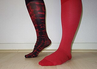
Compression stockings are a specialized hosiery designed to help prevent the occurrence of, and guard against further progression of, venous disorders such as edema, phlebitis and thrombosis. Compression stockings are elastic compression garments worn around the leg, compressing the limb. This reduces the diameter of distended veins and increases venous blood flow velocity and valve effectiveness. Compression therapy helps decrease venous pressure, prevents venous stasis and impairments of venous walls, and relieves heavy and aching legs.
Post-thrombotic syndrome (PTS), also called postphlebitic syndrome and venous stress disorder is a medical condition that may occur as a long-term complication of deep vein thrombosis (DVT).

Chronic venous insufficiency (CVI) is a medical condition in which blood pools in the veins, straining the walls of the vein. The most common cause of CVI is superficial venous reflux which is a treatable condition. As functional venous valves are required to provide for efficient blood return from the lower extremities, this condition typically affects the legs. If the impaired vein function causes significant symptoms, such as swelling and ulcer formation, it is referred to as chronic venous disease. It is sometimes called chronic peripheral venous insufficiency and should not be confused with post-thrombotic syndrome in which the deep veins have been damaged by previous deep vein thrombosis.

Venostasis, or venous stasis, is a condition of slow blood flow in the veins, usually of the legs.
Superficial thrombophlebitis is a thrombosis and inflammation of superficial veins which presents as a painful induration with erythema, often in a linear or branching configuration forming cords.
Pseudothrombophlebitis is a clinical condition where there are signs and symptoms of phlebitis in the absence of a thrombophlebitis lesion. Symptoms include pain, swelling, erythema and tenderness evolving over hours or days. It is often associated with the rupture or dissection of a popliteal cyst otherwise known as a Baker's cyst, although it can be associated with other disorders such as the arthritides. It may also occur as an orthopaedic surgical complication, secondary to trauma or as a presentation of septic arthritis. It is crucial to differentiate this condition from deep vein thrombosis as the treatment for DVT can cause adverse effects in patients with pseudothrombophlebitis.
Lowenberg's sign is a clinical sign found in patients with deep vein thrombosis of the lower leg. The sign is positive when pain is elicited rapidly when a blood pressure cuff is placed around the calf and inflated to 80mmHg. Like other signs of deep vein thrombosis, such as Homans sign and Bancroft's sign, this sign is neither sensitive nor specific for the presence of thrombosis.

Veinoplus is a class IIa medical device with CE marking. It is indicated for the treatment of vascular diseases. This is a neuromuscular stimulator developed by an American scientist, Jozef Cywinski.
Peabody's sign is a clinical sign which may be found in patients with deep vein thrombosis (DVT). The sign is positive when calf muscle spasm occurs on raising the affected leg with the foot extended. The sign is neither sensitive nor specific for the presence of DVT.

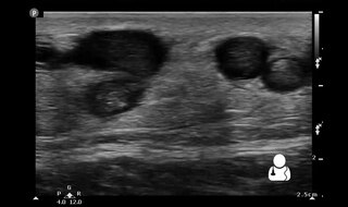
Ultrasonography of suspected or previously confirmed chronic venous insufficiency of leg veins is a risk-free, non-invasive procedure. It gives information about the anatomy, physiology and pathology of mainly superficial veins. As with heart ultrasound (echocardiography) studies, venous ultrasonography requires an understanding of hemodynamics in order to give useful examination reports. In chronic venous insufficiency, sonographic examination is of most benefit; in confirming varicose disease, making an assessment of the hemodynamics, and charting the progression of the disease and its response to treatment. It has become the reference standard for examining the condition and hemodynamics of the lower limb veins. Particular veins of the deep venous system (DVS), and the superficial venous system (SVS) are looked at. The great saphenous vein (GSV), and the small saphenous vein (SSV) are superficial veins which drain into respectively, the common femoral vein and the popliteal vein. These veins are deep veins. Perforator veins drain superficial veins into the deep veins. Three anatomic compartments are described, (N1) containing the deep veins, (N2) containing the perforator veins, and (N3) containing the superficial veins, known as the saphenous compartment. This compartmentalisation makes it easier for the examiner to systematize and map. The GSV can be located in the saphenous compartment where together with the Giacomini vein and the accessory saphenous vein (ASV) an image resembling an eye, known as the 'eye sign' can be seen. The ASV which is often responsible for varicose veins, can be located at the 'alignment sign', where it is seen to align with the femoral vessels.

Thrombosis prevention or thromboprophylaxis is medical treatment to prevent the development of thrombosis in those considered at risk for developing thrombosis. Some people are at a higher risk for the formation of blood clots than others. Prevention measures or interventions are usually begun after surgery as people are at higher risk due to immobility.
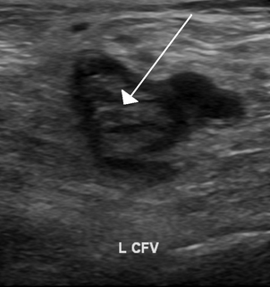
Ultrasonography in suspected deep vein thrombosis focuses primarily on the femoral vein and the popliteal vein, because thrombi in these veins are associated with the greatest risk of harmful pulmonary embolism.
References
- ↑ MOSES WR (February 1946). "The early diagnosis of phlebothrombosis". N. Engl. J. Med. 234 (9): 288–91. doi:10.1056/NEJM194602282340902. PMID 21016458.
- ↑ "Chapter 7: Clinical Assessment of Venous Disease". Venous and Lymphatic Diseases. Nicos Labropoulos, Gerard Stansby. Informa Health Care. 2006. p. 85. ISBN 978-0-8247-2923-3.CS1 maint: others (link)
- ↑ "Chapter 18: History, Physical Examination, and Diagnostic Approach". Manual of Vascular Diseases. Sanjay Rajagopalan, Debabrata Mukherjee, Emile R. Mohler. Lippincott Williams & Wilkins. 2004. p. 258. ISBN 978-0-7817-4499-7.CS1 maint: others (link)
| | This medical sign article is a stub. You can help Wikipedia by expanding it. |