Bennett's fracture

Last updated

Bennett fracture
Bennett-Faktur seitlich cropped.jpg
Bennett fracture on x-ray
Specialty Orthopedic

Bennett fracture is a type of partial broken finger involving the base of the thumb, and extends into the carpometacarpal (CMC) joint. [1]

Contents

Treatment typically requires surgery. [2]

This intra-articular fracture is the most common type of fracture of the thumb, and is nearly always accompanied by some degree of subluxation or frank dislocation of the carpometacarpal joint.[ citation needed ]

Symptoms and signs

Symptoms of Bennett fracture are instability of the CMC joint of the thumb, accompanied by pain and weakness of the pinch grasp. Characteristic signs include pain, swelling, and ecchymosis around the base of the thumb and thenar eminence, and especially over the CMC joint of the thumb. Physical examination demonstrates instability of the CMC joint of the thumb. The patient will often manifest a weakened ability to grasp objects or perform such tasks as tying shoes and tearing a piece of paper. Other complaints include intense pain experienced upon catching the thumb on an object, such as when reaching into a pants pocket.[ citation needed ]

Complications

Many important activities of daily life are dependent on the ability to grasp, pinch, and oppose the thumb. In fact, thumb function constitutes about 50% of overall hand function. These abilities are in turn dependent on an intact and functional thumb CMC joint. The CMC joint of the thumb allows a wide range of motion while maintaining stability for grasp and pinch.[ citation needed ]

With this in mind, failure to properly recognize and treat the Bennett fracture will not only result in an unstable, painful, arthritic CMC joint with diminished range of motion: it will also result in a hand with greatly diminished overall function.[ citation needed ]

In the case of the Bennett fracture, the proximal metacarpal fragment remains attached to the anterior oblique ligament, which in turn is attached to the tubercle of the trapezium bone of the CMC joint. This ligamentous attachment ensures that the proximal fragment remains in its correct anatomical position.[ citation needed ]

The distal fragment of the first metacarpal bone possesses the majority of the articular surface of the first CMC joint. Unlike the proximal fracture fragment, strong ligaments and muscle tendons of the hand tend to pull this fragment out of its correct anatomical position.[ citation needed ]

Specifically:

Tension from the APL and ADP muscles frequently leads to displacement of the fracture fragments, even in cases where the fracture fragments are initially in their proper anatomic position. Because of the aforementioned biomechanical features, Bennett fractures nearly always require some form of intervention to ensure healing in the correct anatomical position and restoration of proper function of the thumb CMC joint.[ citation needed ]

Mechanism

Bennett's fracture BennettFracMark.png
Bennett's fracture

The Bennett fracture is an oblique intraarticular metacarpal fracture dislocation, caused by an axial force directed against the partially flexed metacarpal. This type of compression along the metacarpal bone is often sustained when a person punches a hard object, such as the skull or tibia of an opponent, or a wall. It can also occur as a result of a fall onto the thumb. This is a common injury sustained from bike falls, as the thumb is generally extended while around the handle bars. It is also a common injury in car crashes, especially into fixed objects, from the driver holding the steering wheel during impact. The hand moves forward, while the steering wheel rim hyperextends the thumb.[ citation needed ] Some authors have recently made an assertion against popular belief that the APL tendon is not a deforming force on the Bennett fracture. [3]

Treatment

Bennett's fracture repair BennettFracRepaid.png
Bennett's fracture repair

Though these fractures commonly appear quite subtle or even inconsequential on radiographs, they can result in severe long-term dysfunction of the hand if left untreated. In his original description of this type of fracture in 1882, Bennett stressed the need for early diagnosis and treatment in order to prevent loss of function of the thumb CMC joint, which is critical to the overall function of the hand. [4]

Regardless of which approach is employed (nonsurgical, CRPP, or ORIF), immobilization in a cast or thumb spica splint is required for four to six weeks.[ citation needed ]

Prognosis

If intraarticular trapeziometacarpal fractures (such as the Bennett or Rolando fractures) are allowed to heal in a displaced position, significant post-traumatic osteoarthritis of the base of the thumb is virtually assured. [6] Some form of surgical treatment (typically either a CRPP or an ORIF) is nearly always recommended to ensure a satisfactory outcome for these fractures, if there is significant displacement.[ citation needed ]

The long-term outcome after surgical treatment appears to be similar, whether the CRPP or the ORIF approach is used. Specifically, the overall strength of the affected hand is typically diminished, and post-traumatic osteoarthritis tends to develop in almost all cases. The degree of weakness and the severity of osteoarthritis does however appear to correlate with the quality of reduction of the fracture. Therefore, the goal of treatment of Bennett fracture should be to achieve the most precise reduction possible, whether by the CRPP or the ORIF approach. [7]

Nomenclature

The Bennett fracture is named after Edward Hallaran Bennett, Professor of Surgery (1837–1907) at Trinity College Dublin, who described it in 1882. Bennett said his fracture "passed obliquely across the base of the bone, detaching the greater part of the articular surface, and the separated fragment was very large and the deformity that resulted there-from seemed more a dorsal subluxation of the first metacarpal". [4]

See also

Related Research Articles

<span class="mw-page-title-main">Thumb</span> First digit of the hand

The thumb is the first digit of the hand, next to the index finger. When a person is standing in the medical anatomical position, the thumb is the outermost digit. The Medical Latin English noun for thumb is pollex, and the corresponding adjective for thumb is pollical.

<span class="mw-page-title-main">Wrist</span> Part of the arm between the lower arm and the hand

In human anatomy, the wrist is variously defined as (1) the carpus or carpal bones, the complex of eight bones forming the proximal skeletal segment of the hand; (2) the wrist joint or radiocarpal joint, the joint between the radius and the carpus and; (3) the anatomical region surrounding the carpus including the distal parts of the bones of the forearm and the proximal parts of the metacarpus or five metacarpal bones and the series of joints between these bones, thus referred to as wrist joints. This region also includes the carpal tunnel, the anatomical snuff box, bracelet lines, the flexor retinaculum, and the extensor retinaculum.

<span class="mw-page-title-main">Anatomical snuffbox</span> Indent on back of hand between tendons

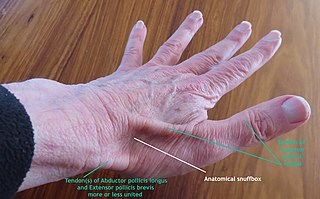
The anatomical snuff box or snuffbox or foveola radialis is a triangular deepening on the radial, dorsal aspect of the hand—at the level of the carpal bones, specifically, the scaphoid and trapezium bones forming the floor. The name originates from the use of this surface for placing and then sniffing powdered tobacco, or "snuff." It is sometimes referred to by its French name tabatière.

<span class="mw-page-title-main">Trapezoid bone</span> Carpal (wrist) bone

The trapezoid bone is a carpal bone in tetrapods, including humans. It is the smallest bone in the distal row of carpal bones that give structure to the palm of the hand. It may be known by its wedge-shaped form, the broad end of the wedge constituting the dorsal, the narrow end the palmar surface; and by its having four articular facets touching each other, and separated by sharp edges. It is homologous with the "second distal carpal" of reptiles and amphibians.

<span class="mw-page-title-main">Metacarpal bones</span> Bones of hand

In human anatomy, the metacarpal bones or metacarpus, also known as the "palm bones", are the appendicular bones that form the intermediate part of the hand between the phalanges (fingers) and the carpal bones, which articulate with the forearm. The metacarpal bones are homologous to the metatarsal bones in the foot.

<span class="mw-page-title-main">Scaphoid bone</span> Bone of the carpus

The scaphoid bone is one of the carpal bones of the wrist. It is situated between the hand and forearm on the thumb side of the wrist. It forms the radial border of the carpal tunnel. The scaphoid bone is the largest bone of the proximal row of wrist bones, its long axis being from above downward, lateralward, and forward. It is approximately the size and shape of a medium cashew nut.

<span class="mw-page-title-main">Colles' fracture</span> Medical condition

A Colles' fracture is a type of fracture of the distal forearm in which the broken end of the radius is bent backwards. Symptoms may include pain, swelling, deformity, and bruising. Complications may include damage to the median nerve.

<span class="mw-page-title-main">Bone fracture</span> Physical damage to the continuity of a bone

A bone fracture is a medical condition in which there is a partial or complete break in the continuity of any bone in the body. In more severe cases, the bone may be broken into several fragments, known as a comminuted fracture. A bone fracture may be the result of high force impact or stress, or a minimal trauma injury as a result of certain medical conditions that weaken the bones, such as osteoporosis, osteopenia, bone cancer, or osteogenesis imperfecta, where the fracture is then properly termed a pathologic fracture.

<span class="mw-page-title-main">Distal radius fracture</span> Fracture of the radius bone near the wrist

A distal radius fracture, also known as wrist fracture, is a break of the part of the radius bone which is close to the wrist. Symptoms include pain, bruising, and rapid-onset swelling. The ulna bone may also be broken.

In human anatomy, the abductor pollicis longus (APL) is one of the extrinsic muscles of the hand. Its major function is to abduct the thumb at the wrist. Its tendon forms the anterior border of the anatomical snuffbox.

<span class="mw-page-title-main">Carpometacarpal joint</span>

The carpometacarpal (CMC) joints are five joints in the wrist that articulate the distal row of carpal bones and the proximal bases of the five metacarpal bones.

<span class="mw-page-title-main">First metacarpal bone</span>

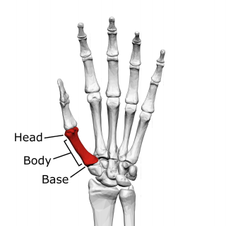
The first metacarpal bone or the metacarpal bone of the thumb is the first bone proximal to the thumb. It is connected to the trapezium of the carpus at the first carpometacarpal joint and to the proximal thumb phalanx at the first metacarpophalangeal joint.

<span class="mw-page-title-main">Triangular fibrocartilage</span> Anatomical feature in the wrist

The triangular fibrocartilage complex (TFCC) is formed by the triangular fibrocartilage discus (TFC), the radioulnar ligaments (RULs) and the ulnocarpal ligaments (UCLs).

<span class="mw-page-title-main">Ulnar collateral ligament injury of the thumb</span> Medical condition

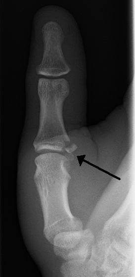
Gamekeeper's thumb is a type of injury to the ulnar collateral ligament (UCL) of the thumb. The UCL may be merely stretched, or it may be torn from its insertion site into the proximal phalanx of the thumb; in approximately 90% of cases part of the bone is actually avulsed from the joint. This condition is commonly observed among gamekeepers and Scottish fowl hunters, as well as athletes. It also occurs among people who sustain a fall onto an outstretched hand while holding a rod, frequently skiers grasping ski poles.

<span class="mw-page-title-main">Scaphoid fracture</span> Fracture of the scaphoid bone in the wrist

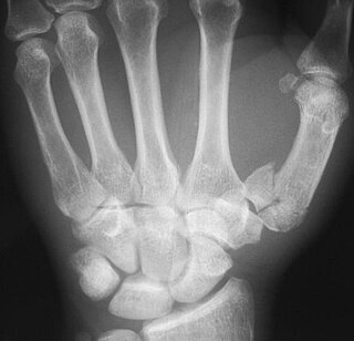
A scaphoid fracture is a break of the scaphoid bone in the wrist. Symptoms generally includes pain at the base of the thumb which is worse with use of the hand. The anatomic snuffbox is generally tender and swelling may occur. Complications may include nonunion of the fracture, avascular necrosis of the proximal part of the bone, and arthritis.

<span class="mw-page-title-main">Rolando fracture</span> Medical condition

The Rolando fracture is a type of broken finger involving the base of the thumb.

<span class="mw-page-title-main">Wrist osteoarthritis</span> Medical condition

Wrist osteoarthritis is gradual loss of articular cartilage and hypertrophic bone changes (osteophytes). While in many joints this is part of normal aging (senescence), in the wrist osteoarthritis usually occurs over years to decades after scapholunate interosseous ligament rupture or an unhealed fracture of the scaphoid. Characteristic symptoms including pain, deformity and stiffness. Pain intensity and incapability are notably variable and do not correspond with arthritis severity on radiographs.

<span class="mw-page-title-main">Trapeziometacarpal osteoarthritis</span> Medical condition

Trapeziometacarpal osteoarthritis (TMC OA) is, also known as osteoarthritis at the base of the thumb, thumb carpometacarpal osteoarthritis, basilar (or basal) joint arthritis, or as rhizarthrosis. This joint is formed by the trapezium bone of the wrist and the metacarpal bone of the thumb. This is one of the joints where most humans develop osteoarthritis with age. Osteoarthritis is age-related loss of the smooth surface of the bone where it moves against another bone (cartilage of the joint). In reaction to the loss of cartilage, the bones thicken at the joint surface, resulting in subchondral sclerosis. Also, bony outgrowths, called osteophytes (also known as “bone spurs”), are formed at the joint margins.

<span class="mw-page-title-main">Muscles of the thumb</span>

The muscles of the thumb are nine skeletal muscles located in the hand and forearm. The muscles allow for flexion, extension, adduction, abduction and opposition of the thumb. The muscles acting on the thumb can be divided into two groups: The extrinsic hand muscles, with their muscle bellies located in the forearm, and the intrinsic hand muscles, with their muscles bellies located in the hand proper.

<span class="mw-page-title-main">Proximal humerus fracture</span> Break of the upper part of the bone of the arm

A proximal humerus fracture is a break of the upper part of the bone of the arm (humerus). Symptoms include pain, swelling, and a decreased ability to move the shoulder. Complications may include axillary nerve or axillary artery injury.

References

  1. White, Timothy O.; Mackenzie, Samuel P.; Gray, Alasdair J. (2016). "13. Hand". McRae's Orthopaedic Trauma and Emergency Fracture Management (3rd ed.). Elsevier. pp. 275–277. ISBN   978-0-7020-5728-1. Archived from the original on 28 August 2021. Retrieved 8 September 2022.
  2. Martel, José; Bueno, Angel (2008). "Fractures with names". In Pope, Thomas; Bloem, Hans L.; Beltran, Javier; Morrison, William B.; John, David (eds.). Musculoskeletal Imaging (2nd ed.). Philadelphia: Elsevier. p. 1232.e2. ISBN   978-1-4557-0813-0.
  3. Edmunds, JO (August 2006). "Traumatic dislocations and instability of the trapeziometacarpal joint of the thumb" (PDF). Hand Clinics. 22 (3): 365–92. doi:10.1016/j.hcl.2006.05.001. PMID   16843802. Archived from the original (PDF) on 17 July 2011.
  4. 1 2 Bennett, EH (1882). "Fractures of the Metacarpal Bones". Dublin Med Sci J. 73: 72–75.
  5. Soyer, AD (November–December 1999). "Fractures of the base of the first metacarpal: current treatment options". The Journal of the American Academy of Orthopaedic Surgeons. 7 (6): 403–12. doi:10.5435/00124635-199911000-00006. PMID   11505928.
  6. Foster, RJ; Hastings H, 2nd (January 1987). "Treatment of Bennett, Rolando, and vertical intraarticular trapezial fractures". Clinical Orthopaedics and Related Research. 214 (214): 121–9. doi:10.1097/00003086-198701000-00018. PMID   3791733.
  7. Timmenga, EJ; Blokhuis, TJ; Maas, M; Raaijmakers, EL (June 1994). "Long-term evaluation of Bennett's fracture. A comparison between open and closed reduction". Journal of Hand Surgery (Edinburgh, Scotland). 19 (3): 373–7. doi:10.1016/0266-7681(94)90093-0. PMID   8077832. S2CID   19601071.