Cyanobacteria, also known as Cyanophyta, are a phylum of gram-negative bacteria that obtain energy via photosynthesis. The name cyanobacteria refers to their color, which similarly forms the basis of cyanobacteria's common name, blue-green algae, although they are not usually scientifically classified as algae. They appear to have originated in a freshwater or terrestrial environment. Sericytochromatia, the proposed name of the paraphyletic and most basal group, is the ancestor of both the non-photosynthetic group Melainabacteria and the photosynthetic cyanobacteria, also called Oxyphotobacteria.

White band disease is a coral disease that affects acroporid corals and is distinguishable by the white band of exposed coral skeleton that it forms. The disease completely destroys the coral tissue of Caribbean acroporid corals, specifically elkhorn coral and staghorn coral. The disease exhibits a pronounced division between the remaining coral tissue and the exposed coral skeleton. These symptoms are similar to white plague, except that white band disease is only found on acroporid corals, and white plague has not been found on any acroporid corals. It is part of a class of similar disease known as "white syndromes", many of which may be linked to species of Vibrio bacteria. While the pathogen for this disease has not been identified, Vibrio carchariae may be one of its factors. The degradation of coral tissue usually begins at the base of the coral, working its way up to the branch tips, but it can begin in the middle of a branch.

Algal mats are one of many types of microbial mat that forms on the surface of water or rocks. They are typically composed of blue-green cyanobacteria and sediments. Formation occurs when alternating layers of blue-green bacteria and sediments are deposited or grow in place, creating dark-laminated layers. Stromatolites are prime examples of algal mats. Algal mats played an important role in the Great Oxidation Event on Earth some 2.3 billion years ago. Algal mats can become a significant ecological problem, if the mats grow so expansive or thick as to disrupt the other underwater marine life by blocking the sunlight or producing toxic chemicals.
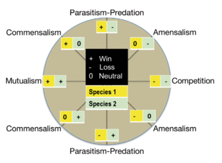
Symbiotic bacteria are bacteria living in symbiosis with another organism or each other. For example, rhizobia living in root nodules of legumes provide nitrogen fixing activity for these plants. Symbiosis was first defined by Marko de Bary in 1869 in a work entitled "Die Erscheinung der Symbiose" in which he defined the term as "namely, the living together of parasite and host". The definition of symbiosis has evolved to encompass a sustained relationship between two or more different organisms "over a considerable fraction of the life of the host." In addition, this relationship is often beneficial for at least one of the organisms involved. There are three main types of symbiotic relationships: commensalism, mutualism, and parasitism. Commensalism is when one organism benefits and the other is neither harmed nor benefits. Mutualism is when both organisms benefit. Lastly, parasitism is when one organism benefits while the other organism is harmed. Organisms can also be involved in multiple of these symbiotic relationships simultaneously.

Beggiatoa is a genus of Gammaproteobacteria belonging to the order Thiotrichales, in the Pseudomonadota phylum. This genus was one of the first bacteria discovered by Ukrainian botanist Sergei Winogradsky. During his research in Anton de Bary's laboratory of botany in 1887, he found that Beggiatoa oxidized hydrogen sulfide (H2S) as an energy source, forming intracellular sulfur droplets, oxygen is the terminal electron acceptor and CO2 is used as a carbon source. Winogradsky named it in honor of the Italian doctor and botanist Francesco Secondo Beggiato (1806 - 1883), from Venice. Winogradsky referred to this form of metabolism as "inorgoxidation" (oxidation of inorganic compounds), today called chemolithotrophy. These organisms live in sulfur-rich environments such as soil, both marine and freshwater, in the deep sea hydrothermal vents and in polluted marine environments. The finding represented the first discovery of lithotrophy. Two species of Beggiatoa have been formally described: the type species Beggiatoa alba and Beggiatoa leptomitoformis, the latter of which was only published in 2017. This colorless and filamentous bacterium, sometimes in association with other sulfur bacteria (for example the genus Thiothrix), can be arranged in biofilm visible to the naked eye formed by a very long white filamentous mat, the white color is due to the stored sulfur. Species of Beggiatoa have cells up to 200 µm in diameter and they are one of the largest prokaryotes on Earth.
Thiothrix is a genus of filamentous sulfur-oxidizing bacteria, related to the genera Beggiatoa and Thioploca. They are usually Gram-negative and rod-shaped. They form ensheathed multicellular filaments that are attached at the base, and form gonidia at their free end. The apical gonidia have gliding motility. Rosettes of the filaments are not always formed but are typical. Sulfur is deposited in invaginations within the cell membrane.
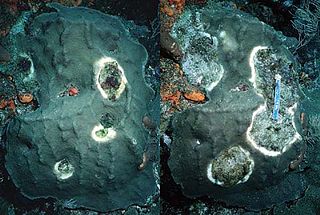
Skeletal eroding band (SEB) is a disease of corals that appears as a black or dark gray band that slowly advances over corals, leaving a spotted region of dead coral in its wake. It is the most common disease of corals in the Indian and Pacific Oceans, and is also found in the Red Sea.

A microbial mat is a multi-layered sheet of microorganisms, mainly bacteria and archaea, or bacteria alone. Microbial mats grow at interfaces between different types of material, mostly on submerged or moist surfaces, but a few survive in deserts. A few are found as endosymbionts of animals.

Microbiota are the range of microorganisms that may be commensal, symbiotic, or pathogenic found in and on all multicellular organisms, including plants. Microbiota include bacteria, archaea, protists, fungi, and viruses, and have been found to be crucial for immunologic, hormonal, and metabolic homeostasis of their host.

Thioploca is a genus of filamentous sulphur-oxidizing bacteria which occurs along 3,000 kilometres (1,900 mi) of coast off the west of South America. Was discovered in 1907 by R. Lauterborn classified as belonging to the order Thiotrichales, part of the Gammaproteobacteria. They inhabit as well marine as freshwater environments, with vast communities present off the Pacific coast of South America and other areas with a high organic matter sedimentation and bottom waters rich in nitrate and poor in oxygen. A large vacuole occupies more than 80% of their cellular volume and is used as a storage for nitrate. This nitrate is used for the sulphur oxidation, an important characteristic of the genus. Due to their unique size in diameters, ranging from 15-40 µm, they are considered part of the largest bacteria known. Because they use both sulfur and nitrogen compounds they may provide an important link between the nitrogen and sulphur cycles. They secrete a sheath of mucus which they use as a tunnel to travel between the sulfide containing sediment and the nitrate containing sea water.

White pox disease, first noted in 1996 on coral reefs near the Florida keys, is a coral disease affecting Elkhorn coral throughout the Caribbean. It causes irregular white patches or blotches on the coral that result from the loss of coral tissue. These patches distinguish white pox disease from white band disease which produces a distinctive white band where the coral skeleton has been denuded. The blotches caused by this disease are also clearly differentiated from coral bleaching and scars caused by coral-eating snails. It is very contagious, spreading to nearby coral.
Yellow-band disease is a coral disease that attacks colonies of coral at a time when coral is already under stress from pollution, overfishing, and climate change. It is characterized by large blotches or patches of bleached, yellowed tissue on Caribbean scleractinian corals.
A microbial consortium or microbial community, is two or more bacterial or microbial groups living symbiotically. Consortiums can be endosymbiotic or ectosymbiotic, or occasionally may be both. The protist Mixotricha paradoxa, itself an endosymbiont of the Mastotermes darwiniensis termite, is always found as a consortium of at least one endosymbiotic coccus, multiple ectosymbiotic species of flagellate or ciliate bacteria, and at least one species of helical Treponema bacteria that forms the basis of Mixotricha protists' locomotion.
The Sippewissett microbial mat is a microbial mat in the Sippewissett Salt Marsh located along the lower eastern Buzzards Bay shoreline of Cape Cod, about 5 miles north of Woods Hole and 1 mile southwest of West Falmouth, Massachusetts, in the United States. The marsh has two regions, the Great Sippewisset Marsh to the north and Little Sippewisset Marsh to the south, separated from each other by a narrow tongue of land. The marsh extends into an estuary in which the intertidal zone provides a dynamic environment that supports a diverse ecology, including threatened and endangered species such as the roseate tern. The ecology of the salt marsh is based in and supported by the microbial mats which cover the ground of the marsh.
Arthrobacter roseus is a species of red-pigmented psychrophilic bacteria first isolated from a cyanobacterial mat.
Microbial symbiosis in marine animals was not discovered until 1981. In the time following, symbiotic relationships between marine invertebrates and chemoautotrophic bacteria have been found in a variety of ecosystems, ranging from shallow coastal waters to deep-sea hydrothermal vents. Symbiosis is a way for marine organisms to find creative ways to survive in a very dynamic environment. They are different in relation to how dependent the organisms are on each other or how they are associated. It is also considered a selective force behind evolution in some scientific aspects. The symbiotic relationships of organisms has the ability to change behavior, morphology and metabolic pathways. With increased recognition and research, new terminology also arises, such as holobiont, which the relationship between a host and its symbionts as one grouping. Many scientists will look at the hologenome, which is the combined genetic information of the host and its symbionts. These terms are more commonly used to describe microbial symbionts.
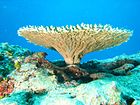
Coral diseases, comprising the diseases that affect corals, injure the living tissues and often result in the death of part or the whole of the colony. These diseases have been occurring more frequently in the twenty-first century as conditions become more stressful for many shallow-water corals. The pathogens causing the diseases include bacteria, fungi and protozoa, but it is not always possible to identify the pathogen involved.

Since the colonization of land by ancestral plant lineages 450 million years ago, plants and their associated microbes have been interacting with each other, forming an assemblage of species that is often referred to as a holobiont. Selective pressure acting on holobiont components has likely shaped plant-associated microbial communities and selected for host-adapted microorganisms that impact plant fitness. However, the high microbial densities detected on plant tissues, together with the fast generation time of microbes and their more ancient origin compared to their host, suggest that microbe-microbe interactions are also important selective forces sculpting complex microbial assemblages in the phyllosphere, rhizosphere, and plant endosphere compartments.

Kimberly B. Ritchie is an American marine biologist. She is an Associate Professor in the Department of Natural Sciences at the University of South Carolina Beaufort. Her research is focused on marine microbiology and how microbes affect animal health in hosts such as corals and sharks.
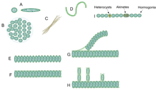
Cyanobacterial morphology refers to the form or shape of cyanobacteria. Cyanobacteria are a large and diverse phylum of bacteria defined by their unique combination of pigments and their ability to perform oxygenic photosynthesis.