Bonnet–Dechaume–Blanc syndrome

Last updated • 5 min readFrom Wikipedia, The Free Encyclopedia
Bonnet–Dechaume–Blanc syndrome
Other namesArteriovenous aneurysm of mid-brain and retina, facial nevi and mental changes, Cerebrofacial arteriovenous metameric syndrome type 2
Intracerebral hemorrage (CT scan).jpg
CT scan showing intracranial hemorrhage
Specialty Medical genetics   OOjs UI icon edit-ltr-progressive.svg

Bonnet–Dechaume–Blanc syndrome, also known as Wyburn-Mason syndrome, is a rare congenital disorder characterized by arteriovenous malformations of the brain, retina or facial nevi. [1] The syndrome has a number of possible symptoms and can, more rarely, affect the skin, bones, kidneys, muscles, and gastrointestinal tract. [2] When the syndrome affects the brain, people can experience severe headaches, seizures, acute stroke, meningism, and progressive neurological deficits due to acute or chronic ischaemia caused by arteriovenous shunting. [2] [3]

Contents

In the retina, the syndrome causes retinocephalic vascular malformations that tend to be present with intracranial hemorrhage and lead to decreased visual acuity, proptosis, pupillary defects, optic atrophy, congestion of bulbar conjunctiva, and visual field defects. [4] [5] Retinal lesions can be unilateral and tortuous, and symptoms begin to appear in the second and third decades of life. [4]

The syndrome can present with cutaneous lesions, or skin with different texture, thickness, and color, usually on the face. [5] The facial features caused by the syndrome vary from slight discoloration to extensive nevi and angiomas of the skin. [3] In some cases, the frontal and maxillary sinus may also be affected. [5]

There have only been 52 reported cases of patients with Bonnet–Dechaume–Blanc syndrome as of 2012. [2] Symptoms are rarely noticed in children and the syndrome is often diagnosed in late childhood or early adulthood when visual impairment is noticed. [3] Fluorescein angiography is commonly used to diagnose the syndrome. [6]

There have been several methods in treating patients with Bonnet–Dechaume–Blanc syndrome. However, the optimal treatment is uncertain. Patients with intracranial lesions have been treated with surgical intervention and in some cases, this procedure has been successful. Other treatments include embolization, radiation therapy, and continued observation. [5]

With limited research on Bonnet–Dechaume–Blanc syndrome, researchers have focused on the clinical and radiological findings rather than how to manage this rare and non-heritable syndrome. [3]

Signs and symptoms

Left eye showing proptosis. 406907P-PA-OCULAR.jpg
Left eye showing proptosis.

Typically not diagnosed until late childhood or later, Bonnet–Dechaume–Blanc syndrome usually presents itself with a combination of central nervous system features (midbrain), ophthalmic features (retina), and facial features. [7] The degree of expression of the syndrome's components varies both clinically and structurally. Common symptoms that lead to diagnosis are headaches, retro-orbital pain, and hemianopia. [5]

Visual field of homonymous hemianopia Visual field homonymous hemianopia.png
Visual field of homonymous hemianopia

The ophthalmic features of Bonnet–Dechaume–Blanc syndrome occur as retinal arteriovenous malformation (AVMs). There are three categories of AVMs based on their severity. The first category consists of the patient having small lesions that usually are asymptomatic. The second category, more severe than the first, is when the patient's malformation is missing a connecting capillary between an artery and a vein; without it, edema, hemorrhage, and visual impairment can result. Category three refers to malformations so severe that their dilated vessels no longer distinguish between artery and vein, and the patient has a significantly increased risk of vision loss. [3] Since the retinal lesions categorized vary from large vascular malformations that affect a majority of the retina to malformations that are barely visible, the lesions can cause a wide range of symptoms, including decrease in visual sharpness, proptosis, pupillary defects, optic nerve degeneration, and visual field defects. [5] The most common type of visual field impairment due to AVMs is homonymous hemianopia, [2] which is usually unilateral. [7]

Central nervous system (CNS) symptoms of Bonnet–Dechaume–Blanc syndrome are highly dependent on the locations and sizes of cerebral AVMs. [2] [5] [7] The most common CNS feature is an intracranial hemangioma in the midbrain. [3] Cerebral malformations can result in severe headaches, cerebral hemorrhages, vomiting, meningism, seizures, acute strokes, and progressive neurological deficits due to acute or chronic ischaemia caused by arteriovenous shunting. [3]

The facial features of Bonnet–Dechaume–Blanc syndrome vary from case to case. A person showing signs of the syndrome may display faint skin discoloration, nevi, or angiomas of the skin. [3] Some patients with this disorder also present with high-flow AVMs in the maxillofacial or mandibular (jaw) regions. [8] Another facial indicator of this disease is malformations affecting the frontal or maxillary sinuses. [5]

Causes

Fluorescein angiography can reveal arteriovenous malformations. Fluorescein angiography.jpg
Fluorescein angiography can reveal arteriovenous malformations.

Bonnet–Dechaume–Blanc syndrome results from arteriovenous malformations. The exact cause of this disorder is unknown, and no specific genetic abnormality has been identified. The syndrome is a congenital disorder that begins to develop around the seventh week of gestation when the maturation of retinal mesenchymal cells do not grow properly. [3] [7] [9] The abnormal development of vascular tissue leads to arteriovenous malformations, which affect both visual and cerebral structures. [9]

Diagnosis

Diagnosis commonly occurs later in childhood and often occurs incidentally in asymptomatic patients or as a cause of visual impairment. [3]

A number of examinations can be used to determine the extent of the syndrome and its severity. Fluorescein angiography is quite useful in diagnosing retinal features of the disease, and the use of ultrasonography and optical coherence tomography (OCT) are helpful in confirming the disease. [6] Neuro-ophthalmic examinations reveal pupillary defects (e.g. Marcus Gunn Pupil). Funduscopic examinations, examinations of the fundus of the eye, allow detection of arteriovenous malformations. [2] Neurological examination can determine neurological deficits such as hemiparesis and paresthesias. [2] MRI scans are used in imaging the brain and can allow visualization of the optic nerve and any possible atrophy. MRI, CT, and cerebral angiography may all be used to investigate the extent and location of any vascular lesions affecting the brain. [2] [5]

Treatment

The treatment for Bonnet–Dechaume–Blanc syndrome is controversial due to a lack of consensus on the different therapeutic procedures for treating arteriovenous malformations. [9] The first successful treatment was performed by Morgan et al., [8] who combined intracranial resection, ligation of the ophthalmic artery, and selective arterial ligature of the external carotid artery. Notably, the patient did not have retinal vascular malformations. [7]

Lesions are watched closely for changes in size. Prognosis is best when lesions are less than 3 cm in length. Most complications occur when the lesions are greater than 6 cm in size. [2] Surgical intervention for intracranial lesions has been done successfully. Nonsurgical treatments include embolization, radiation therapy, and continued observation. [5]

When pursuing treatment, it is important to consider the size of the malformations, their locations, and neurological involvement. [7]

Epidemiology

Related Research Articles

<span class="mw-page-title-main">Arteriovenous malformation</span> Vascular anomaly

An arteriovenous malformation (AVM) is an abnormal connection between arteries and veins, bypassing the capillary system. Usually congenital, this vascular anomaly is widely known because of its occurrence in the central nervous system, but can appear anywhere in the body. The symptoms of AVMs can range from none at all to intense pain or bleeding, and they can lead to other serious medical problems.

<span class="mw-page-title-main">Cerebral arteriovenous malformation</span> Medical condition

A cerebral arteriovenous malformation is an abnormal connection between the arteries and veins in the brain—specifically, an arteriovenous malformation in the cerebrum.

<span class="mw-page-title-main">Micropsia</span> Medical condition

Micropsia is a condition affecting human visual perception in which objects are perceived to be smaller than they actually are. Micropsia can be caused by optical factors, by distortion of images in the eye, by changes in the brain, and from psychological factors. Dissociative phenomena are linked with micropsia, which may be the result of brain-lateralization disturbance.

<span class="mw-page-title-main">Hereditary hemorrhagic telangiectasia</span> Medical condition (genetic disorder)

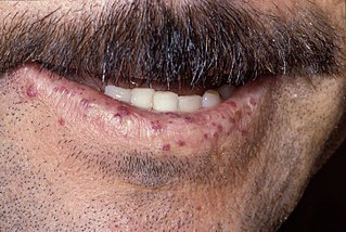
Hereditary hemorrhagic telangiectasia (HHT), also known as Osler–Weber–Rendu disease and Osler–Weber–Rendu syndrome, is a rare autosomal dominant genetic disorder that leads to abnormal blood vessel formation in the skin, mucous membranes, and often in organs such as the lungs, liver, and brain.

<span class="mw-page-title-main">Amaurosis fugax</span> Medical condition

Amaurosis fugax is a painless temporary loss of vision in one or both eyes.

<span class="mw-page-title-main">Cerebral angiography</span> Angiography that produces images of blood vessels in and around the brain

Cerebral angiography is a form of angiography which provides images of blood vessels in and around the brain, thereby allowing detection of abnormalities such as arteriovenous malformations and aneurysms. It was pioneered in 1927 by the Portuguese neurologist Egas Moniz at the University of Lisbon, who also helped develop thorotrast for use in the procedure.

Palinopsia is the persistent recurrence of a visual image after the stimulus has been removed. Palinopsia is not a diagnosis; it is a diverse group of pathological visual symptoms with a wide variety of causes. Visual perseveration is synonymous with palinopsia.

<span class="mw-page-title-main">Sturge–Weber syndrome</span> Medical condition

Sturge–Weber syndrome, sometimes referred to as encephalotrigeminal angiomatosis, is a rare congenital neurological and skin disorder. It is one of the phakomatoses and is often associated with port-wine stains of the face, glaucoma, seizures, intellectual disability, and ipsilateral leptomeningeal angioma. Sturge–Weber syndrome can be classified into three different types. Type 1 includes facial and leptomeningeal angiomas as well as the possibility of glaucoma or choroidal lesions. Normally, only one side of the brain is affected. This type is the most common. Type 2 involvement includes a facial angioma with a possibility of glaucoma developing. There is no evidence of brain involvement. Symptoms can show at any time beyond the initial diagnosis of the facial angioma. The symptoms can include glaucoma, cerebral blood flow abnormalities and headaches. More research is needed on this type of Sturge–Weber syndrome. Type 3 has leptomeningeal angioma involvement exclusively. The facial angioma is absent and glaucoma rarely occurs. This type is only diagnosed via brain scan.

<span class="mw-page-title-main">Intraparenchymal hemorrhage</span> Medical condition

Intraparenchymal hemorrhage (IPH) is one form of intracerebral bleeding in which there is bleeding within brain parenchyma. The other form is intraventricular hemorrhage (IVH).

Optic neuropathy is damage to the optic nerve from any cause. The optic nerve is a bundle of millions of fibers in the retina that sends visual signals to the brain.

<span class="mw-page-title-main">Acute posterior multifocal placoid pigment epitheliopathy</span> Eye disease causing lesions in retina

Acute posterior multifocal placoid pigment epitheliopathy (APMPPE) is an acquired inflammatory uveitis that belongs to the heterogenous group of white dot syndromes in which light-coloured (yellowish-white) lesions begin to form in the macular area of the retina. Early in the course of the disease, the lesions cause acute and marked vision loss that ranges from mild to severe but is usually transient in nature. APMPPE is classified as an inflammatory disorder that is usually bilateral and acute in onset but self-limiting. The lesions leave behind some pigmentation, but visual acuity eventually improves even without any treatment.

The Center for Cerebrovascular Research at the University of California, San Francisco is a collective of faculty and staff investigating matters related to cerebral circulation, particularly cerebrovascular disease resulting from narrowing of major blood vessels in the brain and vascular malformation of the brain. While research offices are located on Parnassus campus, San Francisco General Hospital hosts the center's laboratories and facilities. The center coordinates with additional faculty in various fields of neuroscience and vascular biology. Sponsors include the National Institute of Neurological Disorders and Stroke and the UCSF departments of Anesthesia, Neurological Surgery and Neurology.

The International Classification of Headache Disorders (ICHD) is a detailed hierarchical classification of all headache-related disorders published by the International Headache Society. It is considered the official classification of headaches by the World Health Organization, and, in 1992, was incorporated into the 10th edition of their International Classification of Diseases (ICD-10). Each class of headache contains explicit diagnostic criteria—meaning that the criteria include quantities rather than vague terms like several or usually—that are based on clinical and laboratory observations.

Cobb syndrome is a rare congenital disorder characterized by visible skin lesions and spinal angiomas or arteriovenous malformations (AVMs). The skin lesions of Cobb syndrome typically are present as port wine stains or angiomas, but reports exist of angiokeratomas, angiolipomas, and lymphangioma circumscriptum. The intraspinal lesions may be angiomas or AVMs and occur at levels of the spinal cord corresponding to the affected skin dermatomes. They may in turn produce spinal cord dysfunction and weakness or paralysis.

<span class="mw-page-title-main">Persistent fetal vasculature</span> Medical condition

Persistent fetal vasculature(PFV), also known as persistent fetal vasculature syndrome (PFVS), and until 1997 known primarily as persistent hyperplastic primary vitreous (PHPV), is a rare congenital anomaly which occurs when blood vessels within the developing eye, known as the embryonic hyaloid vasculature network, fail to regress as they normally would in-utero after the eye is fully developed. Defects which arise from this lack of vascular regression are diverse; as a result, the presentation, symptoms, and prognosis of affected patients vary widely, ranging from clinical insignificance to irreversible blindness. The underlying structural causes of PFV are considered to be relatively common, and the vast majority of cases do not warrant additional intervention. When symptoms do manifest, however, they are often significant, causing detrimental and irreversible visual impairment. Persistent fetal vasculature heightens the lifelong risk of glaucoma, cataracts, intraocular hemorrhages, and Retinal detachments, accounting for the visual loss of nearly 5% of the blind community in the developed world. In diagnosed cases of PFV, approximately 90% of patients with a unilateral disease have associated poor vision in the affected eye.

<span class="mw-page-title-main">Parkes Weber syndrome</span> Medical condition

Parkes Weber syndrome (PWS) is a congenital disorder of the vascular system. It is an extremely rare condition, and its exact prevalence is unknown. It is named after British dermatologist Frederick Parkes Weber, who first described the syndrome in 1907.

<span class="mw-page-title-main">Vein of Galen aneurysmal malformations</span> Medical condition

Vein of Galen aneurysmal malformations(VGAMs) and Vein of Galen aneurysmal dilations (VGADs) are the most frequent arteriovenous malformations in infants and fetuses. A VGAM consists of a tangled mass of dilated vessels supplied by an enlarged artery. The malformation increases greatly in size with age, although the mechanism of the increase is unknown. Dilation of the great cerebral vein of Galen is a secondary result of the force of arterial blood either directly from an artery via an arteriovenous fistula or by way of a tributary vein that receives the blood directly from an artery. There is usually a venous anomaly downstream from the draining vein that, together with the high blood flow into the great cerebral vein of Galen causes its dilation. The right sided cardiac chambers and pulmonary arteries also develop mild to severe dilation.

Hallucinatory palinopsia is a subtype of palinopsia, a visual disturbance defined as the persistent or recurrence of a visual image after the stimulus has been removed. Palinopsia is a broad term describing a group of symptoms which is divided into hallucinatory palinopsia and illusory palinopsia. Hallucinatory palinopsia refers to the projection of an already-encoded visual memory and is similar to a complex visual hallucination: the creation of a formed visual image where none exists.

<span class="mw-page-title-main">CLOVES syndrome</span> Medical condition

CLOVES syndrome is a rare overgrowth syndrome with complex vascular anomalies. CLOVES syndrome affects people with various symptoms, ranging from mild fatty soft-tissue tumors to vascular malformations encompassing the spine or internal organs.

<span class="mw-page-title-main">Visual pathway lesions</span> Visual maladies

The visual pathway consists of structures that carry visual information from the retina to the brain. Lesions in that pathway cause a variety of visual field defects. In the visual system of human eye, the visual information processed by retinal photoreceptor cells travel in the following way:
Retina→Optic nerve→Optic chiasma →Optic tract→Lateral geniculate body→Optic radiation→Primary visual cortex

References

  1. Liu, Anthony; Chen, Yi-Wen; Chang, Steven; Liao, Yaping Joyce (March 2012). "Junctional Visual Field Loss in a Case of Wyburn-Mason Syndrome". Journal of Neuro-Ophthalmology. 32 (1): 42–44. doi: 10.1097/WNO.0b013e31821aeefb . PMID   21613961.
  2. 1 2 3 4 5 6 7 8 9 SINGH, A; RUNDLE, P; RENNIE, I (March 2005). "Retinal Vascular Tumors". Ophthalmology Clinics of North America. 18 (1): 167–176. doi:10.1016/j.ohc.2004.07.005. PMID   15763202. S2CID   12853198.
  3. 1 2 3 4 5 6 7 8 9 10 Kim, Jeonghee; Kim, Ok Hwa; Suh, Jung Ho; Lew, Ho Min (20 March 1998). "Wyburn-Mason syndrome: an unusual presentation of bilateral orbital and unilateral brain arteriovenous malformations". Pediatric Radiology. 28 (3): 161. doi:10.1007/s002470050319. PMID   9561534. S2CID   36979750.
  4. 1 2 Muthukumar, N; Sundaralingam, MP (October 1998). "Retinocephalic vascular malformation: case report". British Journal of Neurosurgery. 12 (5): 458–60. doi:10.1080/02688699844718. PMID   10070454.
  5. 1 2 3 4 5 6 7 8 9 10 Dayani, P. N.; Sadun, A. A. (18 January 2007). "A case report of Wyburn-Mason syndrome and review of the literature". Neuroradiology. 49 (5): 445–456. doi:10.1007/s00234-006-0205-x. PMID   17235577. S2CID   11516890.
  6. 1 2 Singh, ArunD; Turell, MaryE (2010). "Vascular tumors of the retina and choroid: Diagnosis and treatment". Middle East African Journal of Ophthalmology. 17 (3): 191–200. doi: 10.4103/0974-9233.65486 . PMC   2934709 . PMID   20844673.
  7. 1 2 3 4 5 6 Lester, Jacobo; Ruano-Calderon, Luis Angel; Gonzalez-Olhovich, Irene (July 2005). "Wyburn-Mason Syndrome". Journal of Neuroimaging. 15 (3): 284–285. doi:10.1111/j.1552-6569.2005.tb00324.x. PMID   15951414. S2CID   1922375.
  8. 1 2 Bhattacharya, JJ; Luo, CB; Suh, DC; Alvarez, H; Rodesch, G; Lasjaunias, P (30 March 2001). "Wyburn-Mason or Bonnet-Dechaume-Blanc as Cerebrofacial Arteriovenous Metameric Syndromes (CAMS). A New Concept and a New Classification". Interventional Neuroradiology. 7 (1): 5–17. doi:10.1177/159101990100700101. PMC   3621461 . PMID   20663326.
  9. 1 2 3 Schmidt, D; Pache, M; Schumacher, M (2008). "The congenital unilateral retinocephalic vascular malformation syndrome (bonnet-dechaume-blanc syndrome or wyburn-mason syndrome): review of the literature". Survey of Ophthalmology. 53 (3): 227–49. doi:10.1016/j.survophthal.2007.10.001. PMID   18501269.