Chondrodystrophy

Last updated
Chondrodystrophy
Specialty Medical genetics   OOjs UI icon edit-ltr-progressive.svg

Chondrodystrophy (literally, "cartilage maldevelopment") refers to a skeletal disorder caused by one of myriad genetic mutations that can affect the development of cartilage. [1] As a very general term, it is only used in the medical literature when a more precise description of the condition is unavailable.

Contents

Presentation

People with chondrodystrophy have a normal-sized trunk and abnormally short limbs and extremities (dwarfism). Those affected with the disorder often call themselves dwarves, little people or short-statured persons. Over 100 specific skeletal dysplasias have been identified. Chondrodystrophy is found in all races and in both females and male and occurs in around one of every 25,000 children. Chondrodystrophy and achondroplasia are the most common forms of genetic hyaline disorders.[ citation needed ]

Hyaline cartilage caps the long bones and the spinal vertebrae. Most childhood limb growth takes place at the ends of the long bones, not in the shaft. Normally, as a child grows, the most interior portion of the joint cartilage converts into bone, and new cartilage forms on the surface to maintain smooth joints. The old joint margins (edges) reabsorb, so that the overall shape of the joint is maintained as growth continues. Failure of this process throughout the body results in skeletal dysplasia. It also leads to very early onset of osteoarthritis, because the defective cartilage is extremely fragile and vulnerable to normal wear and tear.[ citation needed ]

Causes

It is thought that chondrodystrophy [2] is caused by an autosomal, recessive allele. To avoid a potential "lethal dose", both parents can be genetically tested. If a child is conceived with another carrier the outcome may be lethal, or the child may suffer from chondrodystrophy or dwarfism. [3] This means that even though both parents are completely normal in height, the child will have one of the two types of skeletal dysplasia. Type 1 (short limb dysplasia), the more common of the two, is characterised by a long trunk and extremely shortened extremities. Type 2, short-trunk dysplasia, is characterised by a shortened trunk and normal size extremities. Those affected by chondrodystrophy may also experience metabolic and hormonal disorders, both of which may be monitored and controlled by hormonal injections.[ citation needed ]

Animals have been bred specifically to elicit chondrodystrophic traits for research purposes and to more easily allow animals to free-roam without escaping by, for example, jumping over ranch fences. One example of this is the Ancon sheep, which was first bred from a lamb born in 1791 with naturally occurring chondrodystrophy. [4]

Inheritance

Chondrodystrophy has an autosomal recessive pattern of inheritance. Autorecessive.svg
Chondrodystrophy has an autosomal recessive pattern of inheritance.

Chondrodystrophy is an autosomal recessive disorder, meaning that in order for this disease to be expressed, the affected individual must possess two copies of the allele for the disorder. The inheritance of the chondrodystrophy gene is as follows:

Let us name the dominant allele for normal stature "T", and the recessive allele coding for chondrodystrophy "t"; either one or the other is going to be chosen during random selection for a particular "seat" on its chromosome. If both parents are heterozygous for chondrodystrophy, they each possess one copy of the T allele and one copy of the t allele (each person has two copies of every autosomal allele, a paternal and a maternal one). When they reproduce there are then four possible alleles that may be chosen at random, two of them are the T allele (one from the father, one from the mother), and two are t alleles (again, one from the father, and one from the mother). The resulting Mendelian ratio of offspring from this mating would then be:[ citation needed ]

The phenotypes of the offspring would be three unaffected, normal-stature offspring, and one affected chondrodystrophic offspring; there would be a 25% chance of having an affected offspring if both parents were carriers of the recessive allele. Other probabilities for the other possible allele combinations concerning this gene are: 0% chance of affected offspring if only one parent is a carrier, 0% chance of affected offspring if one parent is affected and the other does not carry the allele, and 50% chance of affected offspring if one parent is affected and the other is a carrier. These ratios may be found by drawing up a standard Mendelian punnett square.[ citation needed ]

Percentage risk of inheritance

Diagnosis

There are several ways to determine if a child has chondrodystrophy, including parent testing and x-rays. If the fetus is suspected of having chondrodystrophy, the parents can be tested to find out if the fetus in fact does have the disease. It is not until the baby is born that a diagnosis can be declared. [3] The diagnosis is declared with the help of several x-rays [5] and charted bone growth patterns. Once the child is diagnosed the parents have to monitor the children because of several different factors. As the child gets older, hearing, eyesight and motor skills may be defective. Also, breathing (apnea) and weight problems (obesity) may occur. Structurally, scoliosis, bowed legs (genu varum), and arthritis may result.[ citation needed ]

Treatment

There is no treatment at this time to promote bone growth in chondrodystrophy patients. Certain types of growth hormone seem to increase the rate of growth during the first year of life/treatment, but have no substantial effect in adult patients. Only a few surgical centers in the world perform, experimentally, leg and arm lengthening procedures. Most common therapies are found in seeking help from: family physicians, pediatrics, internists, endocrinologists, geneticists, orthopedists and neurologists.[ citation needed ]

Coping with chondrodystrophy

It is important that the individual experience independence and self-worth. There are several appliances available to help overcome the disadvantages of small stature, including light-switch extenders and longer pedals in cars to enable effective driving. Several organizations that help Little People interact and get involved, such as the Little People of America.[ citation needed ]

Physical education and sports

Having short limbs can limit ball handling, and athletic performance in racquet sports and certain track events, like the long jump and high jump. However, short limbs can be an advantage for a sport like power lifting. Additionally, swimming and bicycling are recommended due to their limited impact on the joints.[ citation needed ]

Modification suggestions

  • Height adjustments for goals and volleyball nets.
  • Modified rules to accommodate size and structure.
  • Modified equipment, such as smaller balls and/or racquets.
  • Lowered swings to allow a feeling of independence on the playground.

The Dwarf Athletic Association of America

The Dwarf Athletic Association was started in 1985 for little people. Common sports are basketball, volleyball, powerlifting, track, field, swimming and bowling. National events are held along with the annual conference of Little People of America. The games accommodate athletes who are 4'10" or less with chondrodystrophy or related disorders. Athletes who meet the above requirements may compete in the Paralympic Games and other events sponsored by the International Paralympic Committee.[ citation needed ]

See also

Related Research Articles

Achondroplasia is a genetic disorder with an autosomal dominant pattern of inheritance whose primary feature is dwarfism. It is the most common cause of dwarfism and affects about 1 in 27,500 people. In those with the condition, the arms and legs are short, while the torso is typically of normal length. Those affected have an average adult height of 131 centimetres for males and 123 centimetres (4 ft) for females. Other features can include an enlarged head with prominent forehead and underdevelopment of the midface. Complications can include sleep apnea or recurrent ear infections. Achondroplasia includes the extremely rare short-limb skeletal dysplasia with severe combined immunodeficiency.

<span class="mw-page-title-main">Dwarfism</span> Small size of an organism, caused by growth deficiency or genetic mutations

Dwarfism is a condition of people and animals marked by unusually small size or short stature. In humans, it is sometimes defined as an adult height of less than 147 centimetres, regardless of sex; the average adult height among people with dwarfism is 120 centimetres (4 ft). Disproportionate dwarfism is characterized by either short limbs or a short torso. In cases of proportionate dwarfism, both the limbs and torso are unusually small. Intelligence is usually normal, and most people with it have a nearly normal life expectancy. People with dwarfism can usually bear children, although there are additional risks to the mother and child depending upon the underlying condition.

Diastrophic dysplasia is an autosomal recessive dysplasia which affects cartilage and bone development. Diastrophic dysplasia is due to mutations in the SLC26A2 gene.

<span class="mw-page-title-main">Atelosteogenesis, type II</span> Medical condition

Atelosteogenesis, type II is a severe disorder of cartilage and bone development. It is rare, and infants with the disorder are usually stillborn; those who survive birth die soon after.

Short stature refers to a height of a human which is below typical. Whether a person is considered short depends on the context. Because of the lack of preciseness, there is often disagreement about the degree of shortness that should be called short. Dwarfism is the condition of being very short, often caused by a medical condition. In a medical context, short stature is typically defined as an adult height that is more than two standard deviations below a population’s mean for age and sex, which corresponds to the shortest 2.3% of individuals in that population.

<span class="mw-page-title-main">Otospondylomegaepiphyseal dysplasia</span> Medical condition

Otospondylomegaepiphyseal dysplasia (OSMED) is an autosomal recessive disorder of bone growth that results in skeletal abnormalities, severe hearing loss, and distinctive facial features. The name of the condition indicates that it affects hearing (oto-) and the bones of the spine (spondylo-), and enlarges the ends of bones (megaepiphyses).

Spondyloepiphyseal dysplasia congenita is a rare disorder of bone growth that results in dwarfism, characteristic skeletal abnormalities, and occasionally problems with vision and hearing. The name of the condition indicates that it affects the bones of the spine (spondylo-) and the ends of bones (epiphyses), and that it is present from birth (congenital). The signs and symptoms of spondyloepiphyseal dysplasia congenita are similar to, but milder than, the related skeletal disorders achondrogenesis type 2 and hypochondrogenesis. Spondyloepiphyseal dysplasia congenita is a subtype of collagenopathy, types II and XI.

<span class="mw-page-title-main">Spondyloepimetaphyseal dysplasia, Strudwick type</span> Medical condition

Spondyloepimetaphyseal dysplasia, Strudwick type is an inherited disorder of bone growth that results in dwarfism, characteristic skeletal abnormalities, and problems with vision. The name of the condition indicates that it affects the bones of the spine (spondylo-) and two regions near the ends of bones. This type was named after the first reported patient with the disorder. Spondyloepimetaphyseal dysplasia, Strudwick type is a subtype of type II collagenopathies.

<span class="mw-page-title-main">Achondrogenesis type 1B</span> Medical condition

Achondrogenesis type 1B is a severe autosomal recessive skeletal disorder, invariably fatal in the perinatal period. It is distinguished by its elongated, spherical midsection, small chest, and exceedingly short limbs. The feet can turn inward and upward (clubfeet), and the fingers and toes are little. Babies affected often have a soft out-pouching at the groin or around the belly button.

<span class="mw-page-title-main">Autosomal recessive multiple epiphyseal dysplasia</span> Medical condition

Autosomal recessive multiple epiphyseal dysplasia (ARMED), also called epiphyseal dysplasia, multiple, 4 (EDM4), multiple epiphyseal dysplasia with clubfoot or –with bilayered patellae, is an autosomal recessive congenital disorder affecting cartilage and bone development. The disorder has relatively mild signs and symptoms, including joint pain, scoliosis, and malformations of the hands, feet, and knees.

An osteochondrodysplasia, or skeletal dysplasia, is a disorder of the development of bone and cartilage. Osteochondrodysplasias are rare diseases. About 1 in 5,000 babies are born with some type of skeletal dysplasia. Nonetheless, if taken collectively, genetic skeletal dysplasias or osteochondrodysplasias comprise a recognizable group of genetically determined disorders with generalized skeletal affection. These disorders lead to disproportionate short stature and bone abnormalities, particularly in the arms, legs, and spine. Skeletal dysplasia can result in marked functional limitation and even mortality.

<span class="mw-page-title-main">Multiple epiphyseal dysplasia</span> Rare genetic disorder

Multiple epiphyseal dysplasia (MED), also known as Fairbank's disease, is a rare genetic disorder that affects the growing ends of bones. Long bones normally elongate by expansion of cartilage in the growth plate near their ends. As it expands outward from the growth plate, the cartilage mineralizes and hardens to become bone (ossification). In MED, this process is defective.

<span class="mw-page-title-main">Pseudoachondroplasia</span> Inherited disorder of bone growth

Pseudoachondroplasia is an inherited disorder of bone growth. It is a genetic autosomal dominant disorder. It is generally not discovered until 2–3 years of age, since growth is normal at first. Pseudoachondroplasia is usually first detected by a drop of linear growth in contrast to peers, a waddling gait or arising lower limb deformities.

<span class="mw-page-title-main">Boomerang dysplasia</span> Medical condition

Boomerang dysplasia is a lethal form of osteochondrodysplasia known for a characteristic congenital feature in which bones of the arms and legs are malformed into the shape of a boomerang. Death usually occurs in early infancy due to complications arising from overwhelming systemic bone malformations.

<span class="mw-page-title-main">Asphyxiating thoracic dysplasia</span> Medical condition

Asphyxiating thoracic dysplasia (ATD), also known as Jeune syndrome, is a rare inherited bone growth disorder that primarily affects the thoracic region. It was first described in 1955 by the French pediatrician Mathis Jeune. Common signs and symptoms can include a narrow chest, short ribs, shortened bones in the arms and legs, short stature, and extra fingers and toes (polydactyly). The restricted growth and expansion of the lungs caused by this disorder results in life-threatening breathing difficulties; occurring in 1 in every 100,000-130,000 live births in the United States.

Langer Mesomelic Dysplasia (LMD) is a rare congenital disorder characterised by altered bone formation, which typically causes affected individuals to experience shortening of the bones of the extremities as well as an abnormally short stature.

Acromesomelic dysplasia is a rare skeletal disorder that causes abnormal bone and cartilage development, leading to shortening of the forearms, lower legs, hands, feet, fingers, and toes. Five different genetic mutations have been implicated in the disorder. Treatment is individualized but is generally aimed at palliating symptoms, for example, treatment of kyphosis and lumbar hyperlordosis.
<span class="mw-page-title-main">Du Pan syndrome</span> Medical condition

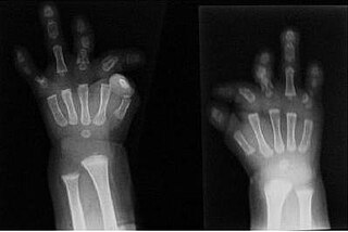
Du Pan syndrome, also known as fibular aplasia-complex brachydactyly syndrome, is an extremely rare genetic condition. Unlike other rare genetic conditions, Du Pan syndrome does not affect brain function or the appearance of the head and trunk. This condition is associated with alterations to the GDF5 gene. The way that this condition is passed on from generation to generation varies, but it is most commonly inherited in an autosomal recessive manner, meaning two copies of the same version of the gene are required to show this condition. Rare cases exist where the mode of inheritance is autosomal dominant, which means having only one version of the gene is enough to cause this condition.

References

  1. "Definition: chondrodystrophy from Online Medical Dictionary" . Retrieved 2007-12-23.
  2. Frankham, Richard; Rideout, Bruce A.; Ballou, Jonathan D.; Ralls, Katherine (May 2000). "CJO - Abstract - Genetic management of chondrodystrophy in California condors". Animal Conservation Forum. 3 (2): 145–153. doi:10.1111/j.1469-1795.2000.tb00239.x. S2CID   26272391 . Retrieved 2007-12-23.
  3. 1 2 "Dwarfism" . Retrieved 2007-12-23.
  4. Gidney, Louisa (May–June 2007). "Earliest Archaeological Evidence of the Ancon Mutation in Sheep from Leicester, UK". International Journal of Osteoarchaeology. 17 (3): 318–321. doi:10.1002/oa.872.
  5. "Dysplasia Epiphysealis Multiplex (DEM) (earlier synonyms: Fairbank's Disease, Ribbing's disease, Epiphyseal dysostosis, Hereditary enchondral dysostosis)" . Retrieved 2007-12-23.