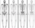
Osteopoikilosis

Last updated
Osteopoikilosis
Osteopoikilie Haende.jpg
Osteopoikilosis on an X-ray of the hands
Specialty Medical genetics   OOjs UI icon edit-ltr-progressive.svg

Osteopoikilosis is a benign, autosomal dominant sclerosing dysplasia of bone characterized by the presence of numerous bone islands in the skeleton. [1]

Contents

Presentation

The radiographic appearance of osteopoikilosis on an X-ray is characterized by a pattern of numerous white densities of similar size spread throughout all the bones. This is a systemic condition. It must be differentiated from blastic metastasis, which can also present radiographically as white densities interspersed throughout bone. Blastic metastasis tends to present with larger and more irregular densities in less of a uniform pattern. Another differentiating factor is age, with blastic metastasis mostly affecting older people, and osteopoikilosis being found in people 20 years of age and younger.

The distribution is variable, though it does not tend to affect the ribs, spine, or skull. [2]

Cause

Epidemiology

Men and women are affected in equal number, [3] reflecting the fact that osteopoikilosis attacks indiscriminately. Additionally, the disease is often associated with melorheostosis, [4] despite the apparent lack of correlation between melorheostosis and genetic heritability.[ citation needed ] It has been tied to LEMD3. [5] Buschke–Ollendorff syndrome is a similar condition, [6] which is also associated with LEMD3. [7]

See also

Related Research Articles

Osteosclerosis Condition in which bones are abnormally hardened, with elevated density

Osteosclerosis is a disorder that is characterized by abnormal hardening of bone and an elevation in bone density. It may predominantly affect the medullary portion and/or cortex of bone. Plain radiographs are a valuable tool for detecting and classifying osteosclerotic disorders. It can manifest in localized or generalized osteosclerosis. Localized osteosclerosis can be caused by Legg–Calvé–Perthes disease, sickle-cell disease and osteoarthritis among others. Osteosclerosis can be classified in accordance with the causative factor into acquired and hereditary.

Osteopetrosis Rare disease of the bones

Osteopetrosis, literally "stone bone", also known as marble bone disease or Albers-Schönberg disease, is an extremely rare inherited disorder whereby the bones harden, becoming denser, in contrast to more prevalent conditions like osteoporosis, in which the bones become less dense and more brittle, or osteomalacia, in which the bones soften. Osteopetrosis can cause bones to dissolve and break.

Palmoplantar keratoderma Medical condition

Palmoplantar keratodermas are a heterogeneous group of disorders characterized by abnormal thickening of the stratum corneum of the palms and soles.

Crouzon syndrome Genetic disorder of the skull and face

Crouzon syndrome is an autosomal dominant genetic disorder known as a branchial arch syndrome. Specifically, this syndrome affects the first branchial arch, which is the precursor of the maxilla and mandible. Since the branchial arches are important developmental features in a growing embryo, disturbances in their development create lasting and widespread effects.

Crouzonodermoskeletal syndrome is a disorder characterized by the premature joining of certain bones of the skull (craniosynostosis) during development and a skin condition called acanthosis nigricans.

Osteochondrodysplasia is a general term for a disorder of the development (dysplasia) of bone ("osteo") and cartilage ("chondro"). Osteochondrodysplasias are rare diseases. About 1 in 5,000 babies are born with some type of skeletal dysplasia. Nonetheless, if taken collectively, genetic skeletal dysplasias or osteochondrodysplasias comprise a recognizable group of genetically determined disorders with generalized skeletal affection. Osteochondrodysplasias can result in marked functional limitation and even mortality.

Pseudoachondroplasia Inherited disorder of bone growth

Pseudoachondroplasia is an inherited disorder of bone growth. It is a genetic autosomal dominant disorder. It is generally not discovered until 2–3 years of age, since growth is normal at first. Pseudoachondroplasia is usually first detected by a drop of linear growth in contrast to peers, a waddling gait or arising lower limb deformities.

Laminopathy Medical condition

Laminopathies are a group of rare genetic disorders caused by mutations in genes encoding proteins of the nuclear lamina. They are included in the more generic term nuclear envelopathies that was coined in 2000 for diseases associated with defects of the nuclear envelope. Since the first reports of laminopathies in the late 1990s, increased research efforts have started to uncover the vital role of nuclear envelope proteins in cell and tissue integrity in animals.

Wallis–Zieff–Goldblatt syndrome Medical condition

Wallis–Zieff–Goldblatt syndrome is a rare condition characterized by inherited skeletal disorders manifested mainly as rhizomelic short stature and lateral clavicular defects. It is also known as Cleidorhizomelic syndrome.

LEM domain-containing protein 3

LEM domain-containing protein 3 (LEMD3), also known as MAN1, is an integral protein in the inner nuclear membrane (INM) of the nuclear envelope. It is encoded by the LEMD3 gene and was first identified after it was isolated from the serum of a patient with a collagen vascular disease.

Buschke–Ollendorff syndrome Medical condition

Buschke–Ollendorff syndrome (BOS)is a rare genetic disorder associated with LEMD3. It is believed to be inherited in an autosomal dominant manner. It is named for Abraham Buschke and Helene Ollendorff Curth, who described it in a 45-year-old woman. Its frequency is almost 1 case per every 20,000 people, and it is equally found in both males and females.

Melorheostosis Medical condition

Melorheostosis is a medical developmental disorder and mesenchymal dysplasia in which the bony cortex widens and becomes hyperdense in a sclerotomal distribution. The condition begins in childhood and is characterized by thickening of the bones. Pain is a frequent symptom and the bone can have the appearance of dripping candle wax.

TECTA

Alpha-tectorin is a protein that in humans is encoded by the TECTA gene.

Boomerang dysplasia Medical condition

Boomerang dysplasia is a lethal form of osteochondrodysplasia known for a characteristic congenital feature in which bones of the arms and legs are malformed into the shape of a boomerang. Death usually occurs in early infancy due to complications arising from overwhelming systemic bone malformations.

Ichthyosis hystrix is a group of rare skin disorders in the ichthyosis family of skin disorders characterized by massive hyperkeratosis with an appearance like spiny scales. This term is also used to refer to a type of epidermal nevi with extensive bilateral distribution.

Worth syndrome Medical condition

Worth syndrome, also known as benign form of Worth hyperostosis corticalis generalisata with torus platinus, autosomal dominant osteosclerosis, autosomal dominant endosteal hyperostosis or Worth disease, is a rare autosomal dominant congenital disorder that is caused by a mutation in the LRP5 gene. It is characterized by increased bone density and benign bony structures on the palate.

Hand-foot-genital syndrome Medical condition

Hand-foot-genital syndrome (HFGS) is characterized by limb malformations and urogenital defects. Mild bilateral shortening of the thumbs and great toes, caused primarily by shortening of the distal phalanx and/or the first metacarpal or metatarsal, is the most common limb malformation and results in impaired dexterity or apposition of the thumbs. Urogenital abnormalities include abnormalities of the ureters and urethra and various degrees of incomplete Müllerian fusion in females and hypospadias of variable severity with or without chordee in males. Vesicoureteral reflux, recurrent urinary tract infections, and chronic pyelonephritis are common; fertility is normal.

Beare–Stevenson cutis gyrata syndrome is a rare genetic disorder characterized by craniosynostosis and a specific skin abnormality, called cutis gyrata, characterized by a furrowed and wrinkled appearance ; thick, dark, velvety areas of skin are sometimes found on the hands and feet and in the groin.

Tricho–dento–osseous syndrome Medical condition

Tricho–dento–osseous syndrome (TDO) is a rare, systemic, autosomal dominant genetic disorder that causes defects in hair, teeth, and bones respectively. This disease is present at birth. TDO has been shown to occur in areas of close geographic proximity and within families; most recent documented cases are in Virginia, Tennessee, and North Carolina. The cause of this disease is a mutation in the DLX3 gene, which controls hair follicle differentiation and induction of bone formation. One-hundred percent of patients with TDO suffer from two co-existing conditions called enamel hypoplasia and taurodontism in which the abnormal growth patterns of the teeth result in severe external and internal defects. The hair defects are characterized as being rough, course, with profuse shedding. Hair is curly and kinky at infancy but later straightens. Dental defects are characterized by dark-yellow/brownish colored teeth, thin and/or possibly pitted enamel, that is malformed. The teeth can also look normal in color, but also have a physical impression of extreme fragility and thinness in appearance. Additionally, severe underbites where the top and bottom teeth fail to correctly align may be present; it is common for the affected individual to have a larger, more pronounced lower jaw and longer bones. The physical deformities that TDO causes become more noticeable with age, and emotional support for the family as well as the affected individual is frequently recommended. Adequate treatment for TDO is a team based approach, mostly involving physical therapists, dentists, and oromaxillofacial surgeons. Genetic counseling is also recommended.

Malignant infantile osteopetrosis, is a rare osteosclerosing type of skeletal dysplasia that typically presents in infancy and is characterized by a unique radiographic appearance of generalized hyperostosis - excessive growth of bone.

References

  1. Bull M, Calderbank P, Ramachandran N (2007). "A cause for concern? Osteopoikilosis found incidentally in the emergency department: a case report". Emerg Med J. 24 (5): e29. doi:10.1136/emj.2006.045765. PMC   2658513 . PMID   17452689.
  2. Balan, Nisha Sharma, Anu (2008). Get through FRCR part 2B : rapid reporting of plain radiographs. London: Royal Society of Medicine. ISBN   978-1853157547.
  3. Serdaroğlu M, Capkin E, Uçüncü F, Tosun M (2007). "Case report of a patient with osteopoikilosis". Rheumatol. Int. 27 (7): 683–6. doi:10.1007/s00296-006-0262-9. PMID   17106662.
  4. Nevin NC, Thomas PS, Davis RI, Cowie GH (1999). "Melorheostosis in a family with autosomal dominant osteopoikilosis". Am. J. Med. Genet. 82 (5): 409–14. doi:10.1002/(SICI)1096-8628(19990219)82:5<409::AID-AJMG10>3.0.CO;2-2. PMID   10069713.
  5. Hellemans J, Preobrazhenska O, Willaert A, et al. (2004). "Loss-of-function mutations in LEMD3 result in osteopoikilosis, Buschke–Ollendorff syndrome and melorheostosis". Nat. Genet. 36 (11): 1213–8. doi: 10.1038/ng1453 . PMID   15489854.
  6. synd/1803 at Who Named It?
  7. Mumm S, Wenkert D, Zhang X, McAlister WH, Mier RJ, Whyte MP (2007). "Deactivating germline mutations in LEMD3 cause osteopoikilosis and Buschke–Ollendorff syndrome, but not sporadic melorheostosis". J. Bone Miner. Res. 22 (2): 243–50. doi:10.1359/jbmr.061102. PMID   17087626.
Classification
D
External resources