Nephrology is a specialty for both adult internal medicine and pediatric medicine that concerns the study of the kidneys, specifically normal kidney function and kidney disease, the preservation of kidney health, and the treatment of kidney disease, from diet and medication to renal replacement therapy. The word "renal" is an adjective meaning "relating to the kidneys", and its roots are French or late Latin. Whereas according to some opinions, "renal" and "nephro" should be replaced with "kidney" in scientific writings such as "kidney medicine" or "kidney replacement therapy", other experts have advocated preserving the use of renal and nephro as appropriate including in "nephrology" and "renal replacement therapy", respectively.

Osteomalacia is a disease characterized by the softening of the bones caused by impaired bone metabolism primarily due to inadequate levels of available phosphate, calcium, and vitamin D, or because of resorption of calcium. The impairment of bone metabolism causes inadequate bone mineralization.

Hyperphosphatemia is an electrolyte disorder in which there is an elevated level of phosphate in the blood. Most people have no symptoms while others develop calcium deposits in the soft tissue. The disorder is often accompanied by low calcium blood levels, which can result in muscle spasms.

Metabolic acidosis is a serious electrolyte disorder characterized by an imbalance in the body's acid-base balance. Metabolic acidosis has three main root causes: increased acid production, loss of bicarbonate, and a reduced ability of the kidneys to excrete excess acids. Metabolic acidosis can lead to acidemia, which is defined as arterial blood pH that is lower than 7.35. Acidemia and acidosis are not mutually exclusive – pH and hydrogen ion concentrations also depend on the coexistence of other acid-base disorders; therefore, pH levels in people with metabolic acidosis can range from low to high.
Renal osteodystrophy is currently defined as an alteration of bone morphology in patients with chronic kidney disease (CKD). It is one measure of the skeletal component of the systemic disorder of chronic kidney disease-mineral and bone disorder (CKD-MBD). The term "renal osteodystrophy" was coined in 1943, 60 years after an association was identified between bone disease and kidney failure.

An osteoma is a new piece of bone usually growing on another piece of bone, typically the skull. It is a benign tumor.

Leontiasis ossea, also known as leontiasis, lion face or lion face syndrome, is a rare medical condition, characterized by an overgrowth of the facial and cranial bones. It is not a disease in itself, but a symptom of other diseases, including Paget's disease, fibrous dysplasia, hyperparathyroidism and renal osteodystrophy.
Osteitis fibrosa cystica is a skeletal disorder resulting in a loss of bone mass, a weakening of the bones as their calcified supporting structures are replaced with fibrous tissue, and the formation of cyst-like brown tumors in and around the bone. Osteitis fibrosis cystica (OFC), also known as osteitis fibrosa, osteodystrophia fibrosa, and von Recklinghausen's disease of bone, is caused by hyperparathyroidism, which is a surplus of parathyroid hormone from over-active parathyroid glands. This surplus stimulates the activity of osteoclasts, cells that break down bone, in a process known as osteoclastic bone resorption. The hyperparathyroidism can be triggered by a parathyroid adenoma, hereditary factors, parathyroid carcinoma, or renal osteodystrophy. Osteoclastic bone resorption releases minerals, including calcium, from the bone into the bloodstream, causing both elevated blood calcium levels, and the structural changes which weaken the bone. The symptoms of the disease are the consequences of both the general softening of the bones and the excess calcium in the blood, and include bone fractures, kidney stones, nausea, moth-eaten appearance in the bones, appetite loss, and weight loss.

Albright's hereditary osteodystrophy is a form of osteodystrophy, and is classified as the phenotype of pseudohypoparathyroidism type 1A; this is a condition in which the body does not respond to parathyroid hormone.
Hypertrophic Osteodystrophy (HOD) is a bone disease that occurs most often in fast-growing large and giant breed dogs; however, it also affects medium breed animals like the Australian Shepherd. The disorder is sometimes referred to as metaphyseal osteopathy, and typically first presents between the ages of 2 and 7 months. HOD is characterized by decreased blood flow to the metaphysis leading to a failure of ossification and necrosis and inflammation of cancellous bone. The disease is usually bilateral in the limb bones, especially the distal radius, ulna, and tibia.
Familial renal disease is an uncommon cause of kidney failure in dogs and cats. Most causes are breed-related (familial) and some are inherited. Some are congenital. Renal dysplasia is a type of familial kidney disease characterized by abnormal cellular differentiation of kidney tissue. Dogs and cats with kidney disease caused by these diseases have the typical symptoms of kidney failure, including weight loss, loss of appetite, depression, and increased water consumption and urination. A list of familial kidney diseases by dog and cat breeds is found below.
Pseudopseudohypoparathyroidism (PPHP) is an inherited disorder, named for its similarity to pseudohypoparathyroidism in presentation. It is more properly Albright hereditary osteodystrophy, although without resistance of parathyroid hormone (PTH), as frequently seen in that affliction. The term is used to describe a condition where the individual has the phenotypic appearance of pseudohypoparathyroidism type 1a, but has normal labs, including calcium and PTH.
Townes–Brocks syndrome (TBS) is a rare genetic disease that has been described in approximately 200 cases in the published literature. It affects both males and females equally. The condition was first identified in 1972. by Philip L. Townes, who was at the time a human geneticists and Professor of Pediatrics, and Eric Brocks, who was at the time a medical student, both at the University of Rochester.

The brown tumor is a bone lesion that arises in settings of excess osteoclast activity, such as hyperparathyroidism. They are a form of osteitis fibrosa cystica. It is not a neoplasm, but rather simply a mass. It most commonly affects the maxilla and mandible, though any bone may be affected. Brown tumours are radiolucent on x-ray.
Bone disease refers to the medical conditions which affect the bone.

A pathologic fracture is a bone fracture caused by weakness of the bone structure that leads to decrease mechanical resistance to normal mechanical loads. This process is most commonly due to osteoporosis, but may also be due to other pathologies such as cancer, infection, inherited bone disorders, or a bone cyst. Only a small number of conditions are commonly responsible for pathological fractures, including osteoporosis, osteomalacia, Paget's disease, Osteitis, osteogenesis imperfecta, benign bone tumours and cysts, secondary malignant bone tumours and primary malignant bone tumours.
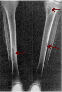
A pseudofracture, also called a Looser zone, is a diagnostic finding in osteomalacia. Pseudofracture also rarely occurs in Paget's disease of bone, hyperparathyroidism, renal osteodystrophy, osteogenesis imperfecta, fibrous dysplasia, and hypophosphatasia. Looser zones are named after Emil Looser, a Swiss physician.
Epiphysiodesis is a pediatric orthopedic surgery procedure that aims at altering or stopping the bone growth naturally occurring through the growth plate also known as the physeal plate. There are two types of epiphysiodesis: temporary hemiepiphysiodesis and permanent epiphysiodesis. Temporary hemiepiphysiodesis is also known as guided growth surgery or growth modulation surgery. Temporary hemiepiphysiodesis is reversible i.e. the metal implants used to achieve epiphysiodesis can be removed after the desired correction is achieved and the growth plate can thus resume its normal growth and function. In contrast, permanent epiphysiodesis is irreversible and the growth plate function cannot be restored after surgery. Both temporary hemiepiphysiodesis and permanent epiphysiodesis are used to treat a diverse array of pediatric orthopedic disorders but the exact indications for each procedure are different.
Calcium acetate/magnesium carbonate is a fixed-dose combination drug that contains 110 mg calcium and 60 mg magnesium ions and is indicated as a phosphate binder for dialysis patients with hyperphosphataemia. It is registered by Fresenius Medical Care under the trade names Renepho (Belgium) and OsvaRen.
Chronic kidney disease–mineral and bone disorder (CKD–MBD) is one of the many complications associated with chronic kidney disease. It represents a systemic disorder of mineral and bone metabolism due to CKD manifested by either one or a combination of the following: