
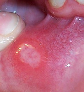
A mouth ulcer is an ulcer that occurs on the mucous membrane of the oral cavity. Mouth ulcers are very common, occurring in association with many diseases and by many different mechanisms, but usually there is no serious underlying cause.

Oral candidiasis, also known as oral thrush among other names, is candidiasis that occurs in the mouth. That is, oral candidiasis is a mycosis of Candida species on the mucous membranes of the mouth.

Lichen planus (LP) is a chronic inflammatory and immune-mediated disease that affects the skin, nails, hair, and mucous membranes. It is characterized by polygonal, flat-topped, violaceous papules and plaques with overlying, reticulated, fine white scale, commonly affecting dorsal hands, flexural wrists and forearms, trunk, anterior lower legs and oral mucosa. Although there is a broad clinical range of LP manifestations, the skin and oral cavity remain as the major sites of involvement. The cause is unknown, but it is thought to be the result of an autoimmune process with an unknown initial trigger. There is no cure, but many different medications and procedures have been used in efforts to control the symptoms.

Pemphigus is a rare group of blistering autoimmune diseases that affect the skin and mucous membranes. The name is derived from the Greek root "pemphix", meaning "pustule".
Aphthous stomatitis is a common condition characterized by the repeated formation of benign and non-contagious mouth ulcers (aphthae) in otherwise healthy individuals. The informal term canker sores is also used, mainly in North America, although this may also refer to any mouth ulcers.

Bullous pemphigoid is an autoimmune pruritic skin disease preferentially in older people, aged over 60, that may involve the formation of blisters (bullae) in the space between the epidermal and dermal skin layers. The disorder is a type of pemphigoid. It is classified as a type II hypersensitivity reaction, with the formation of anti-hemidesmosome antibodies.
The oral mucosa is the mucous membrane lining the inside of the mouth. It comprises stratified squamous epithelium, termed "oral epithelium", and an underlying connective tissue termed lamina propria. The oral cavity has sometimes been described as a mirror that reflects the health of the individual. Changes indicative of disease are seen as alterations in the oral mucosa lining the mouth, which can reveal systemic conditions, such as diabetes or vitamin deficiency, or the local effects of chronic tobacco or alcohol use. The oral mucosa tends to heal faster and with less scar formation compared to the skin. The underlying mechanism remains unknown, but research suggests that extracellular vesicles might be involved.
Nikolsky's sign is a clinical dermatological sign, named after Pyotr Nikolsky (1858–1940), a Russian physician who trained and worked in the Russian Empire. The sign is present when slight rubbing of the skin results in exfoliation of the outermost layer. A typical test would be to place the eraser of a pencil on the roof of a lesion and spin the pencil in a rolling motion between the thumb and forefinger. If the lesion is opened, then the Nikolsky's sign is present/positive.

Pemphigus vulgaris is a rare chronic blistering skin disease and the most common form of pemphigus. Pemphigus was derived from the Greek word pemphix, meaning blister. It is classified as a type II hypersensitivity reaction in which antibodies are formed against desmosomes, components of the skin that function to keep certain layers of skin bound to each other. As desmosomes are attacked, the layers of skin separate and the clinical picture resembles a blister. These blisters are due to acantholysis, or breaking apart of intercellular connections through an autoantibody-mediated response. Over time the condition inevitably progresses without treatment: lesions increase in size and distribution throughout the body, behaving physiologically like a severe burn.
Hailey–Hailey disease, or familial benign chronic pemphigus or familial benign pemphigus, was originally described by the Hailey brothers in 1939. It is a genetic disorder that causes blisters to form on the skin.
Pemphigoid is a group of rare autoimmune blistering diseases of the skin, and mucous membranes. As its name indicates, pemphigoid is similar in general appearance to pemphigus, but, unlike pemphigus, pemphigoid does not feature acantholysis, a loss of connections between skin cells.
Desquamative gingivitis is an erythematous (red), desquamatous (shedding) and ulcerated appearance of the gums. It is a descriptive term and can be caused by several different disorders.

Gestational pemphigoid (GP) is an autoimmune blistering skin disease of pregnancy, typically occurring in the second or third trimester. It was originally called herpes gestationis because of the blistering appearance, although it is not associated with the herpes virus. It is one of the pemphigoid (pemphigus-like) diseases.

Discoid lupus erythematosus is the most common type of chronic cutaneous lupus (CCLE), an autoimmune skin condition on the lupus erythematosus spectrum of illnesses. It presents with red, inflamed, coin-shaped patches of skin with a scaling and crusty appearance, most often on the scalp, cheeks, and ears. Hair loss may occur if the lesions are on the scalp. The lesions can then develop severe scarring, and the centre areas may appear lighter in color with a rim darker than the normal skin. These lesions can last for years without treatment.
Pemphigus foliaceus is an autoimmune blistering disease of the skin. Pemphigus foliaceus causes a characteristic inflammatory attack at the subcorneal layer of epidermis, which results in skin lesions that are scaly or crusted erosions with an erythematous (red) base. Mucosal involvement is absent even with widespread disease.
Paraneoplastic pemphigus (PNP) is an autoimmune disorder stemming from an underlying tumor. It is hypothesized that antigens associated with the tumor trigger an immune response resulting in blistering of the skin and mucous membranes.

Ernst H. Beutner was a German-born microbiologist who discovered the role of autoimmunity in pemphigus and pemphigoid using self-designed immunofluorescent methods. For this achievement, he is often regarded as the "Founder of Immunodermatology". He was the author or co-author of over 10 papers, which were each cited over 100 times.
Autoimmune skin disease in dogs are a group of diseases that occur in dogs that are caused by the body's immune system, where the body's white blood cells or body's antibodies attack its own tissues or extracellular protein of the skin.