DNA adenine methylase

Last updated
Site-specific DNA-methyltransferase (adenine-specific)
Identifiers
EC no. 2.1.1.72
CAS no. 69553-52-2
Databases
IntEnz IntEnz view
BRENDA BRENDA entry
ExPASy NiceZyme view
KEGG KEGG entry
MetaCyc metabolic pathway
PRIAM profile
PDB structures RCSB PDB PDBe PDBsum
Search
PMC articles
PubMed articles
NCBI proteins

DNA adenine methylase, (Dam) [1] (also site-specific DNA-methyltransferase (adenine-specific), EC 2.1.1.72, modification methylase, restriction-modification system) is an enzyme that adds a methyl group to the adenine of the sequence 5'-GATC-3' in newly synthesized DNA. [2] [3] Immediately after DNA synthesis, the daughter strand remains unmethylated for a short time. [4] It is an orphan methyltransferase that is not part of a restriction-modification system and regulates gene expression. [5] [6] [7] [8] This enzyme catalyses the following chemical reaction

Contents

S-adenosyl-L-methionine + DNA adenine S-adenosyl-L-homocysteine + DNA 6-methylaminopurine

This is a large group of enzymes unique to prokaryotes and bacteriophages. [9]

The E. coli DNA adenine methyltransferase enzyme (Dam), is widely used for the chromatin profiling technique DamID, in which the Dam is fused to a DNA-binding protein of interest and expressed as a transgene in a genetically tractable model organism to identify protein binding sites. [10]

Dam methylates adenine of GATC sites after replication. Hemimethylation.svg
Dam methylates adenine of GATC sites after replication.

Role in mismatch repair of DNA

When DNA polymerase makes an error resulting in a mismatched base-pair or a small insertion or deletion during DNA synthesis, the cell will repair the DNA by a pathway called mismatch repair. However, the cell must be able to differentiate between the template strand and the newly synthesized strand. In some bacteria, DNA strands are methylated by Dam methylase, and therefore, immediately after replication, the DNA will be hemimethylated. [4] A repair enzyme, MutS, binds to mismatches in DNA and recruits MutL, which subsequently activates the endonuclease MutH. MutH binds hemimethylated GATC sites and when activated will selectively cleave the unmethylated daughter strand, allowing helicase and exonucleases to excise the nascent strand in the region surrounding the mismatch. [4] [11] The strand is then re-synthesized by DNA polymerase III.

Role in regulation of replication

The firing of the origin of replication (oriC) in bacteria cells is highly controlled to ensure DNA replication occurs only once during each cell division. Part of this can be explained by the slow hydrolysis of ATP by DnaA, a protein that binds to repeats in the oriC to initiate replication. Dam methylase also plays a role because the oriC has 11 5'-GATC-3' sequences (in E. coli). Immediately after DNA replication, the oriC is hemimethylated and sequestered for a period of time. Only after this, the oriC is released and must be fully methylated by Dam methylase before DnaA binding occurs.

Role in regulation of protein expression

Dam also plays a role in the promotion and repression of RNA transcription. In E. coli downstream GATC sequences are methylated, promoting transcription. For example, pyelonephritis-associated pili (PAP) phase variation in uropathogenic E. coli is controlled by Dam through the methylation of the two GATC sites proximal and distal to the PAP promoter. [12] Given its role of protein regulation in E. coli, the Dam methylase gene is nonessential as a knockout of the gene still leaves the bacteria viable. [13] The retainment of viability despite a dam gene knockout is also seen in Salmonella and Aggregatibacter actinomycetemcomitans. [14] [15] However, in organisms like Vibrio cholerae and Yersinia pseudotuberculosis, the dam gene is essential for viability. [16] A knockout of the dam gene in Aggregatibacter actinomycetemcomitans resulted in dysregulated levels of the protein, leukotoxin, and also reduced the microbe's ability to invade oral epithelial cells. [15] Additionally a study on Dam methylase deficient Streptococcus mutans , a dental pathogen, revealed the dysregulation of a 103 genes some of which include cariogenic potential. [16]

Structural features

The similarity in the catalytic domains of C5-cytosine methyltransferases and N6 and N4-adenine methyltransferases provided great interest in understanding the basis for functional similarities and dissimilarities. The methyltransferases or methylases are classified into three groups (Groups α, β, and γ) based on the sequential order of certain 9 motifs and the Target Recognition Domain (TRD). [17] Motif I consists of a Gly-X-Gly tripeptide and is referred to as the G-loop and is implicated in the binding of the S-Adenosyl methionine cofactor. [18] Motif II is highly conserved among N4 and N6-adenine methylases and contains a negatively charged amino acid followed by a hydrophobic side chain in the last positions of the β2 strand to bind the AdoMet. [17] Motif III is also implicated in the binding of Adomet. Motif IV is especially important and well known in methylase characterizations. It consists of a diprolyl component and is highly conserved among N6-adenine methyltransferases as the DPPY motif, however, this motif can vary for N4-adenine and C5-cytosine methyltransferases. The DPPY motif has been found to be essential for AdoMet binding. [19] Motifs IV-VIII play a role in the catalytic activity, while motifs 1-III and X play a role in binding of the cofactor. For N6-adenine methylases the sequential order for these motifs is as such: N-terminal - X - I - II - III - TRD - IV - V - VI - VII - VIII - C-terminal and E. coli Dam methylase follows this structural sequence. [17] A 2015 crystallography experiment showed that E. coli Dam methylase was able to bind non-GATC DNA with the same sequence of motifs discussed; the authors posit that the obtained structure could serve as grounds for repression of transcription that is not methylation based. [20]

The X-ray crystal structure of E. coli Dam methylase shows the enzyme bound to double-stranded DNA and the inhibitor sinefungin. The adenine to be modified is shown as a blue stick flipped out of the double helix and towards the enzyme's interior. Dam Methylase.png
The X-ray crystal structure of E. coli Dam methylase shows the enzyme bound to double-stranded DNA and the inhibitor sinefungin. The adenine to be modified is shown as a blue stick flipped out of the double helix and towards the enzyme's interior.

Orphan bacterial and bacteriophage methylases

Dam methylase is an orphan methyltransferase that is not part of a restriction-modification system but operates independently to regulate gene expression, mismatch repair, and bacterial replication amongst many other functions. This is not the only example of an orphan methyltransferase as there exists the Cell cycle regulated Methyltransferase (CcrM) which methylates 5'-GANTC-'3 hemi-methylated DNA to control the life cycle of Caulobacter crescentus and other related species. [21]

Distinct from their bacterial counterparts, phage orphan methyltransferases also do exist and most notably in the T2, T4, and other T-even bacteriophages that infect E. coli. [5] In a study it was identified that despite sharing any sequence homology, the E. coli and T4 Dam methylase amino acids sequences share sequence identity of up to 64% in four regions of 11 to 33 residues long which suggests a common evolutionary origin for the bacterial and phage methylase genes. [22] The T2 and T4 methylases differ from E. coli Dam methylase in not only their ability to methylate 5-hydroxymethylcytosine but to also methylate non-canonical DNA sites. Despite extensive in vitro characterization of these select few phage orphan methyltransferases their biological purpose is still not clear. [5]

See also

Related Research Articles

A restriction enzyme, restriction endonuclease, REase, ENase orrestrictase is an enzyme that cleaves DNA into fragments at or near specific recognition sites within molecules known as restriction sites. Restriction enzymes are one class of the broader endonuclease group of enzymes. Restriction enzymes are commonly classified into five types, which differ in their structure and whether they cut their DNA substrate at their recognition site, or if the recognition and cleavage sites are separate from one another. To cut DNA, all restriction enzymes make two incisions, once through each sugar-phosphate backbone of the DNA double helix.

<span class="mw-page-title-main">DNA polymerase</span> Form of DNA replication

A DNA polymerase is a member of a family of enzymes that catalyze the synthesis of DNA molecules from nucleoside triphosphates, the molecular precursors of DNA. These enzymes are essential for DNA replication and usually work in groups to create two identical DNA duplexes from a single original DNA duplex. During this process, DNA polymerase "reads" the existing DNA strands to create two new strands that match the existing ones. These enzymes catalyze the chemical reaction

<span class="mw-page-title-main">DNA methyltransferase</span> Class of enzymes

In biochemistry, the DNA methyltransferase family of enzymes catalyze the transfer of a methyl group to DNA. DNA methylation serves a wide variety of biological functions. All the known DNA methyltransferases use S-adenosyl methionine (SAM) as the methyl donor.

<span class="mw-page-title-main">Nuclease</span> Class of enzymes which cleave nucleic acids

In biochemistry, a nuclease is an enzyme capable of cleaving the phosphodiester bonds between nucleotides of nucleic acids. Nucleases variously effect single and double stranded breaks in their target molecules. In living organisms, they are essential machinery for many aspects of DNA repair. Defects in certain nucleases can cause genetic instability or immunodeficiency. Nucleases are also extensively used in molecular cloning.

<span class="mw-page-title-main">DnaA</span>

DnaA is a protein that activates initiation of DNA replication in bacteria. Based on the Replicon Model, a positively active initiator molecule contacts with a particular spot on a circular chromosome called the replicator to start DNA replication. It is a replication initiation factor which promotes the unwinding of DNA at oriC. The DnaA proteins found in all bacteria engage with the DnaA boxes to start chromosomal replication. In addition to the DnaA protein, its concentration, binding to DnaA-boxes, and binding of ATP or ADP, we will cover the regulation of the DnaA gene, the unique characteristics of the DnaA gene expression, promoter strength, and translation efficiency. The onset of the initiation phase of DNA replication is determined by the concentration of DnaA. DnaA accumulates during growth and then triggers the initiation of replication. Replication begins with active DnaA binding to 9-mer (9-bp) repeats upstream of oriC. Binding of DnaA leads to strand separation at the 13-mer repeats. This binding causes the DNA to loop in preparation for melting open by the helicase DnaB.

DNA gyrase, or simply gyrase, is an enzyme within the class of topoisomerase and is a subclass of Type II topoisomerases that reduces topological strain in an ATP dependent manner while double-stranded DNA is being unwound by elongating RNA-polymerase or by helicase in front of the progressing replication fork. It is the only known enzyme to actively contribute negative supercoiling to DNA, while it also is capable of relaxing positive supercoils. It does so by looping the template to form a crossing, then cutting one of the double helices and passing the other through it before releasing the break, changing the linking number by two in each enzymatic step. This process occurs in bacteria, whose single circular DNA is cut by DNA gyrase and the two ends are then twisted around each other to form supercoils. Gyrase is also found in eukaryotic plastids: it has been found in the apicoplast of the malarial parasite Plasmodium falciparum and in chloroplasts of several plants. Bacterial DNA gyrase is the target of many antibiotics, including nalidixic acid, novobiocin, albicidin, and ciprofloxacin.

<span class="mw-page-title-main">DNA methylation</span> Biological process

DNA methylation is a biological process by which methyl groups are added to the DNA molecule. Methylation can change the activity of a DNA segment without changing the sequence. When located in a gene promoter, DNA methylation typically acts to repress gene transcription. In mammals, DNA methylation is essential for normal development and is associated with a number of key processes including genomic imprinting, X-chromosome inactivation, repression of transposable elements, aging, and carcinogenesis.

In molecular biology, endonucleases are enzymes that cleave the phosphodiester bond within a polynucleotide chain. Some, such as deoxyribonuclease I, cut DNA relatively nonspecifically, while many, typically called restriction endonucleases or restriction enzymes, cleave only at very specific nucleotide sequences. Endonucleases differ from exonucleases, which cleave the ends of recognition sequences instead of the middle (endo) portion. Some enzymes known as "exo-endonucleases", however, are not limited to either nuclease function, displaying qualities that are both endo- and exo-like. Evidence suggests that endonuclease activity experiences a lag compared to exonuclease activity.

<span class="mw-page-title-main">Methyltransferase</span> Group of methylating enzymes

Methyltransferases are a large group of enzymes that all methylate their substrates but can be split into several subclasses based on their structural features. The most common class of methyltransferases is class I, all of which contain a Rossmann fold for binding S-Adenosyl methionine (SAM). Class II methyltransferases contain a SET domain, which are exemplified by SET domain histone methyltransferases, and class III methyltransferases, which are membrane associated. Methyltransferases can also be grouped as different types utilizing different substrates in methyl transfer reactions. These types include protein methyltransferases, DNA/RNA methyltransferases, natural product methyltransferases, and non-SAM dependent methyltransferases. SAM is the classical methyl donor for methyltransferases, however, examples of other methyl donors are seen in nature. The general mechanism for methyl transfer is a SN2-like nucleophilic attack where the methionine sulfur serves as the leaving group and the methyl group attached to it acts as the electrophile that transfers the methyl group to the enzyme substrate. SAM is converted to S-Adenosyl homocysteine (SAH) during this process. The breaking of the SAM-methyl bond and the formation of the substrate-methyl bond happen nearly simultaneously. These enzymatic reactions are found in many pathways and are implicated in genetic diseases, cancer, and metabolic diseases. Another type of methyl transfer is the radical S-Adenosyl methionine (SAM) which is the methylation of unactivated carbon atoms in primary metabolites, proteins, lipids, and RNA.

P1 is a temperate bacteriophage that infects Escherichia coli and some other bacteria. When undergoing a lysogenic cycle the phage genome exists as a plasmid in the bacterium unlike other phages that integrate into the host DNA. P1 has an icosahedral head containing the DNA attached to a contractile tail with six tail fibers. The P1 phage has gained research interest because it can be used to transfer DNA from one bacterial cell to another in a process known as transduction. As it replicates during its lytic cycle it captures fragments of the host chromosome. If the resulting viral particles are used to infect a different host the captured DNA fragments can be integrated into the new host's genome. This method of in vivo genetic engineering was widely used for many years and is still used today, though to a lesser extent. P1 can also be used to create the P1-derived artificial chromosome cloning vector which can carry relatively large fragments of DNA. P1 encodes a site-specific recombinase, Cre, that is widely used to carry out cell-specific or time-specific DNA recombination by flanking the target DNA with loxP sites.

<span class="mw-page-title-main">Prokaryotic DNA replication</span> DNA Replication in prokaryotes

Prokaryotic DNA Replication is the process by which a prokaryote duplicates its DNA into another copy that is passed on to daughter cells. Although it is often studied in the model organism E. coli, other bacteria show many similarities. Replication is bi-directional and originates at a single origin of replication (OriC). It consists of three steps: Initiation, elongation, and termination.

Deoxyribonuclease IV (phage-T4-induced) is catalyzes the degradation nucleotides in DsDNA by attacking the 5'-terminal end.

<span class="mw-page-title-main">Protein-glutamate O-methyltransferase</span>

In enzymology, a protein-glutamate O-methyltransferase is an enzyme that catalyzes the chemical reaction

In biology, phase variation is a method for dealing with rapidly varying environments without requiring random mutation. It involves the variation of protein expression, frequently in an on-off fashion, within different parts of a bacterial population. As such the phenotype can switch at frequencies that are much higher than classical mutation rates. Phase variation contributes to virulence by generating heterogeneity. Although it has been most commonly studied in the context of immune evasion, it is observed in many other areas as well and is employed by various types of bacteria, including Salmonella species.

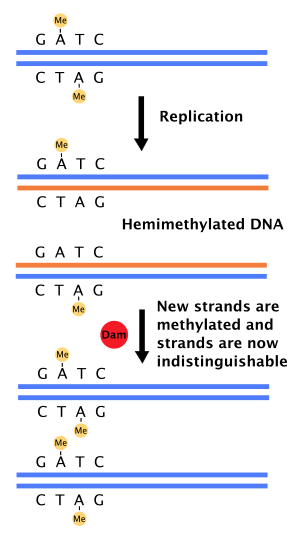
DNA adenine methyltransferase identification, often abbreviated DamID, is a molecular biology protocol used to map the binding sites of DNA- and chromatin-binding proteins in eukaryotes. DamID identifies binding sites by expressing the proposed DNA-binding protein as a fusion protein with DNA methyltransferase. Binding of the protein of interest to DNA localizes the methyltransferase in the region of the binding site. Adenine methylation does not occur naturally in eukaryotes and therefore adenine methylation in any region can be concluded to have been caused by the fusion protein, implying the region is located near a binding site. DamID is an alternate method to ChIP-on-chip or ChIP-seq.

<span class="mw-page-title-main">Circular chromosome</span> Type of chromosome

A circular chromosome is a chromosome in bacteria, archaea, mitochondria, and chloroplasts, in the form of a molecule of circular DNA, unlike the linear chromosome of most eukaryotes.

<span class="mw-page-title-main">SeqA protein</span>

In molecular biology the SeqA protein is found in bacteria and archaea. The function of this protein is highly important in DNA replication. The protein negatively regulates the initiation of DNA replication at the origin of replication, in Escherichia coli, OriC. Additionally the protein plays a further role in sequestration. The importance of this protein is vital, without its help in DNA replication, cell division and other crucial processes could not occur. This protein domain is thought to be part of a much larger protein complex which includes other proteins such as SeqB.

16S rRNA (adenine1518-N6/adenine1519-N6)-dimethyltransferase (EC 2.1.1.182, S-adenosylmethionine-6-N',N'-adenosyl (rRNA) dimethyltransferase, KsgA, ksgA methyltransferase) is an enzyme with systematic name S-adenosyl-L-methionine:16S rRNA (adenine1518-N6/adenine1519-N6)-dimethyltransferase. This enzyme catalyses the following chemical reaction

23S rRNA (guanine745-N1)-methyltransferase (EC 2.1.1.187, Rlma(I), Rlma1, 23S rRNA m1G745 methyltransferase, YebH, RlmAI methyltransferase, ribosomal RNA(m1G)-methylase, rRNA(m1G)methylase, RrmA, 23S rRNA:m1G745 methyltransferase) is an enzyme with systematic name S-adenosyl-L-methionine:23S rRNA (guanine745-N1)-methyltransferase. This enzyme catalyses the following chemical reaction

<span class="mw-page-title-main">Cell cycle regulated Methyltransferase</span> Bacterial enzyme

CcrM is an orphan DNA methyltransferase, that is involved in controlling gene expression in most Alphaproteobacteria. This enzyme modifies DNA by catalyzing the transference of a methyl group from the S-adenosyl-L methionine substrate to the N6 position of an adenine base in the sequence 5'-GANTC-3' with high specificity. In some lineages such as SAR11, the homologous enzymes possess 5'-GAWTC-3' specificity. In Caulobacter crescentus Ccrm is produced at the end of the replication cycle when Ccrm recognition sites are hemimethylated, rapidly methylating the DNA. CcrM is essential in other Alphaproteobacteria but is role is not yet determined. CcrM is a highly specific methyltransferase with a novel DNA recognition mechanism.

References

  1. Brown, Terence (2002). "Chapter 14: Mutation, Repair and Recombination. Section 2.3". Genomes. Garland Science. ISBN   0-471-25046-5.
  2. Marinus MG, Morris NR (June 1973). "Isolation of deoxyribonucleic acid methylase mutants of Escherichia coli K-12". Journal of Bacteriology. 114 (3): 1143–50. doi:10.1128/JB.114.3.1143-1150.1973. PMC   285375 . PMID   4576399.
  3. Geier GE, Modrich P (February 1979). "Recognition sequence of the dam methylase of Escherichia coli K12 and mode of cleavage of Dpn I endonuclease". The Journal of Biological Chemistry. 254 (4): 1408–13. PMID   368070.
  4. 1 2 3 Barras F, Marinus MG (1989). "The great GATC: DNA methylation in E. coli". Trends in Genetics. 5 (5): 139–143. doi:10.1016/0168-9525(89)90054-1. PMID   2667217.
  5. 1 2 3 Murphy J, Mahony J, Ainsworth S, Nauta A, van Sinderen D (December 2013). "Bacteriophage orphan DNA methyltransferases: insights from their bacterial origin, function, and occurrence". Applied and Environmental Microbiology. 79 (24): 7547–55. doi:10.1128/aem.02229-13. PMC   3837797 . PMID   24123737.
  6. Kessler C, Manta V (August 1990). "Specificity of restriction endonucleases and DNA modification methyltransferases a review (Edition 3)". Gene. 92 (1–2): 1–248. doi:10.1016/0378-1119(90)90486-B. PMID   2172084.
  7. Roberts RJ (April 1990). "Restriction enzymes and their isoschizomers". Nucleic Acids Research. 18 Suppl: 2331–65. doi:10.1093/nar/18.suppl.2331. PMC   331877 . PMID   2159140.
  8. Yuan R (1981). "Structure and mechanism of multifunctional restriction endonucleases". Annual Review of Biochemistry. 50: 285–319. doi:10.1146/annurev.bi.50.070181.001441. PMID   6267988.
  9. Roberts RJ, Macelis D (eds.). "The Restriction Enzyme Database". REBASE. Retrieved 22 Feb 2020.
  10. Aughey GN, Southall TD (January 2016). "Dam it's good! DamID profiling of protein-DNA interactions". Wiley Interdisciplinary Reviews. Developmental Biology. 5 (1): 25–37. doi:10.1002/wdev.205. PMC   4737221 . PMID   26383089.
  11. Løbner-Olesen A, Skovgaard O, Marinus MG (April 2005). "Dam methylation: coordinating cellular processes". Current Opinion in Microbiology. 8 (2): 154–60. doi:10.1016/j.mib.2005.02.009. PMID   15802246.
  12. Casadesús J, Low D (September 2006). "Epigenetic gene regulation in the bacterial world". Microbiology and Molecular Biology Reviews. 70 (3): 830–56. doi:10.1128/MMBR.00016-06. PMC   1594586 . PMID   16959970.
  13. Bale A, d'Alarcao M, Marinus MG (February 1979). "Characterization of DNA adenine methylation mutants of Escherichia coli K12". Mutation Research. 59 (2): 157–65. doi:10.1016/0027-5107(79)90153-2. PMID   375073.
  14. Nicholson B, Low D (February 2000). "DNA methylation-dependent regulation of pef expression in Salmonella typhimurium". Molecular Microbiology. 35 (4): 728–42. doi:10.1046/j.1365-2958.2000.01743.x. PMID   10692151.
  15. 1 2 Wu H, Lippmann JE, Oza JP, Zeng M, Fives-Taylor P, Reich NO (August 2006). "Inactivation of DNA adenine methyltransferase alters virulence factors in Actinobacillus actinomycetemcomitans". Oral Microbiology and Immunology. 21 (4): 238–44. doi:10.1111/j.1399-302x.2006.00284.x. PMID   16842508.
  16. 1 2 Julio SM, Heithoff DM, Provenzano D, Klose KE, Sinsheimer RL, Low DA, Mahan MJ (December 2001). "DNA adenine methylase is essential for viability and plays a role in the pathogenesis of Yersinia pseudotuberculosis and Vibrio cholerae". Infection and Immunity. 69 (12): 7610–5. doi:10.1128/iai.69.12.7610-7615.2001. PMC   98854 . PMID   11705940.
  17. 1 2 3 Malone T, Blumenthal RM, Cheng X (November 1995). "Structure-guided analysis reveals nine sequence motifs conserved among DNA amino-methyltransferases, and suggests a catalytic mechanism for these enzymes". Journal of Molecular Biology. 253 (4): 618–32. doi:10.1006/jmbi.1995.0577. PMID   7473738.
  18. Schluckebier G, O'Gara M, Saenger W, Cheng X (March 1995). "Universal catalytic domain structure of AdoMet-dependent methyltransferases". Journal of Molecular Biology. 247 (1): 16–20. doi:10.1006/jmbi.1994.0117. PMID   7897657.
  19. Kossykh VG, Schlagman SL, Hattman S (July 1993). "Conserved sequence motif DPPY in region IV of the phage T4 Dam DNA-[N-adenine]-methyltransferase is important for S-adenosyl-L-methionine binding". Nucleic Acids Research. 21 (15): 3563–6. doi:10.1093/nar/21.15.3563. PMC   331459 . PMID   16617501.
  20. Horton JR, Zhang X, Blumenthal RM, Cheng X (April 2015). "Structures of Escherichia coli DNA adenine methyltransferase (Dam) in complex with a non-GATC sequence: potential implications for methylation-independent transcriptional repression". Nucleic Acids Research. 43 (8): 4296–308. doi:10.1093/nar/gkv251. PMC   4417163 . PMID   25845600.
  21. Zweiger G, Marczynski G, Shapiro L (January 1994). "A Caulobacter DNA methyltransferase that functions only in the predivisional cell". Journal of Molecular Biology. 235 (2): 472–85. doi:10.1006/jmbi.1994.1007. PMID   8289276.
  22. Hattman S, Wilkinson J, Swinton D, Schlagman S, Macdonald PM, Mosig G (November 1985). "Common evolutionary origin of the phage T4 dam and host Escherichia coli dam DNA-adenine methyltransferase genes". Journal of Bacteriology. 164 (2): 932–7. doi:10.1128/JB.164.2.932-937.1985. PMC   214344 . PMID   3902803.