Emanuel syndrome

Last updated
Emanuel syndrome
Other namesderivative 22 syndrome, der(22) syndrome
Human male karyotpe high resolution - Chromosome 11 cropped.png Human male karyotpe high resolution - Chromosome 22 cropped.png
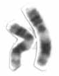
Chromosome 11 (left) and 22 (right) are both involved in causing the syndrome, due to extra genetic material.

Emanuel syndrome, also known as derivative 22 syndrome, or der(22) syndrome, is a rare disorder associated with multiple congenital anomalies, including profound intellectual disability, preauricular skin tags or pits, and conotruncal heart defects. [1] [2] It can occur in offspring of carriers of the constitutional chromosomal translocation t(11;22)(q23;q11), owing to a 3:1 meiotic malsegregation event resulting in partial trisomy of chromosomes 11 and 22. An unbalanced translocation between chromosomes 11 and 22 is described as Emanuel syndrome. It was first described in 1980 by American medical researchers Beverly S. Emanuel and Elaine H. Zackai, and a consortium of European scientists the same year. [3] [4]

Contents

Sign and symptoms

Microcephaly Microcephaly-comparison-500px.jpg
Microcephaly

Infants with Emanuel syndrome have weak muscle tone (hypotonia) and fail to gain weight and grow at the expected rate (failure to thrive). [1] Their development is significantly delayed, and most affected individuals have severe to profound intellectual disability. Other features of Emanuel syndrome include an unusually small head (microcephaly), distinctive facial features, and a small lower jaw (micrognathia). Ear abnormalities are common, including small holes in the skin just in front of the ears (preauricular pits, or sinuses). The external auricle of the ear is typically malformed, with possibly severe microtia with atresia of the external auditory canal present. Deafness may occur, but severe hearing loss is uncommon; the occurrence of mild forms of hearing loss may be underestimated due to difficulties associated with accurately evaluating hearing in individuals with intellectual disabilities. About half of all affected infants are born with an opening in the roof of the mouth (cleft palate) or a high arched palate. Males with Emanuel syndrome often have genital abnormalities. Additional signs of this condition can include heart defects [5] and absent or unusually small (hypoplastic) kidneys; [3] these problems can be life-threatening in infancy or childhood. [1]

Causes

Emanuel syndrome is an inherited chromosome abnormality. It is caused by the presence of extra genetic material from chromosome 11 and chromosome 22 in each cell. In addition to the usual 46 chromosomes, people with Emanuel syndrome have an extra (supernumerary) chromosome consisting of a piece of chromosome 11 attached to a piece of chromosome 22. The extra chromosome is known as a derivative 22, or der(22), chromosome. [6]

Genetics

People with Emanuel syndrome typically inherit the der(22) chromosome from an unaffected parent. The parent carries a chromosomal rearrangement between chromosomes 11 and 22 called a balanced translocation. No genetic material is gained or lost in a balanced translocation, so these chromosomal changes usually do not cause any health problems. As this translocation is passed to the next generation, it can become unbalanced. Individuals with Emanuel syndrome inherit an unbalanced translocation between chromosomes 11 and 22 in the form of a der(22) chromosome. (This der(22) chromosome is classified as one of the small supernumerary marker chromosomes. [7] ) These individuals have two normal copies of chromosome 11, two normal copies of chromosome 22 and extra genetic material from the der(22) chromosome. As a result of the extra chromosome, people with Emanuel syndrome have three copies of some genes in each cell instead of the usual two copies. The excess genetic material disrupts the normal course of development, leading to intellectual disability and birth defects. Researchers are working to determine which genes are included on the der(22) chromosome and what role these genes play in development. [6]

Diagnosis

Emanuel syndrome can be diagnosed with a karyotype, fluorescence in situ hybridization or a chromosomal microarray analysis. [3]

Treatment

Emanuel syndrome has no known cure, but individual symptoms may be treated. Assessments of individual systems, such as the cardiovascular, gastrointestinal, orthopedic, and neurological, may be necessary to determine the extent of impairment and options for treatment.[ citation needed ]

See also

Related Research Articles

Miller–Dieker syndrome, Miller–Dieker lissencephaly syndrome (MDLS), and chromosome 17p13.3 deletion syndrome is a micro deletion syndrome characterized by congenital malformations. Congenital malformations are physical defects detectable in an infant at birth which can involve many different parts of the body including the brain, hearts, lungs, liver, bones, or intestinal tract. MDS is a contiguous gene syndrome – a disorder due to the deletion of multiple gene loci adjacent to one another. The disorder arises from the deletion of part of the small arm of chromosome 17p, leading to partial monosomy. There may be unbalanced translocations, or the presence of a ring chromosome 17.

<span class="mw-page-title-main">Noonan syndrome</span> Genetic condition involving facial, heart, blood and skeletal features

Noonan syndrome (NS) is a genetic disorder that may present with mildly unusual facial features, short height, congenital heart disease, bleeding problems, and skeletal malformations. Facial features include widely spaced eyes, light-colored eyes, low-set ears, a short neck, and a small lower jaw. Heart problems may include pulmonary valve stenosis. The breast bone may either protrude or be sunken, while the spine may be abnormally curved. Intelligence is often normal. Complications of NS can include leukemia.

Jacobsen syndrome is a rare chromosomal disorder resulting from deletion of genes from chromosome 11 that includes band 11q24.1. It is a congenital disorder. Since the deletion takes place on the q arm of chromosome 11, it is also called 11q terminal deletion disorder. The deletion may range from 5 million to 16 million deleted DNA base pairs. The severity of symptoms depends on the number of deletions; the more deletions there are, the more severe the symptoms are likely to be.

<span class="mw-page-title-main">Small supernumerary marker chromosome</span> Abnormal partial or mixed chromosome

A small supernumerary marker chromosome (sSMC) is an abnormal extra chromosome. It contains copies of parts of one or more normal chromosomes and like normal chromosomes is located in the cell's nucleus, is replicated and distributed into each daughter cell during cell division, and typically has genes which may be expressed. However, it may also be active in causing birth defects and neoplasms. The sSMC's small size makes it virtually undetectable using classical cytogenetic methods: the far larger DNA and gene content of the cell's normal chromosomes obscures those of the sSMC. Newer molecular techniques such as fluorescence in situ hybridization, next generation sequencing, comparative genomic hybridization, and highly specialized cytogenetic G banding analyses are required to study it. Using these methods, the DNA sequences and genes in sSMCs are identified and help define as well as explain any effect(s) it may have on individuals.

<span class="mw-page-title-main">Chromosome 22</span> Human chromosome

Chromosome 22 is one of the 23 pairs of chromosomes in human cells. Humans normally have two copies of chromosome 22 in each cell. Chromosome 22 is the second smallest human chromosome, spanning about 51 million DNA base pairs and representing between 1.5 and 2% of the total DNA in cells.

<span class="mw-page-title-main">Greig cephalopolysyndactyly syndrome</span> Medical condition

Greig cephalopolysyndactyly syndrome is a disorder that affects development of the limbs, head, and face. The features of this syndrome are highly variable, ranging from very mild to severe. People with this condition typically have one or more extra fingers or toes (polydactyly) or an abnormally wide thumb or big toe (hallux).

The Pallister–Killian syndrome (PKS), also termed tetrasomy 12p mosaicism or the Pallister mosaic aneuploidy syndrome, is an extremely rare and severe genetic disorder. PKS is due to the presence of an extra and abnormal chromosome termed a small supernumerary marker chromosome (sSMC). sSMCs contain copies of genetic material from parts of virtually any other chromosome and, depending on the genetic material they carry, can cause various genetic disorders and neoplasms. The sSMC in PKS consists of multiple copies of the short arm of chromosome 12. Consequently, the multiple copies of the genetic material in the sSMC plus the two copies of this genetic material in the two normal chromosome 12's are overexpressed and thereby cause the syndrome. Due to a form of genetic mosaicism, however, individuals with PKS differ in the tissue distributions of their sSMC and therefore show different syndrome-related birth defects and disease severities. For example, individuals with the sSMC in their heart tissue are likely to have cardiac structural abnormalities while those without this sSMC localization have a structurally normal heart.

<span class="mw-page-title-main">1p36 deletion syndrome</span> Medical condition

1p36 deletion syndrome is a congenital genetic disorder characterized by moderate to severe intellectual disability, delayed growth, hypotonia, seizures, limited speech ability, malformations, hearing and vision impairment, and distinct facial features. The symptoms may vary, depending on the exact location of the chromosomal deletion.

<span class="mw-page-title-main">Rhizomelic chondrodysplasia punctata</span> Recessive genetic condition

Rhizomelic chondrodysplasia punctata is a rare developmental brain disorder characterized by abnormally short arms and legs (rhizomelia), seizures, recurrent respiratory tract infections and congenital cataracts.

<span class="mw-page-title-main">Isodicentric 15</span> Condition caused by two joined and mirrored duplications of part of chromosome 15

Isodicentric 15, also called marker chromosome 15 syndrome, idic(15), partial tetrasomy 15q, or inverted duplication 15, is a chromosome abnormality in which a child is born with extra genetic material from chromosome 15. People with idic(15) are typically born with 47 chromosomes in their body cells, instead of the normal 46. The extra chromosome, which is classified as a small supernumerary marker chromosome, is made up of a piece of chromosome 15 that has been duplicated end-to-end like a mirror image. It is the presence of this extra genetic material that is thought to account for the symptoms seen in some people with idic(15). Individuals with idic(15) have a total of four copies of this chromosome 15 region instead of the usual two copies. The term isodicentric refers to a duplication and inversion of a centromere-containing chromosomal segment.

<span class="mw-page-title-main">Branchio-oto-renal syndrome</span> Medical condition

Branchio-oto-renal syndrome (BOR) is an autosomal dominant genetic disorder involving the kidneys, ears, and neck. It is also known as Melnick-Fraser syndrome.

A marker chromosome (mar) is a small fragment of a chromosome which generally cannot be identified without specialized genomic analysis due to the size of the fragment. The significance of a marker is variable as it depends on what material is contained within the marker. The large majority of these marker chromosomes are smaller than one of the smaller human chromosomes, chromosome 20, and by definition are termed small supernumerary marker chromosomes.

<span class="mw-page-title-main">Tetrasomy 18p</span> Presence of four copies of the short arm of chromosome 18

Tetrasomy 18p is a genetic condition that is caused by the presence of an isochromosome composed of two copies of the short arm of chromosome 18 in addition to the two normal copies of the chromosome. It is characterized by multiple medical and developmental concerns.

<span class="mw-page-title-main">Cat eye syndrome</span> Genetic partial duplication of chromosome 22

Cat-eye syndrome (CES) or Schmid–Fraccaro syndrome is a rare condition caused by an abnormal extra chromosome, i.e. a small supernumerary marker chromosome. This chromosome consists of the entire short arm and a small section of the long arm of chromosome 22. In consequence, individuals with the cat-eye syndrome have three (trisomic) or four (tetrasomic) copies of the genetic material contained in the abnormal chromosome instead of the normal two copies. The prognosis for patients with CES varies depending on the severity of the condition and their associated signs and symptoms, especially when heart or kidney abnormalities are seen.

<span class="mw-page-title-main">Trisomy 22</span> Medical condition

Trisomy 22 is a chromosomal disorder in which three copies of chromosome 22 are present rather than two. It is a frequent cause of spontaneous abortion during the first trimester of pregnancy. Progression to the second trimester and live birth are rare. This disorder is found in individuals with an extra copy or a variation of chromosome 22 in some or all cells of their bodies.

<span class="mw-page-title-main">Tetrasomy 9p</span> Presence of four copies of the short arm of chromosome 9

Tetrasomy 9p is a rare chromosomal disorder characterized by the presence of two extra copies of the short arm of chromosome 9, in addition to the usual two. Symptoms of tetrasomy 9p vary widely among affected individuals but typically include varying degrees of delayed growth, abnormal facial features and intellectual disability. Symptoms of the disorder are comparable to those of trisomy 9p.

<span class="mw-page-title-main">FG syndrome</span> Rare genetic disease

FG syndrome (FGS) is a rare genetic syndrome caused by one or more recessive genes located on the X chromosome and causing physical anomalies and developmental delays. FG syndrome was named after the first letters of the surnames of the first patients noted with the disease. First reported by American geneticists John M. Opitz and Elisabeth G. Kaveggia in 1974, its major clinical features include intellectual disability, hyperactivity, hypotonia, and a characteristic facial appearance including macrocephaly.

Genitopatellar syndrome is a rare disorder consisting of congenital flexion contractures of the lower extremities, abnormal or missing patellae, and urogenital anomalies. Additional symptoms include microcephaly, severe psychomotor disability. In 2012, it was shown that mutations in the gene KAT6B cause the syndrome. Genitopatellar syndrome (GTPTS) can be caused by heterozygous mutation in the KAT6B gene on chromosome 10q22. The Say-Barber-Biesecker variant of Ohdo syndrome, which has many overlapping features with GTPTS, can also be caused by heterozygous mutation in the KAT6B gene.

<span class="mw-page-title-main">Okamoto syndrome</span> Rare genetic condition involving urinary, heart, facial and neurological features

Okamoto syndrome (OS), also known as Au–Kline syndrome (AKS), is a very rare autosomal dominant genetic condition characterised by congenital hydronephrosis, low muscle tone, heart defects, intellectual disability and characteristic facial features. Those affected often have neurological and skeletal abnormalities, as well as frequent urinary tract infections. Language and walking are usually delayed. Facial features include prominent, downturned ears, an open, downturned mouth and drooping eyelids (ptosis).

<span class="mw-page-title-main">CDK13-related disorder</span> Rare genetic disorder involving heart, facial and neurological features

CDK13-related disorder, also known as congenital heart defects, dysmorphic facial features and intellectual developmental disorder (CHDFIDD), is a very rare autosomal dominant genetic condition characterised by congenital heart defects, intellectual disability and characteristic facial features. Those affected typically have motor and language delays, low muscle tone and gastrointestinal dysmotility. Facial features include a wide nasal bridge, widely-spaced eyes, prominent, low-set ears, a flat nose tip and a small mouth. Less common features include congenital spinal abnormalities, hearing loss or seizures.

References

  1. 1 2 3 Reference, Genetics Home. "Emanuel syndrome". Genetics Home Reference. Retrieved 2017-02-24.
  2. "Emanuel syndrome | Genetic and Rare Diseases Information Center (GARD) – an NCATS Program". rarediseases.info.nih.gov. Retrieved 2017-02-25.
  3. 1 2 3 Emanuel, Beverly S.; Zackai, Elaine H.; Medne, Livija (1993). "Emanuel Syndrome". In Pagon, Roberta A.; Adam, Margaret P.; Ardinger, Holly H.; Wallace, Stephanie E.; Amemiya, Anne; Bean, Lora J.H.; Bird, Thomas D.; Ledbetter, Nikki; Mefford, Heather C. (eds.). GeneReviews. Seattle (WA): University of Washington, Seattle. PMID   20301440. NBK1263.
  4. "What is Emanuel Syndrome?". www.emanuelsyndrome.org. Archived from the original on 2020-08-12. Retrieved 2018-03-05.
  5. McDonald-McGinn DM, Hain HS, Emanuel BS, et al. (1993–2020). "22q11.2 Deletion Syndrome". In Adam MP, Ardinger HH, Pagon RA, et al. (eds.). GeneReviews. University of Washington. PMID   20301696. NBK1523.
  6. 1 2 "Chromosome 22". Genetics Home Reference. U.S. National Library of Medicine. 2016.
  7. Jafari-Ghahfarokhi H, Moradi-Chaleshtori M, Liehr T, Hashemzadeh-Chaleshtori M, Teimori H, Ghasemi-Dehkordi P (2015). "Small supernumerary marker chromosomes and their correlation with specific syndromes". Advanced Biomedical Research. 4: 140. doi: 10.4103/2277-9175.161542 . PMC   4544121 . PMID   26322288.

Further reading