Galactose-1-phosphate uridylyltransferase deficiency

Last updated
Galactose-1-phosphate uridylyltransferase deficiency
Other namesGalactosemia type 1, Classic galactosemia or GALT deficiency
Beta-D-Galactopyranose.svg
Galactose
Specialty Endocrinology   OOjs UI icon edit-ltr-progressive.svg

Galactose-1-phosphate uridylyltransferase deficiency(classic galactosemia) is the most common type of galactosemia, an inborn error of galactose metabolism, caused by a deficiency of the enzyme galactose-1-phosphate uridylyltransferase. [1] It is an autosomal recessive metabolic disorder that can cause liver disease and death if untreated. Treatment of galactosemia is most successful if initiated early and includes dietary restriction of lactose intake. Because early intervention is key, galactosemia is included in newborn screening programs in many areas. On initial screening, which often involves measuring the concentration of galactose in blood, classic galactosemia may be indistinguishable from other inborn errors of galactose metabolism, including galactokinase deficiency and galactose epimerase deficiency. Further analysis of metabolites and enzyme activities are needed to identify the specific metabolic error.

Contents

Symptoms and signs

In undiagnosed and untreated children, the accumulation of precursor metabolites due to the deficient activity of galactose 1-phosphate uridylyltransferase (GALT) can lead to feeding problems, failure to thrive, liver damage, bleeding, and infections. The first presenting symptom in an infant is often prolonged jaundice. Without intervention in the form of galactose restriction, infants can develop hyperammonemia and sepsis, possibly leading to shock. The accumulation of galactitol and subsequent osmotic swelling can lead to cataracts which are similar to those seen in galactokinase deficiency. [2] Long-term consequences of continued galactose intake can include developmental delay, developmental verbal dyspraxia, and motor abnormalities. Galactosemic females frequently suffer from ovarian failure, regardless of treatment in the form of galactose restriction. [2]

Cause

Lactose is a disaccharide consisting of glucose and galactose. After the ingestion of lactose, most commonly from breast milk for an infant or cow milk and any milk from an animal, the enzyme lactase hydrolyzes the sugar into its monosaccharide constituents, glucose and galactose. In the first step of galactose metabolism, galactose is converted to galactose-1-phosphate (Gal-1-P) by the enzyme galactokinase. Gal-1-P is converted to uridine diphosphate galactose (UDP-galactose) by the enzyme galactose-1-phosphate uridylyltransferase, with UDP-glucose acting as the UDP donor. UDP-galactose can then be converted to lactose, by the enzyme lactose synthase or to UDP-glucose by UDP-galactose epimerase (GALE). [3]

Normal metabolic pathway for galactose in humans Galactose metabolism.png
Normal metabolic pathway for galactose in humans

In classic galactosemia, galactose-1-phosphate uridylyltransferase activity is reduced or absent; leading to an accumulation of the precursors, galactose, galactitol, and Gal-1-P. [3] The elevation of precursors can be used to differentiate GALT deficiency from galactokinase deficiency, as Gal-1-P is typically not elevated in galactokinase deficiency.

If the activity of the enzyme galactose 1-phosphate uridyltransferase is decreased, there is a buildup of the precursors in the pathway, mainly galactose 1-phosphate and galactose. Galactose Metabolism GALT.png
If the activity of the enzyme galactose 1-phosphate uridyltransferase is decreased, there is a buildup of the precursors in the pathway, mainly galactose 1-phosphate and galactose.

Genetics

Classic galactosemia has an autosomal recessive pattern of inheritance. Autorecessive.svg
Classic galactosemia has an autosomal recessive pattern of inheritance.

All forms of galactosemia are inherited in an autosomal recessive manner, meaning individuals affected with classic galactosemia must have inherited a mutated copy of the GALT gene from both parents. Each child from two carrier parents would have a 25% chance of being affected, a 50% chance of being a carrier, and a 25% chance of inheriting normal versions of the gene from each parent.[ citation needed ]

There are several variants in the GALT gene, which have different levels of residual enzyme activity. A patient homozygous for one of the severe mutations in the GALT gene (commonly referred to as G/G) will typically have less than 5% of the enzyme activity expected in an unaffected patient. [2] Duarte galactosemia is caused by mutations that produce an unstable form of the GALT enzyme, with reduced promoter expression. Patients who are homozygous for Duarte mutations (D/D) will have reduced levels of enzyme activity compared to normal controls, but can often maintain a normal diet. Compound heterozygotes (D/G) will often be detected by newborn screening and treatment is based on the extent of residual enzyme activity. [2]

Diagnosis

In most regions, galactosemia is diagnosed as a result of newborn screening, most commonly by determining the concentration of galactose in a dried blood spot. Some regions will perform a second-tier test of GALT enzyme activity on samples with elevated galactose, while others perform both GALT and galactose measurements. While awaiting confirmatory testing for classic galactosemia, the infant is typically fed a soy-based formula, as human and cow milk contains galactose as a component of lactose. [4] Confirmatory testing would include measurement of enzyme activity in red blood cells, determination of Gal-1-P levels in the blood, and mutation testing. The differential diagnosis for elevated galactose concentrations in blood on a newborn screening result can include other disorders of galactose metabolism, including galactokinase deficiency and galactose epimerase deficiency. Enzyme assays are commonly done using fluorometric detection or older radioactively labeled substrates.

Treatment

There is no cure for GALT deficiency, in the most severely affected patients, treatment involves a galactose free diet for life. Early identification and implementation of a modified diet greatly improves the outcome for patients. The extent of residual GALT enzyme activity determines the degree of dietary restriction. Patients with higher levels of residual enzyme activity can typically tolerate higher levels of galactose in their diets. As patients get older, dietary restriction is often relaxed. [2] With the increased identification of patients and their improving outcomes, the management of patients with galactosemia in adulthood is still being understood.[ citation needed ]

After diagnosis, patients are often supplemented with calcium and vitamin D3. Long-term manifestations of the disease including ovarian failure in females, ataxia, and growth delays are not fully understood. [2] Routine monitoring of patients with GALT deficiency includes determining metabolite levels (galactose 1-phosphate in red blood cells and galactitol in urine) to measure the effectiveness of and adherence to dietary therapy, ophthalmologic examination for the detection of cataracts and assessment of speech, with the possibility of speech therapy if developmental verbal dyspraxia is evident. [2]

Animal models

Gal-1-P is assumed as to be a toxic agent, since the inhibition of the Galactokinase prevents toxicity in disease's models, [5] [6] although this is controversial for Drosophila models. [7] Phosphate depletion as a consequence of Gal-1-P is also proposed as a mechanism of toxicity in yeast models. [8]

Related Research Articles

<span class="mw-page-title-main">Galactose</span> Monosaccharide sugar

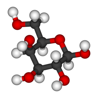
Galactose, sometimes abbreviated Gal, is a monosaccharide sugar that is about as sweet as glucose, and about 65% as sweet as sucrose. It is an aldohexose and a C-4 epimer of glucose. A galactose molecule linked with a glucose molecule forms a lactose molecule.

<span class="mw-page-title-main">Galactosemia</span> Medical condition

Galactosemia is a rare genetic metabolic disorder that affects an individual's ability to metabolize the sugar galactose properly. Galactosemia follows an autosomal recessive mode of inheritance that confers a deficiency in an enzyme responsible for adequate galactose degradation.

<span class="mw-page-title-main">Transferase</span> Class of enzymes which transfer functional groups between molecules

In biochemistry, a transferase is any one of a class of enzymes that catalyse the transfer of specific functional groups from one molecule to another. They are involved in hundreds of different biochemical pathways throughout biology, and are integral to some of life's most important processes.

<span class="mw-page-title-main">Galactokinase</span> Enzyme

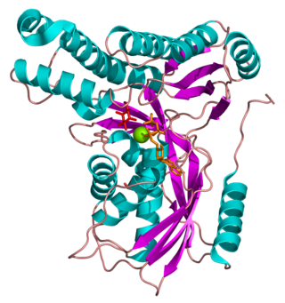
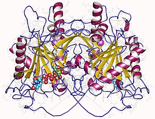
Galactokinase is an enzyme (phosphotransferase) that facilitates the phosphorylation of α-D-galactose to galactose 1-phosphate at the expense of one molecule of ATP. Galactokinase catalyzes the second step of the Leloir pathway, a metabolic pathway found in most organisms for the catabolism of α-D-galactose to glucose 1-phosphate. First isolated from mammalian liver, galactokinase has been studied extensively in yeast, archaea, plants, and humans.

<span class="mw-page-title-main">Galactose-1-phosphate uridylyltransferase</span> Mammalian protein found in Homo sapiens

Galactose-1-phosphate uridyltransferase is an enzyme responsible for converting ingested galactose to glucose.

<span class="mw-page-title-main">Galactokinase deficiency</span> Medical condition

Galactokinase deficiency is an autosomal recessive metabolic disorder marked by an accumulation of galactose and galactitol secondary to the decreased conversion of galactose to galactose-1-phosphate by galactokinase. The disorder is caused by mutations in the GALK1 gene, located on chromosome 17q24. Galactokinase catalyzes the first step of galactose phosphorylation in the Leloir pathway of intermediate metabolism. Galactokinase deficiency is one of the three inborn errors of metabolism that lead to hypergalactosemia. The disorder is inherited as an autosomal recessive trait. Unlike classic galactosemia, which is caused by deficiency of galactose-1-phosphate uridyltransferase, galactokinase deficiency does not present with severe manifestations in early infancy. Its major clinical symptom is the development of cataracts during the first weeks or months of life, as a result of the accumulation, in the lens, of galactitol, a product of an alternative route of galactose utilization. The development of early cataracts in homozygous affected infants is fully preventable through early diagnosis and treatment with a galactose-restricted diet. Some studies have suggested that, depending on milk consumption later in life, heterozygous carriers of galactokinase deficiency may be prone to presenile cataracts at 20–50 years of age.

<span class="mw-page-title-main">UTP—glucose-1-phosphate uridylyltransferase</span> Class of enzymes

UTP—glucose-1-phosphate uridylyltransferase also known as glucose-1-phosphate uridylyltransferase is an enzyme involved in carbohydrate metabolism. It synthesizes UDP-glucose from glucose-1-phosphate and UTP; i.e.,

<span class="mw-page-title-main">Galactose epimerase deficiency</span> Medical condition

Galactose epimerase deficiency, also known as GALE deficiency, Galactosemia III and UDP-galactose-4-epimerase deficiency, is a rare, autosomal recessive form of galactosemia associated with a deficiency of the enzyme galactose epimerase.

<span class="mw-page-title-main">Galactose 1-phosphate</span> Chemical compound

D-Galactose-1-phosphate is an intermediate in the intraconversion of glucose and uridine diphosphate galactose. It is formed from galactose by galactokinase.The improper metabolism of galactose-1-phosphate is a characteristic of galactosemia. The Leloir pathway is responsible for such metabolism of galactose and its intermediate, galactose-1-phosphate. Deficiency of enzymes found in this pathway can result in galactosemia; therefore, diagnosis of this genetic disorder occasionally involves measuring the concentration of these enzymes. One of such enzymes is galactose-1-phosphate uridylytransferase (GALT). The enzyme catalyzes the transfer of a UDP-activator group from UDP-glucose to galactose-1-phosphate. Although the cause of enzyme deficiency in the Leloir pathway is still disputed amongst researchers, some studies suggest that protein misfolding of GALT, which may lead to an unfavorable conformational change that impacts its thermal stability and substrate-binding affinity, may play a role in the deficiency of GALT in Type 1 galactosemia. Increase in galactitol concentration can be seen in patients with galactosemia; putting patients at higher risk for presenile cataract.

<span class="mw-page-title-main">UDP-glucose 4-epimerase</span> Class of enzymes

The enzyme UDP-glucose 4-epimerase, also known as UDP-galactose 4-epimerase or GALE, is a homodimeric epimerase found in bacterial, fungal, plant, and mammalian cells. This enzyme performs the final step in the Leloir pathway of galactose metabolism, catalyzing the reversible conversion of UDP-galactose to UDP-glucose. GALE tightly binds nicotinamide adenine dinucleotide (NAD+), a co-factor required for catalytic activity.

<span class="mw-page-title-main">UDP-glucose—hexose-1-phosphate uridylyltransferase</span> Class of enzymes

In enzymology, an UDP-glucose—hexose-1-phosphate uridylyltransferase is an enzyme that catalyzes the chemical reaction

<span class="mw-page-title-main">UTP—hexose-1-phosphate uridylyltransferase</span> Class of enzymes

In enzymology, an UTP—hexose-1-phosphate uridylyltransferase is an enzyme that catalyzes the chemical reaction

The gal operon is a prokaryotic operon, which encodes enzymes necessary for galactose metabolism. Repression of gene expression for this operon works via binding of repressor molecules to two operators. These repressors dimerize, creating a loop in the DNA. The loop as well as hindrance from the external operator prevent RNA polymerase from binding to the promoter, and thus prevent transcription. Additionally, since the metabolism of galactose in the cell is involved in both anabolic and catabolic pathways, a novel regulatory system using two promoters for differential repression has been identified and characterized within the context of the gal operon.

A galactosemic cataract is cataract which is associated with the consequences of galactosemia.

<span class="mw-page-title-main">Inborn errors of carbohydrate metabolism</span> Medical condition

Inborn errors of carbohydrate metabolism are inborn error of metabolism that affect the catabolism and anabolism of carbohydrates.

<span class="mw-page-title-main">Galactitol</span> Chemical compound

Galactitol (dulcitol) is a sugar alcohol, the reduction product of galactose. It has a slightly sweet taste. In people with galactokinase deficiency, a form of galactosemia, excess dulcitol forms in the lens of the eye leading to cataracts.

Galactolysis refers to the catabolism of galactose.

<span class="mw-page-title-main">Leloir pathway</span>

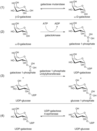
The Leloir pathway is a metabolic pathway for the catabolism of D-galactose. It is named after Luis Federico Leloir, who first described it.

<span class="mw-page-title-main">Duarte galactosemia</span> Medical condition

Duarte galactosemia is an inherited condition associated with diminished ability to metabolize galactose due to a partial deficiency of the enzyme galactose-1-phosphate uridylyltransferase. DG differs from classic galactosemia in that patients with Duarte galactosemia have partial GALT deficiency whereas patients with classic galactosemia have complete, or almost complete, GALT deficiency. Duarte galactosemia (DG) is much more common than classic galactosemia, and is estimated to affect close to one in 4,000 infants born in the United States. Historically, most healthcare professionals have considered DG to be clinically mild based on pilot studies and anecdotal experience, and in 2019 a large study confirmed that children with DG are not at increased risk for developmental problems relative to children who do not have DG. Due to regional variations in newborn screening (NBS) protocols, some infants with DG are identified by NBS but others are not.

References

  1. Online Mendelian Inheritance in Man (OMIM): Galactosemia - 230400
  2. 1 2 3 4 5 6 7 Elsas LJ (1993). "Classic Galactosemia and Clinical Variant Galactosemia". Galactosemia. University of Washington, Seattle. PMID   20301691. NBK1518. In Pagon RA, Bird TD, Dolan CR, et al., eds. (1993). GeneReviews [Internet]. Seattle WA: University of Washington, Seattle.
  3. 1 2 Salway JG (2013). "Chart 47.2 Galactose and galactitol metabolism". Metabolism at a Glance (3rd ed.). John Wiley & Sons. p. 102. ISBN   978-1-118-68207-4.
  4. "Newborn Screening ACT Sheet [Absent/Reduced Galactose-1-Phosphate Uridyltransferase (GALT)] Classical Galactosemia" (PDF). American College of Medical Genetics. Retrieved 2011-11-05.
  5. De-Souza EA, Pimentel FS, Machado CM, Martins LS, da-Silva WS, Montero-Lomelí M, Masuda CA (January 2014). "The unfolded protein response has a protective role in yeast models of classic galactosemia". Disease Models & Mechanisms. 7 (1): 55–61. doi:10.1242/dmm.012641. PMC   3882048 . PMID   24077966.
  6. Mumma JO, Chhay JS, Ross KL, Eaton JS, Newell-Litwa KA, Fridovich-Keil JL (February 2008). "Distinct roles of galactose-1P in galactose-mediated growth arrest of yeast deficient in galactose-1P uridylyltransferase (GALT) and UDP-galactose 4'-epimerase (GALE)". Molecular Genetics and Metabolism. 93 (2): 160–71. doi:10.1016/j.ymgme.2007.09.012. PMC   2253667 . PMID   17981065.
  7. Daenzer JM, Jumbo-Lucioni PP, Hopson ML, Garza KR, Ryan EL, Fridovich-Keil JL (November 2016). "Acute and long-term outcomes in a Drosophila melanogaster model of classic galactosemia occur independently of galactose-1-phosphate accumulation". Disease Models & Mechanisms. 9 (11): 1375–1382. doi:10.1242/dmm.022988. PMC   5117221 . PMID   27562100.
  8. Machado CM, De-Souza EA, De-Queiroz AL, Pimentel FS, Silva GF, Gomes FM, Montero-Lomelí M, Masuda CA (June 2017). "The galactose-induced decrease in phosphate levels leads to toxicity in yeast models of galactosemia". Biochimica et Biophysica Acta (BBA) - Molecular Basis of Disease. 1863 (6): 1403–1409. doi: 10.1016/j.bbadis.2017.02.014 . PMID   28213126.