Microsporum fulvum

Last updated

Contents

Microsporum fulvum
Scientific classification OOjs UI icon edit-ltr.svg
Domain: Eukaryota
Kingdom: Fungi
Division: Ascomycota
Class: Eurotiomycetes
Order: Onygenales
Family: Arthrodermataceae
Genus: Microsporum
Species:
M. fulvum
Binomial name
Microsporum fulvum
Uriburu (1909)
Synonyms
  • Closterosporia fulvaGrigoraki (1925)
  • Sabouraudites fulvisM Ota & Langeron (1923)

Microsporum fulvum is a wildly-distributed dermatophyte species in the Fungi Kingdom. [1] It is known to be a close relative to other dermatophytes such as Trichophyton and Epidermophyton. [2] The fungus is common within soil environments and grows well on keratinized material, such as hair, nails and dead skin. [1] It is recognized as an opportunistic fungal pathogen capable of causing cutaneous mycoses in humans and animals. Originally, the fungus was thought to be Microsporum gypseum until enhanced genetic examination separated the two as distinct species in 1963. [3]

History and taxonomy

Microsporum fulvum was first documented in 1909 as Microsporum gypseum by Weitzman et al. (Argentina Medical Society). [4] The fungus was thought to be the imperfect state of the anamorphic, asexually reproducing, M. gypseum. [4] However, in Stockdale (1963) M. fulvum was considered and described as its own species, Nannizzia fulva , [4] the perfect state of the fungus. In the present, M. fulvum is still referred to as part of the Microsporum gypseum complex. [5] This complex includes the three distinct species: Microsporum gypseum, Microsporum fulvum and Microsporum incurvatum. [3] Each of these fungi possess a high degree of morphological similarity but do have significant genetic differences accounting for the speciation.

Growth

Microsporum fulvum will grow well on soil in a wide-variety of climate conditions and is found in world-wide distribution. [6] The fungus tends to colonize keratin-rich environments and will grow rapidly in culture or in nature at diverse temperatures. [5] [6] M. fulvum commonly occupies materials such as dead skin cells and fragmented hair filaments. [4] In culture, growth will occur within 4–5 days and forms floccose, wooly colonies. [4] The clustered cells appear granular with a sandy-brown tinge where most compact and a less dense, whiter periphery. [4] [5]

Morphology

M. fulvum looks morphologically similar to Microsporum gypseum. The fungus propagates sexually reproductive asci that are 5-7 microns large with up to 8 spores, which are densely packed with ascospores. [6] Asexual macroconidia are also formed and appear in large clusters (up to 8 microns in diameter) with hyphae branches. [4] The macroconidia are fusoid-shaped with tapered ends and have very thin walls. [5] Microconidia are drop-shaped and are observed with sparse, irregular hyphae. [5] When grown on soil combined with horse hair, closed fruiting bodies called cleistothecia form. [6]

Reproduction

Microsporum fulvum is classified as a teleomorph ic species as it sexually reproduces. [4] This sexual state has been referred to as Arthroderma fulvum. [4] Two mating types have been distinguished for the sexual reproduction of the fungus and are characterized with (+) and (-). [1] The (+) mating type synthesizes the elastase enzyme, where as the (-) mating type is unable to produce the elastase. Experiments involving the mating types suggest that the (+) mating type has higher virulence and causes more cases of infection. [4] However, both strains demonstrate a positive result for their ability to hydrolyze the urea molecule, indicating the presence of the urease enzyme. [5] A conducted study showed that a majority of tested isolates (>50%) of M. fulvum tested positive for urea hydrolysis within 0–7 days, and almost all isolates tested positive within 10–12 days, suggesting rapid growth of the organism. [7]

Habitat and ecology

Like many other dermatophytes, M. fulvum is a soil-loving, geophilic organism. [4] The species is commonly found invading obsolete hair fibres and skin molecules which are made of keratin proteins. [2] It can be found growing on the keratinized material of many different mammalian species including humans, horses, rodents, dogs, etc. without perceived preference. [6] However, a higher incidence of colonization of M. fulvum has been documented on the epidermis of hedgehogs without causing any serious skin lesions, suggesting a commensal relationship between the organisms. [8] Isolates of the fungus have been found in skin and lung samples of healthy rodents without indication of impairment. [8] As well, other Microsporum species have arthrospores that are known to remain on human scalp hairs for months without causing skin lesions. [2] Due to the similar behavioural description of species in the Microsporum genus, it is believed that penetration of the mucous membrane is necessary for the fungus to become infectious to the host, otherwise the fungal organism can coexist without harm. [1]

Geographically, Microporum fulvum has been found in diverse climate regions world-wide. The distribution of the fungus seems to be very broad and indiscriminate to temperature fluctuations. [4] Isolates have been documented in Germany, Spain, France, Gabon, Iran and many more geographically distinct areas. [9] The fungus is commonly isolated in Canadian soils, however few infections have been reported. [9] After a 20-year-old man in Iran was diagnosed with a M. fulvum fungal infection, [9] a study was conducted on Iranian soil cultures from regions of varying temperatures. Experiments concluded that M. fulvum is colonized in the soils of a wide variety of environmental regions. [10]

Detection

A mechanism termed baiting can be used to attract Microsporum fulvum to specific soil samples. The method involves preparing the desired soil with keratinized molecules (hair fragments, nail clippings, etc.) and waiting for colonization of fungus onto the material. [8] Once fungal collections populate the sample, morphological examination can be conducted and completed. Morphological analysis is used to distinguish Microsporum species from other keratin-loving dermatophytes. [7] To accurately decipher Microsporum gypseum andMicrosporum fulvum from one another, many researchers use genetic analysis procedures. [9] PCR amplification and genetic testing can be used to uncover information about the mitochondrial DNA, which differs between the two species. [9] In clinical reports, researchers have used methods referred to as Internal Transcribed Spacer (ITS) Sequencing and Matrix-assisted laser desorption/ionization (Maldi-TOF) Mass Spectrometry to differentiate the species of the Microsporum gypseum complex. [11]

Disease

Pathogenicity

Microsporum fulvum causes infection in humans, similar to the ringworm infection caused by Microsporum gypseum that can lead to disease progression. [4] M. fulvum has been reported in cases of dermatophytosis, cutaneous mycoses on any keratinized tissue (dead skin). [1] Tinea corporis refers to the generic ringworm infection that can occur on any area of the body. [12] Tinea pedis is commonly referred to as Athlete's Foot and involves Microsporum fungi infecting the feet. [8] Tinea capitis refers to Microsporum fungi infection of the scalp. [12] Each of these can be caused by Microsporum fulvum, as well as other fungi, and elicit a similar immune response leading to dry patches, rashes, redness, edema, etc. in a localized area. [13] Research shows that most infections from M. fulvum will have a duration of approximately one month and regress on their own, with the exception of more severe cases that require treatment. [13]

These mycoses are restricted to keratinized tissue and remain localized as non-systemic infections. [8] The causative agent of disease are fungal keratinases, which degrade dead material and break down tissue. However, defense mechanisms are present within mammalian blood serum that inhibit these molecules and do not allow the fungus to travel throughout the body in the circulatory system. [5] If the fungus gains access to mucosal membranes, it begins to release the elastase enzyme as well, which is responsible for causing an inflammatory response in the host. [4]

Transmission

Initial infections with Microsporum fulvum are most likely from indirect sources where the fungus inhabited soil that came into contact with the mucous membrane of an animal. [8] New infections can arise through direct or indirect contact of animals and humans who have been introduced to another contaminated source. [8] Often transmission involves the shared use of clothing (shoes- tinea pedis, hats- capitis, etc.). [5] Incidence of infection cases increase in winter months with cold climates due to continued use of closed-toed footwear which provide a good environment for fungal growth. [6] Symptoms of infection are also exacerbated and more severe in these conditions. [8]

Initial infections are common in those with high contact to soil and plant material, such as gardeners. [11] Secondary contamination of M. fulvum often occurs in bursts with multiple infections. In 1949, there were 8 cases of microsporosis caused by M. fulvum that were all in children under the age of 10 who lived in the same neighbourhood of Indiana. [14] Thus, the fungus can spread through contact transmission.

Treatment and prevention

Due to the biological properties and transmission methods of M. fulvum, proper hygienic practices assist with prevention of infection, especially regarding tinea pedis (infection of the feet). [8] Health officials recommend the avoidance of contaminated flooring, such as locker rooms where fungi grow rapidly. [8]

In past reports, treatment methods for Microsporum gypseum infections have been effective for M. fulvum infections. Anti-fungal drugs may be prescribed. As few infections have been confirmed to be caused by M. fulvum, little data has become available on treatment options. However, one confirmed infection in an Iranian man had been treated with oral terbinafine (antifungal drug) and the infection cleared. [9]

Biotechnology applications

Keratinase is an enzyme that functions to break down and degrade keratin molecules. [15] Microsporum fulvum has the ability to synthesize keratinase and utilize the enzyme to inhabit and exploit the environment in which it resides. Poultry industries have become interested in organisms which intrinsically produce keratinase for the reasoning that chicken feathers, which have high levels of dense keratin, are tough to break down and utilize. Fungal keratinase could be cultivated to break down chicken feathers into smaller products which could be converted into other material, such as animal feed. [15]

Related Research Articles

Dermatophyte is a common label for a group of fungus of Arthrodermataceae that commonly causes skin disease in animals and humans. Traditionally, these anamorphic mold genera are: Microsporum, Epidermophyton and Trichophyton. There are about 40 species in these three genera. Species capable of reproducing sexually belong in the teleomorphic genus Arthroderma, of the Ascomycota. As of 2019 a total of nine genera are identified and new phylogenetic taxonomy has been proposed.

<span class="mw-page-title-main">Tinea capitis</span> Cutaneous fungal infection of the scalp

Tinea capitis is a cutaneous fungal infection (dermatophytosis) of the scalp. The disease is primarily caused by dermatophytes in the genera Trichophyton and Microsporum that invade the hair shaft. The clinical presentation is typically single or multiple patches of hair loss, sometimes with a 'black dot' pattern, that may be accompanied by inflammation, scaling, pustules, and itching. Uncommon in adults, tinea capitis is predominantly seen in pre-pubertal children, more often boys than girls.

<span class="mw-page-title-main">Fungal infection</span> Disease caused by fungi to animals or humans

Fungal infection, also known as mycosis, is a disease caused by fungi. Different types are traditionally divided according to the part of the body affected; superficial, subcutaneous, and systemic. Superficial fungal infections include common tinea of the skin, such as tinea of the body, groin, hands, feet and beard, and yeast infections such as pityriasis versicolor. Subcutaneous types include eumycetoma and chromoblastomycosis, which generally affect tissues in and beneath the skin. Systemic fungal infections are more serious and include cryptococcosis, histoplasmosis, pneumocystis pneumonia, aspergillosis and mucormycosis. Signs and symptoms range widely. There is usually a rash with superficial infection. Fungal infection within the skin or under the skin may present with a lump and skin changes. Pneumonia-like symptoms or meningitis may occur with a deeper or systemic infection.

<span class="mw-page-title-main">Dermatophytosis</span> Fungal infection of the skin

Dermatophytosis, also known as ringworm, is a fungal infection of the skin (Dermatomycosis). Typically it results in a red, itchy, scaly, circular rash. Hair loss may occur in the area affected. Symptoms begin four to fourteen days after exposure. Multiple areas can be affected at a given time.

<i>Trichophyton rubrum</i> Species of fungus

Trichophyton rubrum is a dermatophytic fungus in the phylum Ascomycota. It is an exclusively clonal, anthropophilic saprotroph that colonizes the upper layers of dead skin, and is the most common cause of athlete's foot, fungal infection of nail, jock itch, and ringworm worldwide. Trichophyton rubrum was first described by Malmsten in 1845 and is currently considered to be a complex of species that comprises multiple, geographically patterned morphotypes, several of which have been formally described as distinct taxa, including T. raubitschekii, T. gourvilii, T. megninii and T. soudanense.

<i>Trichophyton</i> Genus of Fungi

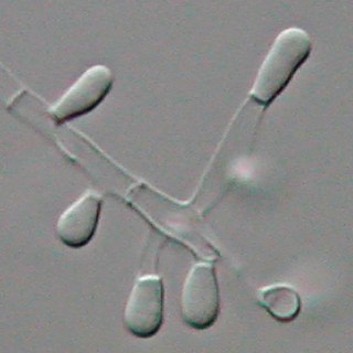
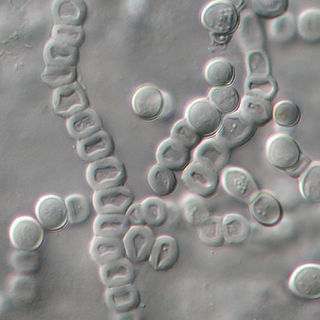
Trichophyton is a genus of fungi, which includes the parasitic varieties that cause tinea, including athlete's foot, ringworm, jock itch, and similar infections of the nail, beard, skin and scalp. Trichophyton fungi are molds characterized by the development of both smooth-walled macro- and microconidia. Macroconidia are mostly borne laterally directly on the hyphae or on short pedicels, and are thin- or thick-walled, clavate to fusiform, and range from 4 to 8 by 8 to 50 μm in size. Macroconidia are few or absent in many species. Microconidia are spherical, pyriform to clavate or of irregular shape, and range from 2 to 3 by 2 to 4 μm in size.

<i>Microsporum</i> Genus of fungi

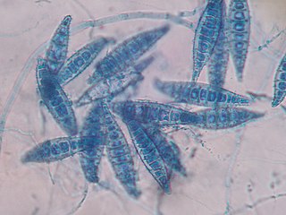
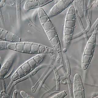
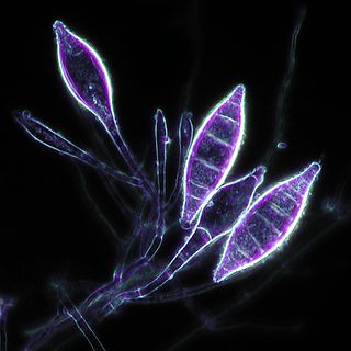
Microsporum is a genus of fungi that causes tinea capitis, tinea corporis, ringworm, and other dermatophytoses. Microsporum forms both macroconidia and microconidia on short conidiophores. Macroconidia are hyaline, multiseptate, variable in form, fusiform, spindle-shaped to obovate, 7–20 by 30–160 um in size, with thin or thick echinulate to verrucose cell walls. Their shape, size and cell wall features are important characteristics for species identification. Microconidia are hyaline, single-celled, pyriform to clavate, smooth-walled, 2.5–3.5 by 4–7 um in size and are not diagnostic for any one species.

<i>Microsporum audouinii</i> Species of fungus

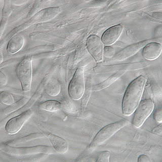
Microsporum audouinii is an anthropophilic fungus in the genus Microsporum. It is a type of dermatophyte that colonizes keratinized tissues causing infection. The fungus is characterized by its spindle-shaped macroconidia, clavate microconidia as well as its pitted or spiny external walls.

<i>Trichophyton tonsurans</i> Species of fungus

Trichophyton tonsurans is a fungus in the family Arthrodermataceae that causes ringworm infection of the scalp. It was first recognized by David Gruby in 1844. Isolates are characterized as the "–" or negative mating type of the Arthroderma vanbreuseghemii complex. This species is thought to be conspecific with T. equinum, although the latter represents the "+" mating strain of the same biological species Despite their biological conspecificity, clones of the two mating types appear to have undergone evolutionary divergence with isolates of the T. tonsurans-type consistently associated with Tinea capitis whereas the T. equinum-type, as its name implies, is associated with horses as a regular host. Phylogenetic relationships were established in isolates from Northern Brazil, through fingerprinting polymorphic RAPD and M13 markers. There seems to be lower genomic variability in the T. tonsurans species due to allopatric divergence. Any phenotypic density is likely due to environmental factors, not genetic characteristics of the fungus.

<i>Microsporum gypseum</i> Species of fungus

Microsporum gypseum is a soil-associated dermatophyte that occasionally is known to colonise and infect the upper dead layers of the skin of mammals. The name refers to an asexual "form-taxon" that has been associated with four related biological species of fungi: the pathogenic taxa Arthroderma incurvatum, A. gypsea, A. fulva and the non-pathogenic saprotroph A. corniculata. More recent studies have restricted M. gypseum to two teleomorphic species A. gypseum and A. incurvatum. The conidial states of A. fulva and A. corniculata have been assigned to M. fulvum and M. boullardii. Because the anamorphic states of these fungi are so similar, they can be identified reliably only by mating. Two mating strains have been discovered, "+" and "–". The classification of this species has been based on the characteristically rough-walled, blunt, club-shaped, multicelled macroconidia. Synonyms include Achorion gypseum, Microsporum flavescens, M. scorteum, and M. xanthodes. There has been past nomenclatural confusion in the usage of the generic names Microsporum and Microsporon.

<i>Trichophyton mentagrophytes</i> Species of fungus

Trichophyton mentagrophytes is a species in the fungal genus Trichophyton. It is one of three common fungi which cause ringworm in companion animals. It is also the second-most commonly isolated fungus causing tinea infections in humans, and the most common or one of the most common fungi that cause zoonotic skin disease. Trichophyton mentagrophytes is being frequently isolated from dogs, cats, rabbits, guinea pigs and other rodents, though at least some genetic variants possess the potential of human-to-human transmission, e.g. Type VII and Type VIII. Particular genetic variants of the fungus have distinct geographic ranges.

<i>Microsporum canis</i> Species of fungus

Microsporum canis is a pathogenic, asexual fungus in the phylum Ascomycota that infects the upper, dead layers of skin on domesticated cats, and occasionally dogs and humans. The species has a worldwide distribution.

Microsporum nanum is a pathogenic fungus in the family Arthrodermataceae. It is a type of dermatophyte that causes infection in dead keratinized tissues such as skin, hair, and nails. Microsporum nanum is found worldwide and is both zoophilic and geophilic. Animals such as pigs and sheep are the natural hosts for the fungus; however, infection of humans is also possible. Majority of the human cases reported are associated with pig farming. The fungus can invade the skin of the host; if it is scratched off by the infected animal, the fungus is still capable of reproducing in soil.

<i>Aphanoascus fulvescens</i> Species of fungus

Aphanoascus fulvescens is a mould fungus that behaves as a keratinophilic saprotroph and belongs to the Ascomycota. It is readily isolated from soil and dung containing keratin-rich tissues that have been separated from their animal hosts. This organism, distributed worldwide, is most commonly found in areas of temperate climate, in keeping with its optimal growth temperature of 28 °C (82 °F). While A. fulvescens is recognized as a geophilic fungal species, it is also a facultative opportunistic pathogen. Although it is not a dermatophyte, A. fulvescens has occasionally been shown to cause onychomycosis infections in humans. Its recognition in the laboratory is clinically important for correct diagnosis and treatment of human dermal infections.

<i>Microsporum gallinae</i> Species of fungus

Microsporum gallinae is a fungus of the genus Microsporum that causes dermatophytosis, commonly known as ringworm. Chickens represent the host population of Microsporum gallinae but its opportunistic nature allows it to enter other populations of fowl, mice, squirrels, cats, dogs and monkeys. Human cases of M. gallinae are rare, and usually mild, non-life-threatening superficial infections.

<i>Chrysosporium keratinophilum</i> Species of fungus

Chrysosporium keratinophilum is a mold that is closely related to the dermatophytic fungi and is mainly found in soil and the coats of wild animals to break down keratin. Chrysosporium keratinophilum is one of the more commonly occurring species of the genus Chrysosporium in nature. It is easily detected due to its characteristic "light-bulb" shape and flat base. Chrysosporium keratinophilum is most commonly found in keratin-rich, dead materials such as feathers, skin scales, hair, and hooves. Although not identified as pathogenic, it is a regular contaminant of cutaneous specimens which leads to the common misinterpretation that this fungus is pathogenic.

<span class="mw-page-title-main">Favus</span> Medical condition

Favus or tinea favosa is the severe form of tinea capitis, a skin infectious disease caused by the dermatophyte fungus Trichophyton schoenleinii. Typically the species affects the scalp, but occasionally occurs as onychomycosis, tinea barbae, or tinea corporis.

<i>Trichophyton verrucosum</i> Species of fungus

Trichophyton verrucosum, commonly known as the cattle ringworm fungus, is a dermatophyte largely responsible for fungal skin disease in cattle, but is also a common cause of ringworm in donkeys, dogs, goat, sheep, and horses. It has a worldwide distribution, however human infection is more common in rural areas where contact with animals is more frequent, and can cause severe inflammation of the afflicted region. Trichophyton verrucosum was first described by Emile Bodin in 1902.

<i>Epidermophyton floccosum</i> Species of fungus

Epidermophyton floccosum is a filamentous fungus that causes skin and nail infections in humans. This anthropophilic dermatophyte can lead to diseases such as tinea pedis, tinea cruris, tinea corporis and onychomycosis. Diagnostic approaches of the fungal infection include physical examination, culture testing, and molecular detection. Topical antifungal treatment, such as the use of terbinafine, itraconazole, voriconazole, and ketoconazole, is often effective.

References

  1. 1 2 3 4 5 Chandler, Francis W.; Kaplan, William; Ajello, Libero (1980). A Colour Atlas and Textbook of the Histopathology of Mycotic Diseases. Weert, Netherlands: Wolfe Medical Pub. ISBN   9780723407546.
  2. 1 2 3 Roberts, Stephen O. B.; Hay, R. J.; Mackenzie, D. W.R. (1984). A Clinician's Guide to Fungal Diseases. New York: Marcel Dekker, Inc. ISBN   0-8247-7190-7.
  3. 1 2 Dolenc-Voljč, Mateja; Gasparič, Jurij (2017). "Human Infections with Microsporum gypseum Complex (Nannizzia gypsea) in Slovenia". Mycopathologia. 182 (11–12): 1069–1075. doi:10.1007/s11046-017-0194-9. PMID   28840426. S2CID   21156729.
  4. 1 2 3 4 5 6 7 8 9 10 11 12 13 14 Rippon, John Willard (1988). Medical Mycology: the Pathogenic Fungi and the Pathogenic Actinomycetes (1st ed.). Philadelphia, PA: Saunders. ISBN   0721624448.
  5. 1 2 3 4 5 6 7 8 Versalovic, James; Jorgensen, James H.; Carroll, Karen C.; Funke, Guido; Landry, Marie Louise; Warnock, David W. (2011). "Manual of Clinical Microbiology". Washington: American Society for Microbiology. pp. 1919–1924. ISBN   978-1-55581-463-2.
  6. 1 2 3 4 5 6 Wilson, J. Walter; Plunkett, Orda A. (1965). The Fungous Diseases of Man. Los Angeles, California: University of California Press. ISBN   9780520013445.
  7. 1 2 McGinnis, Michael R. (1980). Laboratory Handbook of Medical Mycology. New York: Academic Press, Inc. ISBN   0-12-482850-7.
  8. 1 2 3 4 5 6 7 8 9 10 Emmons, Chester W.; Binford, Chapman H.; Utz, John P.; Kwon-Chung, K. J. (1977). Medical Mycology (3rd ed.). Philadelphia: Lea & Febiger. ISBN   978-0812105667.
  9. 1 2 3 4 5 6 Nouripour-Sasakht, Sadegh; Rezaei-Matehkolaei, Ali; Abastabar, Mahdi; Najafzadeh, Mohammad; Satoh, Kazuo; Ahmadi, Bahram; Hosseinpour, Lela (2013). "Microsporum fulvum, an Ignored Pathogenic Dermatophyte: A New Clinical Isolation from Iran". Mycopathologia. 176 (1–2): 157–160. doi:10.1007/s11046-013-9665-9. PMID   23700315. S2CID   6420478.
  10. Rezaei-Matehkolaei, Ali; Jahangiri, Abbas; Mahmoudabadi, Ali Zarei; Najafzadeh, Mohammad; Nouripour-Sisakht, Sadegh; Makimura, Koichi (2017). "Morpho-Molecular Characterization of Soil Inhabitant Dermatophytes from Ahvaz, Southwest of Iran, a High Occurrence of Microsporum fulvum". Mycopathologia. 182 (7–8): 691–699. doi:10.1007/s11046-017-0116-x. PMID   28108868. S2CID   3860164.
  11. 1 2 Seyfarth, F.; Goetze, S.; Erhard, M.; Burmester, A.; Elsner, P; Hipler, U. C. (2009). "Infection with a rare, geophilic dermatophyte". The Dermatologist. 61 (8): 694–699. doi:10.1007/s00105-009-1819-9. PMID   19680615.
  12. 1 2 Beneke, E. S.; Rogers, A. L. (1970). Medical Mycology Manual. Minneapolis: Burgess Publishing Company. ISBN   08087-0263-7.
  13. 1 2 Lewis, George M.; Hopper, Mary E. (1939). An Introduction to Medical Mycology. Chicago: An Introduction to Medical Mycology.
  14. Dalton, John; Slaughter, Robert; Jenkins, Robert; Phelps, Stephen; Hackney, Victor (1950). "Microsporosis Due to Microsporum fulvum". Journal of Investigative Dermatology. 15 (6): 421–423. doi: 10.1038/jid.1950.124 . PMID   14794995.
  15. 1 2 Darah, I.; Nur-Diyana, A.; Nurul-Husna, S.; Jain, K.; Lim, Sheh-Hong (2013). "Microsporum fulvum IBRL SD3: As Novel Isolate for Chicken Feathers Degradation". Appl Biochem Biotechnol. 171 (7): 1900–1910. doi:10.1007/s12010-013-0496-4. PMID   24013862. S2CID   941109.