Neurogastroenterology

Last updated

Neurogastroenterology encompasses the study of the brain, the gut, and their interactions with relevance to the understanding and management of gastrointestinal motility and functional gastrointestinal disorders. Specifically, neurogastroenterology focuses on the functions, malfunctions, and the malformations of the sympathetic, parasympathetic, and enteric divisions of the digestive tract. [1]

Motility Ability to move spontaneously and actively, consuming energy in the process

Motility is the ability of an organism to move independently, using metabolic energy. This is in contrast to mobility, which describes the ability of an object to be moved. Motility is genetically determined, but may be affected by environmental factors. For instance, muscles give animals motility but the consumption of hydrogen cyanide would adversely affect muscle physiology, causing them to stiffen, leading to rigor mortis. In addition to animal locomotion, most animals are motile. The term applies to bacteria and other microorganisms, and to some multicellular organisms, as well as to some mechanisms of fluid flow in multicellular organs and tissue. Motile marine animals are commonly called free-swimming, and motile non-parasitic organisms are called free-living.

Sympathetic nervous system

The sympathetic nervous system (SNS) is one of the two main divisions of the autonomic nervous system, the other being the parasympathetic nervous system.

Parasympathetic nervous system one of the three divisions of the autonomic nervous system, the others being the sympathetic nervous system and enteric nervous system.

The parasympathetic nervous system (PSNS) is one of the two divisions of the autonomic nervous system, the other being the sympathetic nervous system. The autonomic nervous system is responsible for regulating the body's unconscious actions. The parasympathetic system is responsible for stimulation of "rest-and-digest" or "feed and breed" activities that occur when the body is at rest, especially after eating, including sexual arousal, salivation, lacrimation (tears), urination, digestion and defecation. Its action is described as being complementary to that of the sympathetic nervous system, which is responsible for stimulating activities associated with the fight-or-flight response.

Contents

Function of neurons in the gastrointestinal tract

A simplified image showing peristalsis Peristalsis.gif
A simplified image showing peristalsis

The peristaltic reflex

Peristalsis is a series of radially symmetrical contractions and relaxations of muscles which propagate down a muscular tube. In humans and other mammals, peristalsis is found in the smooth muscles of the digestive tract to propel contents through the digestive system. The word is derived from New Latin and comes from the Greek peristallein, "to wrap around," from peri-, "around" + stallein, "to place". Peristalsis was discovered in 1899 by the work of physiologists William Bayliss and Ernest Starling. Working on the small intestines of dogs, they found that the response of increasing the pressure in the intestine caused the contraction of the muscle wall above the point of stimulation and the relaxation of the muscle wall below the point of stimulation. [2] [3]

William Bayliss British physiologist

Sir William Maddock Bayliss was an English physiologist.

Ernest Starling British biologist

Ernest Henry Starling was a British physiologist who contributed many fundamental ideas to this subject. These ideas were important parts of the British contribution to physiology, which at that time led the world.

Segmentation

Segmentation contractions are the contractions in intestines carried out by the smooth muscle walls. Unlike peristalsis, which involves the contraction and relaxation of muscles in one direction, segmentation occurs simultaneously in both directions as the circular muscles alternatively contract. This allows for thorough mixing of intestinal contents, known as chyme, to allow greater absorption.

Chyme or chymus is the semi-fluid mass of partly digested food that is expelled by the stomach, through the pyloric valve, into the duodenum.

Secretion

The secretion of gastrointestinal enzymes, such as gastrin and secretin, is regulated through cholinergic neurons residing in the walls of the digestive tract. Hormone secretion is controlled by the vagovagal reflex, where the neurons in the digestive tract communicate through both afferent and efferent pathways with the vagus nerve. [4]

Digestive enzymes are a group of enzymes that break down polymeric macromolecules into their smaller building blocks, in order to facilitate their absorption by the body. Digestive enzymes are found in the digestive tracts of animals and in the traps of carnivorous plants, where they aid in the digestion of food, as well as inside cells, especially in their lysosomes, where they function to maintain cellular survival. Digestive enzymes of diverse specificities are found in the saliva secreted by the salivary glands, in the secretions of cells lining

Gastrin protein-coding gene in the species Homo sapiens

Gastrin is a peptide hormone that stimulates secretion of gastric acid (HCl) by the parietal cells of the stomach and aids in gastric motility. It is released by G cells in the pyloric antrum of the stomach, duodenum, and the pancreas.

Secretin protein-coding gene in the species Homo sapiens

Secretin is a hormone that regulates water homeostasis throughout the body and influences the environment of the duodenum by regulating secretions in the stomach, pancreas, and liver. It is a peptide hormone produced in the S cells of the duodenum, which are located in the intestinal glands. In humans, the secretin peptide is encoded by the SCT gene.

Anatomy

Enteric nervous system

The organization of the lining of the gastrointestinal system. GI Organization.svg
The organization of the lining of the gastrointestinal system.

The enteric nervous system is one of the main divisions of the nervous system and is the main focus of neurogastroenterology. The enteric nervous system refers to the entire system of neurons that govern the gastrointestinal system. [5] It is capable of operating independently of the brain and spinal cord, [3] but does rely on innervation from the autonomic nervous system via the vagus nerve and prevertebral ganglia in healthy subjects. However, studies have shown that the system is operable with a severed vagus nerve. [6] The neurons of the enteric nervous system control the motor functions of the system, in addition to the secretion of gastrointestinal enzymes. These neurons communicate through many neurotransmitters similar to the CNS, including acetylcholine, dopamine, and serotonin. The large presence of serotonin and dopamine in the gut are key areas of research for neurogastroenterologists. [7] [8] [9]

Nervous system Control the whole body function

The nervous system is the part of an animal that coordinates its actions by transmitting signals to and from different parts of its body. The nervous system detects environmental changes that impact the body, then works in tandem with the endocrine system to respond to such events. Nervous tissue first arose in wormlike organisms about 550 to 600 million years ago. In vertebrates it consists of two main parts, the central nervous system (CNS) and the peripheral nervous system (PNS). The CNS consists of the brain and spinal cord. The PNS consists mainly of nerves, which are enclosed bundles of the long fibers or axons, that connect the CNS to every other part of the body. Nerves that transmit signals from the brain are called motor or efferent nerves, while those nerves that transmit information from the body to the CNS are called sensory or afferent. Spinal nerves serve both functions and are called mixed nerves. The PNS is divided into three separate subsystems, the somatic, autonomic, and enteric nervous systems. Somatic nerves mediate voluntary movement. The autonomic nervous system is further subdivided into the sympathetic and the parasympathetic nervous systems. The sympathetic nervous system is activated in cases of emergencies to mobilize energy, while the parasympathetic nervous system is activated when organisms are in a relaxed state. The enteric nervous system functions to control the gastrointestinal system. Both autonomic and enteric nervous systems function involuntarily. Nerves that exit from the cranium are called cranial nerves while those exiting from the spinal cord are called spinal nerves.

Vagus nerve tenth cranial nerve

The vagus nerve, historically cited as the pneumogastric nerve, is the tenth cranial nerve or CN X, and interfaces with the parasympathetic control of the heart, lungs, and digestive tract. The vagus nerves are paired but are normally referred to in the singular. It is the longest nerve of the autonomic nervous system in the human body.

Prevertebral ganglia

Prevertebral ganglia are sympathetic ganglia which lie between the paravertebral ganglia and the target organ.

Auerbach's plexus

Auerbach's plexus, also known as the myenteric plexus, is a collection of unmyelinated fibers and postganglionic autonomic cell bodies that lie between the circular and longitudinal layers of the muscularis externa in the gastrointestinal tract.[ citation needed ] It was discovered and named by German neuropathologist Leopold Auerbach. These neurons provide motor inputs to both layers of the muscularis externa, and provide both parasympathetic and sympathetic input. The anatomy of the plexus is similar to the anatomy of the central nervous system. The plexus includes sensory receptors, such as chemoreceptors and mechanoreceptors, that are used to provide sensory input to the interneurons in the enteric nervous system. The plexus is the parasympathetic nucleus of origin for the vagus nerve, and communicate with the medulla oblongata through both the anterior and posterior vagal nerves.

Leopold Auerbach German anatomist and neuropathologist

Leopold Auerbach was a German anatomist and neuropathologist born in Breslau.

Central nervous system part of the nervous system consisting of the brain and spinal cord

The central nervous system (CNS) is the part of the nervous system consisting of the brain and spinal cord. The CNS is so named because it integrates the received information and coordinates and influences the activity of all parts of the bodies of bilaterally symmetric animals—that is, all multicellular animals except sponges and radially symmetric animals such as jellyfish—and it contains the majority of the nervous system. Many consider the retina and the optic nerve, as well as the olfactory nerves and olfactory epithelium as parts of the CNS, synapsing directly on brain tissue without intermediate ganglia. As such, the olfactory epithelium is the only central nervous tissue in direct contact with the environment, which opens up for therapeutic treatments. The CNS is contained within the dorsal body cavity, with the brain housed in the cranial cavity and the spinal cord in the spinal canal. In vertebrates, the brain is protected by the skull, while the spinal cord is protected by the vertebrae. The brain and spinal cord are both enclosed in the meninges. Within the CNS, the interneuronal space is filled with a large amount of supporting non-nervous cells called neuroglial cells.

Meissner's plexus

Meissner's plexus is a collection of the plexuses of parasympathetic nerves that run from Auerbach's plexus to the muscularis mucosae of the gastrointestinal wall. It was discovered and named by German physiologist Georg Meissner. It functions as a pathway for the innervation in the mucosa layer of the gastrointestinal wall.

Disorders

Functional gastrointestinal disorders

Functional gastrointestinal (GI) disorders are a class of gastrointestinal disorders where there is a malfunction in the normal activities of the gastrointestinal tract, but there are no structural abnormalities that can explain the cause. There are rarely any tests that can detect the presence of these disorders. Clinical research in neurogastroenterology focuses mainly on the study of common functional gastrointestinal disorders such as irritable bowel syndrome, the most common functional GI disorder. [10]

Motility disorders

Motility disorders are the second classification of gastrointestinal disorder studied by neurogastroenterologists. Motility disorders are divided by what they affect, with four regions: The esophagus, the stomach, the small intestines, and the large intestines. Clinical research in neurogastroenterology focuses mainly on the study of common motility disorders such as gastroesophageal reflux disease, the damage of the mucosa of the esophagus caused by rising stomach acid through the lower esophageal sphincter. [11]

Neurogastroenterology societies

See also

Additional images

Related Research Articles

Peripheral nervous system part of the nervous system

The peripheral nervous system (PNS) is one of two components that make up the nervous system of bilateral animals, with the other part being the central nervous system (CNS). The PNS consists of the nerves and ganglia outside the brain and spinal cord. The main function of the PNS is to connect the CNS to the limbs and organs, essentially serving as a relay between the brain and spinal cord and the rest of the body. Unlike the CNS, the PNS is not protected by the vertebral column and skull, or by the blood–brain barrier, which leaves it exposed to toxins and mechanical injuries.

Stomach digestive organ

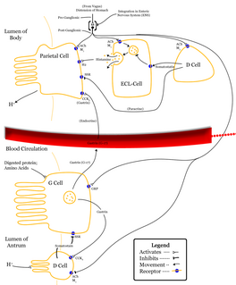
The stomach is a muscular, hollow organ in the gastrointestinal tract of humans and many other animals, including several invertebrates. The stomach has a dilated structure and functions as a vital digestive organ. In the digestive system the stomach is involved in the second phase of digestion, following mastication (chewing).

Gastrointestinal tract organ system within humans and other animals pertaining the stomach and intestines

The gastrointestinal tract is an organ system within humans and other animals which takes in food, digests it to extract and absorb energy and nutrients, and expels the remaining waste as feces. The mouth, esophagus, stomach and intestines are part of the gastrointestinal tract. Gastrointestinal is an adjective meaning of or pertaining to the stomach and intestines. A tract is a collection of related anatomic structures or a series of connected body organs.

Autonomic nervous system division of the peripheral nervous system

The autonomic nervous system (ANS), formerly the vegetative nervous system, is a division of the peripheral nervous system that supplies smooth muscle and glands, and thus influences the function of internal organs. The autonomic nervous system is a control system that acts largely unconsciously and regulates bodily functions such as the heart rate, digestion, respiratory rate, pupillary response, urination, and sexual arousal. This system is the primary mechanism in control of the fight-or-flight response.

Esophagus organ in vertebrates

The esophagus or oesophagus, commonly known as the food pipe or gullet, is an organ in vertebrates through which food passes, aided by peristaltic contractions, from the pharynx to the stomach. The esophagus is a fibromuscular tube, about 25 centimetres long in adults, which travels behind the trachea and heart, passes through the diaphragm and empties into the uppermost region of the stomach. During swallowing, the epiglottis tilts backwards to prevent food from going down the larynx and lungs. The word oesophagus is the Greek word οἰσοφάγος oisophagos, meaning "gullet".

Enteric nervous system

The enteric nervous system (ENS) or intrinsic nervous system is one of the main divisions of the autonomic nervous system (ANS) and consists of a mesh-like system of neurons that governs the function of the gastrointestinal tract. It is capable of acting independently of the sympathetic and parasympathetic nervous systems, although it may be influenced by them. The ENS is also called the second brain.

Nucleus ambiguus nucleus

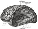
The nucleus ambiguus is a group of large motor neurons, situated deep in the medullary reticular formation named by Jacob Clarke. The nucleus ambiguus contains the cell bodies of nerves that innervate the muscles of the soft palate, pharynx, and larynx which are strongly associated with speech and swallowing. As well as motor neurons, the nucleus ambiguus in its "external formation" contains cholinergic preganglionic parasympathetic neurons for the heart.

Myenteric plexus part of the enteric nervous system

The myenteric plexus provides motor innervation to both layers of the muscular layer of the gut, having both parasympathetic and sympathetic input, whereas the submucous plexus has only parasympathetic fibers and provides secretomotor innervation to the mucosa nearest the lumen of the gut.

Nerve plexus

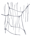
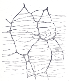
A nerve plexus is a plexus of intersecting nerves. A nerve plexus is composed of afferent and efferent fibers that arise from the merging of the anterior rami of spinal nerves and blood vessels. There are five spinal nerve plexuses, except in the thoracic region, as well as other forms of autonomic plexuses, many of which are a part of the enteric nervous system. The nerves that arise from the plexuses have both sensory and motor functions. These functions include muscle contraction, the maintenance of body coordination and control, and the reaction to sensations such as heat, cold, pain, and pressure. There are several plexuses in the body, including:

An electrogastrogram (EGG) is a graphic produced by an electrogastrograph, which records the electrical signals that travel through the stomach muscles and control the muscles' contractions. An electrogastroenterogram is a similar procedure, which writes down electric signals not only from the stomach, but also from intestines.

Submucous plexus part of the enteric nervous system

The submucous plexus lies in the submucosa of the intestinal wall. The nerves of this plexus are derived from the myenteric plexus which itself is derived from the plexuses of parasympathetic nerves around the superior mesenteric artery. Branches from the myenteric plexus perforate the circular muscle fibers to form the submucous plexus. Ganglia from the plexus extend into the muscularis mucosae and to the mucous membrane.

Vagovagal reflex

Vagovagal reflex refers to gastrointestinal tract reflex circuits where afferent and efferent fibers of the vagus nerve coordinate responses to gut stimuli via the dorsal vagal complex in the brain. The vagovagal reflex controls contraction of the gastrointestinal muscle layers in response to distension of the tract by food. This reflex also allows for the accommodation of large amounts of food in the gastrointestinal tracts.

Dorsal nucleus of vagus nerve

The dorsal nucleus of the vagus nerve is a cranial nerve nucleus for the vagus nerve in the medulla that lies ventral to the floor of the fourth ventricle. It mostly serves parasympathetic vagal functions in the gastrointestinal tract, lungs, and other thoracic and abdominal vagal innervations. The cell bodies for the preganglionic parasympathetic vagal neurons that innervate the heart reside in the nucleus ambiguus.

Gastrointestinal physiology is the branch of human physiology that addresses the physical function of the gastrointestinal (GI) tract. The function of the GI tract is to process ingested food by mechanical and chemical means, extract nutrients and excrete waste products. The GI tract is composed of the alimentary canal, that runs from the mouth to the anus, as well as the associated glands, chemicals, hormones, and enzymes that assist in digestion.The major processes that occur in the GI tract are: motility, secretion, regulation, digestion and circulation. The proper function and coordination of these processes are vital for maintaining good health by providing for the effective digestion and uptake of nutrients.

Segmentation contractions are a type of intestinal motility.

The basal or basic electrical rhythm (BER) or electrical control activity (ECA) is the spontaneous depolarization and repolarization of pacemaker cells in the smooth muscle of the stomach, small intestine, and large intestine. This electrical rhythm is spread through gap junctions in the smooth muscle of the GI tract. These pacemaker cells, also called the interstitial cells of Cajal, control the frequency of contractions in the gastrointestinal tract. The cells can be located in either the circular or longitudinal layer of the smooth muscle in the GI tract; circular for the small and large intestine, longitudinal for the stomach. The frequency of contraction differs at each location in the GI tract beginning with 3 per minute in the stomach, then 12 per minute in the duodenum, 9 per minute in the ileum, and a normally low one contraction per 30 minutes in the large intestines that increases 3 to 4 times a day due to a phenomenon called mass movement. The basal electrical rhythm controls the frequency of contraction but additional neuronal and hormonal controls regulate the strength of each contraction.

Gastrointestinal wall

The gastrointestinal wall surrounding the lumen of the gastrointestinal tract is made up of four layers of specialised tissue – from the lumen outwards:

Human digestive system combination of anatomical organs that are responsible for digestive function

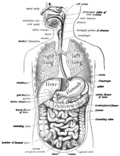
The human digestive system consists of the gastrointestinal tract plus the accessory organs of digestion. Digestion involves the breakdown of food into smaller and smaller components, until they can be absorbed and assimilated into the body. The process of digestion has many stages. The first stage is the cephalic phase of digestion which begins with gastric secretions in response to the sight and smell of food. The next stage starts in the mouth.

References

  1. Wood, JD; DH Alpers; PLR Andrews (1999). "Fundamentals of Neurogastroenterology". Gut. 45: 6–16. doi:10.1136/gut.45.2008.ii6. PMC   1766686 Lock-green.svg.
  2. Keet, A. D. "The Pyloric Sphincteric Cylinder in health and disease" . Retrieved 18 November 2013.
  3. 1 2 Gershon, Michael (1998). The Second Brain. New York: HarperCollins. pp. 2–7. ISBN   0-06-018252-0.
  4. Herman MA, Cruz MT, Sahibzada N, Verbalis J, Gillis RA (January 2009). "GABA signaling in the nucleus tractus solitarius sets the level of activity in dorsal motor nucleus of the vagus cholinergic neurons in the vagovagal circuit". Am. J. Physiol. Gastrointest. Liver Physiol. 296 (1): G101–11. doi:10.1152/ajpgi.90504.2008. PMC   2636929 Lock-green.svg. PMID   19008339.
  5. John Barton Furness (15 April 2008). The Enteric Nervous System. John Wiley & Sons. ISBN   978-1-4051-7344-5 . Retrieved 16 January 2013.
  6. Li,Ying; Owyang,Chung (September 2003). "Musings on the Wanderer: What's New in Our Understanding of Vago-Vagal Reflexes? V. Remodeling of vagus and enteric neural circuitry after vagal injury". American Journal of Physiology. Gastrointestinal and Liver Physiology. 285 (3): G461–9. doi:10.1152/ajpgi.00119.2003.
  7. Pasricha, Pankaj Jay. "Stanford Hospital: Brain in the Gut - Your Health".
  8. Martinucci I et al. Genetics and pharmacogenetics of aminergic transmitter pathways in functional gastrointestinal disorders. Pharmacogenomics. 2015;16(5):523-39. Review. PMID   25916523
  9. Smitka K, et al. The role of "mixed" orexigenic and anorexigenic signals and autoantibodies reacting with appetite-regulating neuropeptides and peptides of the adipose tissue-gut-brain axis: relevance to food intake and nutritional status in patients with anorexia nervosa and bulimia nervosa. Int J Endocrinol. 2013;2013:483145. Review. PMID   24106499 Free full text PMC 3782835
  10. Kumar, A.; Rinwa P.; Sharma N. (2012). "Irritable Bowel Syndrome: A Review". J Phys Pharm Adv. 2 (2): 97–108.
  11. DeVault KR, Castell DO (1999). "Updated guidelines for the diagnosis and treatment of gastroesophageal reflux disease. The Practice Parameters Committee of the American College of Gastroenterology". Am J Gastroenterol . 94 (6): 1434–42. doi:10.1111/j.1572-0241.1999.1123_a.x. PMID   10364004.
  12. ANMS - American Neurogastroenterology and Motility Society
  13. ESNM - European Society for Neurogastroenterology & Motility