Pioneer factor

Last updated

Pioneer factors are transcription factors that can directly bind condensed chromatin. They can have positive and negative effects on transcription and are important in recruiting other transcription factors and histone modification enzymes as well as controlling DNA methylation. They were first discovered in 2002 as factors capable of binding to target sites on nucleosomal DNA in compacted chromatin and endowing competency for gene activity during hepatogenesis. [1] Pioneer factors are involved in initiating cell differentiation and activation of cell-specific genes. This property is observed in histone fold-domain containing transcription factors (fork head box (FOX) [2] and NF-Y [3] ) and other transcription factors that use zinc finger(s) for DNA binding (Groucho TLE, Gal4, and GATA). [2] [4]

Contents

The eukaryotic cell condenses its genome into tightly packed chromatin and nucleosomes. This ability saves space in the nucleus for only actively transcribed genes and hides unnecessary or detrimental genes from being transcribed. Access to these condensed regions is done by chromatin remodelling by either balancing histone modifications or directly with pioneer factors that can loosen the chromatin themselves or as a flag recruiting other factors. Pioneer factors are not necessarily required for assembly of the transcription apparatus and may dissociate after being replaced by other factors.

Active rearrangement

Opening of condensed chromatin by a pioneer factor to initiate transcription. The pioneer factor binds to tightly packed chromatin and causes a nucleosomal rearrangement. This new configuration allows space for other transcription factors to bind and initiate transcription. Pioneer Factor rearrange the nucleosome.jpg
Opening of condensed chromatin by a pioneer factor to initiate transcription. The pioneer factor binds to tightly packed chromatin and causes a nucleosomal rearrangement. This new configuration allows space for other transcription factors to bind and initiate transcription.

Pioneer factors can also actively affect transcription by directly opening up condensed chromatin in an ATP-independent process. [2] [3] This is a common trait of fork head box factors (which contain a winged helix DNA-binding domain that mimics the DNA-binding domain of the linker H1 histone [5] ), and NF-Y (whose NF-YB and NF-YC subunits contain histone-fold domains similar to those of the core histones H2A/H2B [6] ).

Fork head box factors

The similarity to histone H1 explains how fork head factors are able to bind chromatin by interacting with the major groove of only the one available side of DNA wrapped around a nucleosome. [5] [7] Fork head domains also have a helix that confers sequence specificity unlike linker histone. [5] [8] The C terminus is associated with higher mobility around the nucleosome than linker histone, displacing it and rearranging nucleosomal landscapes effectively. [7] This active re-arrangement of the nucleosomes allows for other transcription factors to bind the available DNA. In thyroid cell differentiation FoxE binds to compacted chromatin of the thyroid peroxidase promoter and opens it for NF1 binding. [9]

NF-Y

NF-Y is a heterotrimeric complex composed of NF-YA, NF-YB, and NF-YC subunits. The key structural feature of the NF-Y/DNA complex is the minor-groove interaction of its DNA binding domain-containing subunit NF-YA, which induces an ~80° bend in the DNA. NF-YB and NF-YC interact with DNA through non-specific histone-fold domain-DNA contacts. [6] NF-YA's unique DNA-binding mode and NF-YB/NF-YC's nucleosome-like properties of non-specific DNA binding impose sufficient spatial constraints to induce flanking nucleosomes to slide outward, making nearby recognition sites for other transcription factors accessible. [3]

Passive factors

An example of the cell 'priming' for rapidly induced transcription. The pioneer factor, FoxA1 binds the enhancer in the first step but can not initiate transcription. Next when the signal, estrogen, is present the estrogen receptor can quickly find the 'bookmark' pioneer factor. When the estrogen receptor is bound transcription is initiated. Pioneer Factor's role in response of the external signal.jpg
An example of the cell ‘priming’ for rapidly induced transcription. The pioneer factor, FoxA1 binds the enhancer in the first step but can not initiate transcription. Next when the signal, estrogen, is present the estrogen receptor can quickly find the ‘bookmark’ pioneer factor. When the estrogen receptor is bound transcription is initiated.

Pioneer factors can function passively, by acting as a bookmark for the cell to recruit other transcription factors to specific genes in condensed chromatin. This can be important for priming the cell for a rapid response as the enhancer is already bound by a pioneer transcription factor giving it a head start towards assembling the transcription preinitiation complex. Hormone responses are often quickly induced in the cell using this priming method such as with the estrogen receptor. [10] Another form of priming is when an enhancer is simultaneously bound by activating and repressing pioneer factors. This balance can be tipped by dissociation of one of the factors. In hepatic cell differentiation the activating pioneer factor FOXA1 recruits a repressor, grg3, that prevents transcription until the repressor is down-regulated later on in the differentiation process. [11]
In a direct role pioneer factors can bind an enhancer and recruit activation complex that will modify the chromatin directly. The change in the chromatin changes the affinity, decreasing the affinity of the pioneer factor such that it is replaced by a transcription factor that has a higher affinity. This is a mechanism for the cell to switch a gene on was observed with glucocorticoid receptor recruiting modification factors that then modify the site to bind activated estrogen receptor which was coined as a “bait and switch” mechanism. [12]

Epigenetic effects

Pioneer factor, PU.1, binding cell-specific gene regulation in hematopoietic differentiation. In hematopoietic stem cells PU.1 binds different lineage-specific enhancers and recruits histone modification enzymes that mark these enhancers with H3K4me1. These modified histones are then recognized by cell-specific transcription factors that activate genes leading to the differentiation of B-cells or macrophages. Pioneer Factor in the Cell Differentiation.jpg
Pioneer factor, PU.1, binding cell-specific gene regulation in hematopoietic differentiation. In hematopoietic stem cells PU.1 binds different lineage-specific enhancers and recruits histone modification enzymes that mark these enhancers with H3K4me1. These modified histones are then recognized by cell-specific transcription factors that activate genes leading to the differentiation of B-cells or macrophages.

Pioneer factors can exhibit their greatest range of effects on transcription through the modulation of epigenetic factors by recruiting activating or repressing histone modification enzymes and controlling CpG methylation by protecting specific cysteine residues. This has effects on controlling the timing of transcription during cell differentiation processes.

Histone modification

Histone modification is a well-studied mechanism to transiently adjust chromatin density. Pioneer factors can play a role in this by binding specific enhancers and flagging histone modification enzymes to that specific gene. Repressive pioneer factors can inhibit transcription by recruiting factors that modify histones that further tighten the chromatin. This is important to limit gene expression to specific cell types and has to be removed only when cell differentiation begins. FoxD3 has been associated as a repressor of both B-cell and melanocytic cell differentiation pathways, maintaining repressive histone modifications where bound, that have to be overcome to start differentiation. [13] [14] Pioneer factors can also be associated with recruiting transcription-activating histone modifications. Enzymes that modify H3K4 with mono and di-methylation are associated with increasing transcription and have been shown to bind pioneer factors. [10] In B cell differentiation PU.1 is necessary to signal specific histones for activating H3K4me1 modifications that differentiate hematopoietic stem cells into either the B-cell or macrophage lineage. [15] FoxA1 binding induces HSK4me2 during neuronal differentiation of pluripotent stem cells [16] as well as the loss of DNA methylation. [17] SOX9 recruits histone modification enzymes MLL3 and MLL4 to deposit H3K4me1 prior to the opening of enhancers in developing hair follicle and basal cell carcinoma. [18]

DNA methylation

Pioneer factors can also affect transcription and differentiation through the control of DNA methylation. Pioneer factors that bind to CpG islands and cytosine residues block access to methyltransferases. Many eukaryotic cells have CpG islands in their promoters that can be modified by methylation having adverse effects on their ability to control transcription. [19] This phenomenon is also present in promoters without CpG islands where single cytosine residues are protected from methylation until further cell differentiation. An example is FoxD3 preventing methylation of a cytosine residue in Alb1 enhancer, acting as a place holder for FoxA1 later in hepatic [20] as well as in CpG islands of genes in chronic lymphocytic leukemia. [21] For stable control of methylation state the cytosine residues are covered during mitosis, unlike most other transcription factors, to prevent methylation. Studies have shown that during mitosis 15% of all interphase FoxA1 binding sites were bound. [22] The protection of cytosine methylation can be quickly removed allowing for rapid induction when a signal is present.

Other pioneer factors

A well studied pioneer factor family is the Groucho-related (Gro/TLE/Grg) transcription factors that often have a negative effect on transcription. These chromatin binding domains can span up to 3-4 nucleosomes. These large domains are scaffolds for further protein interactions and also modify the chromatin for other pioneer factors such as FoxA1 which has been shown to bind to Grg3. [23] Transcription factors with zinc finger DNA binding domains, such as the GATA family and glucocorticoid receptor. [10] The zinc finger domains do not appear to bind nucleosomes well and can be displaced by FOX factors. [22]

In the skin epidermis, SOX family transcription factor, SOX9, also behaves as a pioneer factor that governs hair follicle cell fate and can reprogram epidermal stem cells to a hair follicle fate. [24]

Role in cancer

The ability of pioneer factors to respond to extracellular signals to differentiate cell type has been studied as a potential component of hormone-dependent cancers. Hormones such as estrogen and IGFI are shown to increase pioneer factor concentration leading to a change in transcription. [25] Known pioneer factors such as FoxA1, PBX1, TLE, AP2ɣ, GATA factors 2/3/4, and PU.1 have been associated with hormone-dependent cancer . FoxA1 is necessary for estrogen and androgen mediated hepatocarcinogenesis and is a defining gene for ER+ luminal breast cancer, as is another pioneer factor GATA3. [10] [25] FOXA1 particularly is expressed in 90% of breast cancer metastases and 89% of metastic prostate cancers. [25] [26] In the breast cancer cell line, MCF-7, it was found that FoxA1 was bound to 50% of estrogen receptor binding sites independent of estrogen presence. High expression of pioneer factors is associated with poor prognosis with the exception of breast cancer where FoxA1 is associated with a stronger outcome. [25]
The correlation between pioneer factors and cancer has led to prospective therapeutic targeting. In knockdown studies in the MCF-7 breast cancer cell line it was found that decreasing pioneer factors FoxA1 and AP2ɣ decreased ER signalling. [4] [25] Other fork head proteins have been associated with cancer, including FoxO3 and FoxM that repress the cell survival pathways Ras and PPI3K/AKT/IKK. [27] Drugs such as Paclitaxel, Imatinib, and doxorubicin which activate FoxO3a or its targets are being used. Modification to modulate related factors with pioneer activity is a topic of interest in the early stages as knocking down pioneer factors may have toxic effects through alteration of the lineage pathways of healthy cells. [25]

Related Research Articles

<span class="mw-page-title-main">Histone</span> Protein family around which DNA winds to form nucleosomes

In biology, histones are highly basic proteins abundant in lysine and arginine residues that are found in eukaryotic cell nuclei and in most Archaeal phyla. They act as spools around which DNA winds to create structural units called nucleosomes. Nucleosomes in turn are wrapped into 30-nanometer fibers that form tightly packed chromatin. Histones prevent DNA from becoming tangled and protect it from DNA damage. In addition, histones play important roles in gene regulation and DNA replication. Without histones, unwound DNA in chromosomes would be very long. For example, each human cell has about 1.8 meters of DNA if completely stretched out; however, when wound about histones, this length is reduced to about 90 micrometers (0.09 mm) of 30 nm diameter chromatin fibers.

<span class="mw-page-title-main">Transcription factor</span> Protein that regulates the rate of DNA transcription

In molecular biology, a transcription factor (TF) is a protein that controls the rate of transcription of genetic information from DNA to messenger RNA, by binding to a specific DNA sequence. The function of TFs is to regulate—turn on and off—genes in order to make sure that they are expressed in the desired cells at the right time and in the right amount throughout the life of the cell and the organism. Groups of TFs function in a coordinated fashion to direct cell division, cell growth, and cell death throughout life; cell migration and organization during embryonic development; and intermittently in response to signals from outside the cell, such as a hormone. There are approximately 1600 TFs in the human genome. Transcription factors are members of the proteome as well as regulome.

<span class="mw-page-title-main">Cellular differentiation</span> Developmental biology

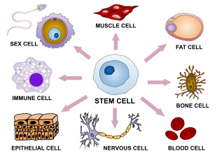
Cellular differentiation is the process in which a stem cell changes from one type to a differentiated one. Usually, the cell changes to a more specialized type. Differentiation happens multiple times during the development of a multicellular organism as it changes from a simple zygote to a complex system of tissues and cell types. Differentiation continues in adulthood as adult stem cells divide and create fully differentiated daughter cells during tissue repair and during normal cell turnover. Some differentiation occurs in response to antigen exposure. Differentiation dramatically changes a cell's size, shape, membrane potential, metabolic activity, and responsiveness to signals. These changes are largely due to highly controlled modifications in gene expression and are the study of epigenetics. With a few exceptions, cellular differentiation almost never involves a change in the DNA sequence itself. However, metabolic composition does get altered quite dramatically where stem cells are characterized by abundant metabolites with highly unsaturated structures whose levels decrease upon differentiation. Thus, different cells can have very different physical characteristics despite having the same genome.

In molecular biology and genetics, transcriptional regulation is the means by which a cell regulates the conversion of DNA to RNA (transcription), thereby orchestrating gene activity. A single gene can be regulated in a range of ways, from altering the number of copies of RNA that are transcribed, to the temporal control of when the gene is transcribed. This control allows the cell or organism to respond to a variety of intra- and extracellular signals and thus mount a response. Some examples of this include producing the mRNA that encode enzymes to adapt to a change in a food source, producing the gene products involved in cell cycle specific activities, and producing the gene products responsible for cellular differentiation in multicellular eukaryotes, as studied in evolutionary developmental biology.

<span class="mw-page-title-main">Histone acetyltransferase</span> Enzymes that catalyze acyl group transfer from acetyl-CoA to histones

Histone acetyltransferases (HATs) are enzymes that acetylate conserved lysine amino acids on histone proteins by transferring an acetyl group from acetyl-CoA to form ε-N-acetyllysine. DNA is wrapped around histones, and, by transferring an acetyl group to the histones, genes can be turned on and off. In general, histone acetylation increases gene expression.

HMGN proteins are members of the broader class of high mobility group (HMG) chromosomal proteins that are involved in regulation of transcription, replication, recombination, and DNA repair.

Histone H2B is one of the 5 main histone proteins involved in the structure of chromatin in eukaryotic cells. Featuring a main globular domain and long N-terminal and C-terminal tails, H2B is involved with the structure of the nucleosomes.

Histone methylation is a process by which methyl groups are transferred to amino acids of histone proteins that make up nucleosomes, which the DNA double helix wraps around to form chromosomes. Methylation of histones can either increase or decrease transcription of genes, depending on which amino acids in the histones are methylated, and how many methyl groups are attached. Methylation events that weaken chemical attractions between histone tails and DNA increase transcription because they enable the DNA to uncoil from nucleosomes so that transcription factor proteins and RNA polymerase can access the DNA. This process is critical for the regulation of gene expression that allows different cells to express different genes.

<span class="mw-page-title-main">Histone acetylation and deacetylation</span> Biological processes used in gene regulation

Histone acetylation and deacetylation are the processes by which the lysine residues within the N-terminal tail protruding from the histone core of the nucleosome are acetylated and deacetylated as part of gene regulation.

<span class="mw-page-title-main">Histone-modifying enzymes</span> Type of enzymes

Histone-modifying enzymes are enzymes involved in the modification of histone substrates after protein translation and affect cellular processes including gene expression. To safely store the eukaryotic genome, DNA is wrapped around four core histone proteins, which then join to form nucleosomes. These nucleosomes further fold together into highly condensed chromatin, which renders the organism's genetic material far less accessible to the factors required for gene transcription, DNA replication, recombination and repair. Subsequently, eukaryotic organisms have developed intricate mechanisms to overcome this repressive barrier imposed by the chromatin through histone modification, a type of post-translational modification which typically involves covalently attaching certain groups to histone residues. Once added to the histone, these groups elicit either a loose and open histone conformation, euchromatin, or a tight and closed histone conformation, heterochromatin. Euchromatin marks active transcription and gene expression, as the light packing of histones in this way allows entry for proteins involved in the transcription process. As such, the tightly packed heterochromatin marks the absence of current gene expression.

Chromatin remodeling is the dynamic modification of chromatin architecture to allow access of condensed genomic DNA to the regulatory transcription machinery proteins, and thereby control gene expression. Such remodeling is principally carried out by 1) covalent histone modifications by specific enzymes, e.g., histone acetyltransferases (HATs), deacetylases, methyltransferases, and kinases, and 2) ATP-dependent chromatin remodeling complexes which either move, eject or restructure nucleosomes. Besides actively regulating gene expression, dynamic remodeling of chromatin imparts an epigenetic regulatory role in several key biological processes, egg cells DNA replication and repair; apoptosis; chromosome segregation as well as development and pluripotency. Aberrations in chromatin remodeling proteins are found to be associated with human diseases, including cancer. Targeting chromatin remodeling pathways is currently evolving as a major therapeutic strategy in the treatment of several cancers.

<span class="mw-page-title-main">NFYA</span> Protein-coding gene in the species Homo sapiens

Nuclear transcription factor Y subunit alpha is a protein that in humans is encoded by the NFYA gene.

Epigenomics is the study of the complete set of epigenetic modifications on the genetic material of a cell, known as the epigenome. The field is analogous to genomics and proteomics, which are the study of the genome and proteome of a cell. Epigenetic modifications are reversible modifications on a cell's DNA or histones that affect gene expression without altering the DNA sequence. Epigenomic maintenance is a continuous process and plays an important role in stability of eukaryotic genomes by taking part in crucial biological mechanisms like DNA repair. Plant flavones are said to be inhibiting epigenomic marks that cause cancers. Two of the most characterized epigenetic modifications are DNA methylation and histone modification. Epigenetic modifications play an important role in gene expression and regulation, and are involved in numerous cellular processes such as in differentiation/development and tumorigenesis. The study of epigenetics on a global level has been made possible only recently through the adaptation of genomic high-throughput assays.

<span class="mw-page-title-main">FOXA2</span> Mammalian protein found in Homo sapiens

Forkhead box protein A2 (FOXA2), also known as hepatocyte nuclear factor 3-beta (HNF-3B), is a transcription factor that plays an important role during development, in mature tissues and, when dysregulated or mutated, also in cancer.

Epigenetics of human development is the study of how epigenetics effects human development.

H3K4me3 is an epigenetic modification to the DNA packaging protein Histone H3 that indicates tri-methylation at the 4th lysine residue of the histone H3 protein and is often involved in the regulation of gene expression. The name denotes the addition of three methyl groups (trimethylation) to the lysine 4 on the histone H3 protein.

H3K27me3 is an epigenetic modification to the DNA packaging protein Histone H3. It is a mark that indicates the tri-methylation of lysine 27 on histone H3 protein.

H3K9me3 is an epigenetic modification to the DNA packaging protein Histone H3. It is a mark that indicates the tri-methylation at the 9th lysine residue of the histone H3 protein and is often associated with heterochromatin.

H4K20me is an epigenetic modification to the DNA packaging protein Histone H4. It is a mark that indicates the mono-methylation at the 20th lysine residue of the histone H4 protein. This mark can be di- and tri-methylated. It is critical for genome integrity including DNA damage repair, DNA replication and chromatin compaction.

H4K16ac is an epigenetic modification to the DNA packaging protein Histone H4. It is a mark that indicates the acetylation at the 16th lysine residue of the histone H4 protein.

References

  1. Cirillo LA, Lin FR, Cuesta I, Friedman D, Jarnik M, Zaret KS (February 2002). "Opening of compacted chromatin by early developmental transcription factors HNF3 (FoxA) and GATA-4". Molecular Cell. 9 (2): 279–89. doi: 10.1016/S1097-2765(02)00459-8 . PMID   11864602.
  2. 1 2 3 Zaret, Kenneth S.; Carroll, Jason S. (2011-11-01). "Pioneer transcription factors: establishing competence for gene expression". Genes & Development. 25 (21): 2227–2241. doi:10.1101/gad.176826.111. ISSN   1549-5477. PMC   3219227 . PMID   22056668.
  3. 1 2 3 Oldfield, Andrew J.; Yang, Pengyi; Conway, Amanda E.; Cinghu, Senthilkumar; Freudenberg, Johannes M.; Yellaboina, Sailu; Jothi, Raja (2014-09-04). "Histone-fold domain protein NF-Y promotes chromatin accessibility for cell type-specific master transcription factors". Molecular Cell. 55 (5): 708–722. doi:10.1016/j.molcel.2014.07.005. ISSN   1097-4164. PMC   4157648 . PMID   25132174.
  4. 1 2 Magnani L, Eeckhoute J, Lupien M (November 2011). "Pioneer factors: directing transcriptional regulators within the chromatin environment". Trends in Genetics. 27 (11): 465–74. doi:10.1016/j.tig.2011.07.002. PMID   21885149.
  5. 1 2 3 Clark KL, Halay ED, Lai E, Burley SK (July 1993). "Co-crystal structure of the HNF-3/fork head DNA-recognition motif resembles histone H5". Nature. 364 (6436): 412–20. Bibcode:1993Natur.364..412C. doi:10.1038/364412a0. PMID   8332212. S2CID   4363526.
  6. 1 2 Nardini, Marco; Gnesutta, Nerina; Donati, Giacomo; Gatta, Raffaella; Forni, Claudia; Fossati, Andrea; Vonrhein, Clemens; Moras, Dino; Romier, Christophe (2013-01-17). "Sequence-specific transcription factor NF-Y displays histone-like DNA binding and H2B-like ubiquitination" (PDF). Cell. 152 (1–2): 132–143. doi: 10.1016/j.cell.2012.11.047 . ISSN   1097-4172. PMID   23332751. S2CID   17899925.
  7. 1 2 Zaret KS, Caravaca JM, Tulin A, Sekiya T (2010). "Nuclear mobility and mitotic chromosome binding: similarities between pioneer transcription factor FoxA and linker histone H1". Cold Spring Harbor Symposia on Quantitative Biology. 75: 219–26. doi: 10.1101/sqb.2010.75.061 . PMID   21502411.
  8. Sekiya T, Muthurajan UM, Luger K, Tulin AV, Zaret KS (April 2009). "Nucleosome-binding affinity as a primary determinant of the nuclear mobility of the pioneer transcription factor FoxA". Genes & Development. 23 (7): 804–9. doi:10.1101/gad.1775509. PMC   2666343 . PMID   19339686.
  9. Cuesta I, Zaret KS, Santisteban P (October 2007). "The forkhead factor FoxE1 binds to the thyroperoxidase promoter during thyroid cell differentiation and modifies compacted chromatin structure". Molecular and Cellular Biology. 27 (20): 7302–14. doi:10.1128/MCB.00758-07. PMC   2168900 . PMID   17709379.
  10. 1 2 3 4 Zaret KS, Carroll JS (November 2011). "Pioneer transcription factors: establishing competence for gene expression". Genes & Development. 25 (21): 2227–41. doi:10.1101/gad.176826.111. PMC   3219227 . PMID   22056668.
  11. Xu CR, Cole PA, Meyers DJ, Kormish J, Dent S, Zaret KS (May 2011). "Chromatin "prepattern" and histone modifiers in a fate choice for liver and pancreas". Science. 332 (6032): 963–6. Bibcode:2011Sci...332..963X. doi:10.1126/science.1202845. PMC   3128430 . PMID   21596989.
  12. Voss TC, Schiltz RL, Sung MH, Yen PM, Stamatoyannopoulos JA, Biddie SC, Johnson TA, Miranda TB, John S, Hager GL (August 2011). "Dynamic exchange at regulatory elements during chromatin remodeling underlies assisted loading mechanism". Cell. 146 (4): 544–54. doi:10.1016/j.cell.2011.07.006. PMC   3210475 . PMID   21835447.
  13. Liber D, Domaschenz R, Holmqvist PH, Mazzarella L, Georgiou A, Leleu M, Fisher AG, Labosky PA, Dillon N (July 2010). "Epigenetic priming of a pre-B cell-specific enhancer through binding of Sox2 and Foxd3 at the ESC stage". Cell Stem Cell. 7 (1): 114–26. doi: 10.1016/j.stem.2010.05.020 . PMID   20621055.
  14. Katiyar P, Aplin AE (May 2011). "FOXD3 regulates migration properties and Rnd3 expression in melanoma cells". Molecular Cancer Research. 9 (5): 545–52. doi:10.1158/1541-7786.MCR-10-0454. PMC   3096755 . PMID   21478267.
  15. Heinz S, Benner C, Spann N, Bertolino E, Lin YC, Laslo P, Cheng JX, Murre C, Singh H, Glass CK (May 2010). "Simple combinations of lineage-determining transcription factors prime cis-regulatory elements required for macrophage and B cell identities". Molecular Cell. 38 (4): 576–89. doi:10.1016/j.molcel.2010.05.004. PMC   2898526 . PMID   20513432.
  16. Sérandour AA, Avner S, Percevault F, Demay F, Bizot M, Lucchetti-Miganeh C, Barloy-Hubler F, Brown M, Lupien M, Métivier R, Salbert G, Eeckhoute J (April 2011). "Epigenetic switch involved in activation of pioneer factor FOXA1-dependent enhancers". Genome Research. 21 (4): 555–65. doi:10.1101/gr.111534.110. PMC   3065703 . PMID   21233399.
  17. Taube JH, Allton K, Duncan SA, Shen L, Barton MC (May 2010). "Foxa1 functions as a pioneer transcription factor at transposable elements to activate Afp during differentiation of embryonic stem cells". The Journal of Biological Chemistry. 285 (21): 16135–44. doi: 10.1074/jbc.M109.088096 . PMC   2871482 . PMID   20348100.
  18. Yang, Yihao; Gomez, Nicholas; Infarinato, Nicole; Adam, Rene C.; Sribour, Megan; Baek, Inwha; Laurin, Mélanie; Fuchs, Elaine (2023-07-24). "The pioneer factor SOX9 competes for epigenetic factors to switch stem cell fates". Nature Cell Biology. 25 (8): 1185–1195. doi: 10.1038/s41556-023-01184-y . ISSN   1476-4679. PMC   10415178 . PMID   37488435.
  19. Smale ST (October 2010). "Pioneer factors in embryonic stem cells and differentiation". Current Opinion in Genetics & Development. 20 (5): 519–26. doi:10.1016/j.gde.2010.06.010. PMC   2943026 . PMID   20638836.
  20. Xu J, Watts JA, Pope SD, Gadue P, Kamps M, Plath K, Zaret KS, Smale ST (December 2009). "Transcriptional competence and the active marking of tissue-specific enhancers by defined transcription factors in embryonic and induced pluripotent stem cells". Genes & Development. 23 (24): 2824–38. doi:10.1101/gad.1861209. PMC   2800090 . PMID   20008934.
  21. Chen SS, Raval A, Johnson AJ, Hertlein E, Liu TH, Jin VX, Sherman MH, Liu SJ, Dawson DW, Williams KE, Lanasa M, Liyanarachchi S, Lin TS, Marcucci G, Pekarsky Y, Davuluri R, Croce CM, Guttridge DC, Teitell MA, Byrd JC, Plass C (August 2009). "Epigenetic changes during disease progression in a murine model of human chronic lymphocytic leukemia". Proceedings of the National Academy of Sciences of the United States of America. 106 (32): 13433–8. Bibcode:2009PNAS..10613433C. doi: 10.1073/pnas.0906455106 . PMC   2726368 . PMID   19666576.
  22. 1 2 Caravaca JM, Donahue G, Becker JS, He X, Vinson C, Zaret KS (February 2013). "Bookmarking by specific and nonspecific binding of FoxA1 pioneer factor to mitotic chromosomes". Genes & Development. 27 (3): 251–60. doi:10.1101/gad.206458.112. PMC   3576511 . PMID   23355396.
  23. Sekiya T, Zaret KS (October 2007). "Repression by Groucho/TLE/Grg proteins: genomic site recruitment generates compacted chromatin in vitro and impairs activator binding in vivo". Molecular Cell. 28 (2): 291–303. doi:10.1016/j.molcel.2007.10.002. PMC   2083644 . PMID   17964267.
  24. Yang, Yihao; Gomez, Nicholas; Infarinato, Nicole; Adam, Rene C.; Sribour, Megan; Baek, Inwha; Laurin, Mélanie; Fuchs, Elaine (2023-07-24). "The pioneer factor SOX9 competes for epigenetic factors to switch stem cell fates". Nature Cell Biology. 25 (8): 1185–1195. doi: 10.1038/s41556-023-01184-y . ISSN   1476-4679. PMC   10415178 . PMID   37488435.
  25. 1 2 3 4 5 6 Jozwik KM, Carroll JS (May 2012). "Pioneer factors in hormone-dependent cancers". Nature Reviews. Cancer. 12 (6): 381–5. doi:10.1038/nrc3263. PMID   22555282. S2CID   25004425.
  26. Ross-Innes CS, Stark R, Teschendorff AE, Holmes KA, Ali HR, Dunning MJ, Brown GD, Gojis O, Ellis IO, Green AR, Ali S, Chin SF, Palmieri C, Caldas C, Carroll JS (January 2012). "Differential oestrogen receptor binding is associated with clinical outcome in breast cancer". Nature. 481 (7381): 389–93. Bibcode:2012Natur.481..389R. doi:10.1038/nature10730. PMC   3272464 . PMID   22217937.
  27. Yang JY, Hung MC (February 2009). "A new fork for clinical application: targeting forkhead transcription factors in cancer". Clinical Cancer Research. 15 (3): 752–7. doi:10.1158/1078-0432.CCR-08-0124. PMC   2676228 . PMID   19188143.