Betulinic acid

Last updated
Betulinic acid
Betulinic acid.svg
Names
IUPAC name
3β-Hydroxylup-20(29)-en-28-oic acid
Systematic IUPAC name
(1R,3aS,5aR,5bR,7aR,9S,11aR,11bR,13aR,13bR)-9-Hydroxy-5a,5b,8,8,11a-pentamethyl-1-(prop-1-en-1-yl)icosahydro-3aH-cyclopenta[a]chrysene-3a-carboxylic acid
Other names
Betulic acid
Mairin
Identifiers
3D model (JSmol)
ChEBI
ChEMBL
ChemSpider
ECHA InfoCard 100.006.773 OOjs UI icon edit-ltr-progressive.svg
EC Number
  • 207-448-8
PubChem CID
UNII
  • InChI=1S/C30H48O3/c1-18(2)19-10-15-30(25(32)33)17-16-28(6)20(24(19)30)8-9-22-27(5)13-12-23(31)26(3,4)21(27)11-14-29(22,28)7/h19-24,31H,1,8-17H2,2-7H3,(H,32,33)/t19-,20+,21-,22+,23-,24+,27-,28+,29+,30-/m0/s1 Yes check.svgY
    Key: QGJZLNKBHJESQX-FZFNOLFKSA-N Yes check.svgY
  • InChI=1/C30H48O3/c1-18(2)19-10-15-30(25(32)33)17-16-28(6)20(24(19)30)8-9-22-27(5)13-12-23(31)26(3,4)21(27)11-14-29(22,28)7/h19-24,31H,1,8-17H2,2-7H3,(H,32,33)/t19-,20+,21-,22+,23-,24+,27-,28+,29+,30-/m0/s1
    Key: QGJZLNKBHJESQX-FZFNOLFKBI
  • O=C(O)[C@@]54[C@@H]([C@@H]3[C@@]([C@]1([C@@H]([C@]2(C)[C@@H](CC1)C(C)(C)[C@@H](O)CC2)CC3)C)(C)CC4)[C@H](C(=C)C)CC5
Properties
C30H48O3
Molar mass 456.711 g·mol−1
Melting point 316 to 318 °C (601 to 604 °F; 589 to 591 K)
Except where otherwise noted, data are given for materials in their standard state (at 25 °C [77 °F], 100 kPa).
X mark.svgN  verify  (what is  Yes check.svgYX mark.svgN ?)

Betulinic acid is a naturally occurring pentacyclic triterpenoid which has antiretroviral, antimalarial, and anti-inflammatory properties, as well as a more recently discovered potential as an anticancer agent, by inhibition of topoisomerase. [1] It is found in the bark of several species of plants, principally the white birch (Betula pubescens) [2] from which it gets its name, but also the ber tree ( Ziziphus mauritiana ), selfheal ( Prunella vulgaris ), the tropical carnivorous plants Triphyophyllum peltatum and Ancistrocladus heyneanus , Diospyros leucomelas , a member of the persimmon family, Tetracera boiviniana , the jambul ( Syzygium formosanum), [3] flowering quince ( Pseudocydonia sinensis , former Chaenomeles sinensis KOEHNE), [4] rosemary, [5] and Pulsatilla chinensis. [6]

Contents

Antitumor activity

In 1995, betulinic acid was reported as a selective inhibitor of human melanoma. [7] Then it was demonstrated to induce apoptosis in human neuroblastoma in vitro and in vivo in model systems. [8] At one time, it was undergoing drug development with assistance from the Rapid Access to Intervention Development program of the National Cancer Institute. [2] Also, betulinic acid was found active in vitro against neuroectodermal (neuroblastoma, medulloblastoma, Ewing's sarcoma [9] ) and malignant brain tumors, [3] [10] ovarian carcinoma, [3] in human leukemia HL-60 cells, [6] and malignant head and neck squamous cell carcinoma SCC25 and SCC9 cell lines. [11] In contrast, epithelial tumors, such as breast, colon, small cell lung and renal cell carcinomas, as well as T-cell leukemia cells, were completely unresponsive to treatment with betulinic acid. [9]

The effects of betulinic acid as an anticancer agent in breast cancer is found to be cannabinoid receptor dependent. Betulinic acid behaves as a CB1 antagonist and CB2 agonist. [12]

Mode of action

Regarding the mode of action of betulinic acid, little is known about its antiproliferative and apoptosis-inducing mechanisms. In neuroectodermal tumor cells, betulinic acid–induced apoptosis is accompanied by caspase activation, mitochondrial membrane alterations and DNA fragmentation. [9] [11] Caspases are produced as inactive proenzymes, which are proteolytically processed to their active forms. These proteases can cooperate in proteolytic cascades, in which caspases activate themselves and each other. The initiation of the caspases cascade may lead to the activation of endonucleases such as caspase-activated DNAase (CAD). After activation, CAD contributes to DNA degradation. [11] Betulinic acid induces apoptosis by direct effects on mitochondria, leading to cytochrome-C release, which in turn regulates the "downstream" caspase activation. [11] Betulinic acid bypasses resistance to CD95 and doxorubicin-mediated apoptosis, due to different molecular mechanism of betulinic acid-induced apoptosis.

The role of p53 in betulinic acid-induced apoptosis is controversial. Fulda suggested a p53-independent mechanism of the apoptosis, based on no accumulation of wild-type p53 detected upon treatment with the betulinic acid, whereas wild-type p53 protein strongly increased after treatment with doxorubicin. [9] The suggestion is supported by study of Raisova. [13] Alternatively, Rieber suggested betulinic acid exerts its inhibitory effect on human metastatic melanoma partly by increasing p53. [14]

The study also demonstrated preferential apoptotic effect of betulinic acid on C8161 metastatic melanoma cells, with greater DNA fragmentation and growth arrest and earlier loss of viability than their nonmetastatic C8161/neo 6.3 counterpart. [14] Comparing betulinic acid with other treatment modes, Zuco demonstrated it was less than 10% as potent as doxorubicin and showed an in vitro antiproliferative activity against melanoma and nonmelanoma cell lines, including those resistant to doxorubicin. On the human normal dermatoblast cell line, betulinic acid was one-half to one-fifth as toxic as doxorubicin. [3] The ability of betulinic acid to induce two different effects (cytotoxic and cytostatic) on two clones derived from the same human melanoma metastasis suggests the development of clones resistant to this agent will be more unlikely, than that to conventional cytotoxic drugs. Moreover, in spite of the lower potency compared with doxorubicin, betulinic acid seems to be selective for tumor cells with minimal toxicity against normal cells. [3] The effect of betulinic acid on melanoma cell lines is stronger than its growth-inhibitory effect on primary melanocytes. [15] A study of a combination of betulinic acid with γ-irradiation showed clearly additive effects, and indicated they differ in their modes of action. [15]

C-3 esterification of betulinic acid led to the discovery of bevirimat, an HIV-1 maturation inhibitor patented by Rhone-Poulenc (now Sanofi-Aventis). The clinical development, however, was stopped due to poor pharmacodynamic properties. [16]

Use in cosmetics

There has been great emphasis on the use of betulinic acid as an antioxidative additive. Creams containing betulinic acid have been proven to help against highly reactive radicals that might cause skin DNA damage. Furthermore, betulinic acid was able to counteract the effects of ionizing radiation like UV. This makes betulinic acid a great additive for sunscreems and sunblocks and also creams for anti-aging purposes. [17]

Biosynthesis

Saccharomyces cerevisiae has been engineered to produce betulinic acid from the mevalonate pathway, with squalene 2,3-epoxide as an intermediate. Acetyl-CoA is converted to squalene through use of the 3-hydroxyl-3-methylglutaryl-CoA reductase (HMGR) and the bifunctional farnesyl-diphosphate farnesyltransferase and squalene synthase (ERG9) and oxidation of NADPH to NADP+. This is then further oxygenated by the squalene monooxygenase (ERG1) to squalene 2,3-epoxide. This is cyclized to lupeol by the Arabidopsis thaliana lupeol synthase (AtLUP1). Finally, lupeol is converted to betulinic acid through the Catharanthus roseus P450 monooxygenase (CrAO) with the oxidation of NADPH to NADP+. [18]

Biosynthetic pathway of betulinic acid, with all of the enzymes, structures, and cofactors as described by Li et al. in "Modulating betulinic acid production in Saccharomyces cerevisiae by managing the intracellular supplies of the co-factor NADPH and oxygen". Betulinic Acid Biosynthesis.png
Biosynthetic pathway of betulinic acid, with all of the enzymes, structures, and cofactors as described by Li et al. in "Modulating betulinic acid production in Saccharomyces cerevisiae by managing the intracellular supplies of the co-factor NADPH and oxygen".

Anticancer derivatives

A major inconvenience for the future clinical development of betulinic acid and analogues resides in their poor solubility in aqueous media such as blood serum and polar solvents used for bioassays. To circumvent this problem of hydrosolubility and to enhance pharmacological properties, many derivatives were synthesized and evaluated for cytotoxic activity. One study showed C-20 modifications involve the loss of cytotoxicity. Another study demonstrated the importance of the presence of the -COOH group, since compounds substituted at this position, such as lupeol and methyl betulinate, were less active on human melanoma than betulinic acid. Moreover, some C-28 amino acids and C-3 phthalates derivatives exhibited higher cytotoxic activity against cancer cell lines with improved selective toxicity and water solubility. Chatterjee et al. obtained the 28-O-β-D-glucopyranoside of betulinic acid by microbial transformation with Cunninghamella species, while Baglin et al. obtained it by organic synthesis. This glucoside did not exhibit any significant in vitro activity on human melanoma (MEL-2) and human colorectal adenocarcinoma (HT-29) cell lines, which confirms the importance of the carboxylic acid function to preserve the cytotoxicity. Recently, Gauthier et al. synthesized a series of 3-O-glycosides of betulinic acid which exhibited a strongly potent in vitro anticancer activity against human cancer cell lines. [19]

See also

Related Research Articles

<span class="mw-page-title-main">Apoptosis</span> Programmed cell death in multicellular organisms

Apoptosis is a form of programmed cell death that occurs in multicellular organisms and in some eukaryotic, single-celled microorganisms such as yeast. Biochemical events lead to characteristic cell changes (morphology) and death. These changes include blebbing, cell shrinkage, nuclear fragmentation, chromatin condensation, DNA fragmentation, and mRNA decay. The average adult human loses between 50 and 70 billion cells each day due to apoptosis. For an average human child between eight and fourteen years old, each day the approximate loss is 20 to 30 billion cells.

Granzymes are serine proteases released by cytoplasmic granules within cytotoxic T cells and natural killer (NK) cells. They induce programmed cell death (apoptosis) in the target cell, thus eliminating cells that have become cancerous or are infected with viruses or bacteria. Granzymes also kill bacteria and inhibit viral replication. In NK cells and T cells, granzymes are packaged in cytotoxic granules along with perforin. Granzymes can also be detected in the rough endoplasmic reticulum, golgi complex, and the trans-golgi reticulum. The contents of the cytotoxic granules function to permit entry of the granzymes into the target cell cytosol. The granules are released into an immune synapse formed with a target cell, where perforin mediates the delivery of the granzymes into endosomes in the target cell, and finally into the target cell cytosol. Granzymes are part of the serine esterase family. They are closely related to other immune serine proteases expressed by innate immune cells, such as neutrophil elastase and cathepsin G.

<span class="mw-page-title-main">Fas ligand</span> Protein-coding gene in the species Homo sapiens

Fas ligand is a type-II transmembrane protein expressed on cytotoxic T lymphocytes and natural killer (NK) cells. Its binding with Fas receptor (FasR) induces programmed cell death in the FasR-carrying target cell. Fas ligand/receptor interactions play an important role in the regulation of the immune system and the progression of cancer.

p14ARF is an alternate reading frame protein product of the CDKN2A locus. p14ARF is induced in response to elevated mitogenic stimulation, such as aberrant growth signaling from MYC and Ras (protein). It accumulates mainly in the nucleolus where it forms stable complexes with NPM or Mdm2. These interactions allow p14ARF to act as a tumor suppressor by inhibiting ribosome biogenesis or initiating p53-dependent cell cycle arrest and apoptosis, respectively. p14ARF is an atypical protein, in terms of its transcription, its amino acid composition, and its degradation: it is transcribed in an alternate reading frame of a different protein, it is highly basic, and it is polyubiquinated at the N-terminus.

<span class="mw-page-title-main">Fas receptor</span> Protein found in humans

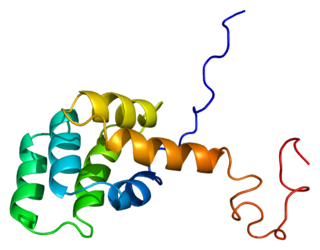
The Fas receptor, also known as Fas, FasR, apoptosis antigen 1, cluster of differentiation 95 (CD95) or tumor necrosis factor receptor superfamily member 6 (TNFRSF6), is a protein that in humans is encoded by the FAS gene. Fas was first identified using a monoclonal antibody generated by immunizing mice with the FS-7 cell line. Thus, the name Fas is derived from FS-7-associated surface antigen.

<span class="mw-page-title-main">Death-inducing signaling complex</span>

The death-inducing signaling complex or DISC is a multi-protein complex formed by members of the death receptor family of apoptosis-inducing cellular receptors. A typical example is FasR, which forms the DISC upon trimerization as a result of its ligand (FasL) binding. The DISC is composed of the death receptor, FADD, and caspase 8. It transduces a downstream signal cascade resulting in apoptosis.

<span class="mw-page-title-main">BH3 interacting-domain death agonist</span> Protein-coding gene in the species Homo sapiens

The BH3 interacting-domain death agonist, or BID, gene is a pro-apoptotic member of the Bcl-2 protein family. Bcl-2 family members share one or more of the four characteristic domains of homology entitled the Bcl-2 homology (BH) domains, and can form hetero- or homodimers. Bcl-2 proteins act as anti- or pro-apoptotic regulators that are involved in a wide variety of cellular activities.

p53 upregulated modulator of apoptosis Protein-coding gene in the species Homo sapiens

The p53 upregulated modulator of apoptosis (PUMA) also known as Bcl-2-binding component 3 (BBC3), is a pro-apoptotic protein, member of the Bcl-2 protein family. In humans, the Bcl-2-binding component 3 protein is encoded by the BBC3 gene. The expression of PUMA is regulated by the tumor suppressor p53. PUMA is involved in p53-dependent and -independent apoptosis induced by a variety of signals, and is regulated by transcription factors, not by post-translational modifications. After activation, PUMA interacts with antiapoptotic Bcl-2 family members, thus freeing Bax and/or Bak which are then able to signal apoptosis to the mitochondria. Following mitochondrial dysfunction, the caspase cascade is activated ultimately leading to cell death.

<span class="mw-page-title-main">Survivin</span> Mammalian protein

Survivin, also called baculoviral inhibitor of apoptosis repeat-containing 5 or BIRC5, is a protein that, in humans, is encoded by the BIRC5 gene.

<span class="mw-page-title-main">PAC-1</span> Chemical compound

PAC-1 is a synthesized chemical compound that selectively induces apoptosis, in cancerous cells. It was granted orphan drug status by the FDA in 2016.

<span class="mw-page-title-main">Bcl-xL</span> Transmembrane molecule in the mitochondria

B-cell lymphoma-extra large (Bcl-xL), encoded by the BCL2-like 1 gene, is a transmembrane molecule in the mitochondria. It is a member of the Bcl-2 family of proteins, and acts as an anti-apoptotic protein by preventing the release of mitochondrial contents such as cytochrome c, which leads to caspase activation and ultimately, programmed cell death.

<span class="mw-page-title-main">Caspase 3</span> Protein-coding gene in the species Homo sapiens

Caspase-3 is a caspase protein that interacts with caspase-8 and caspase-9. It is encoded by the CASP3 gene. CASP3 orthologs have been identified in numerous mammals for which complete genome data are available. Unique orthologs are also present in birds, lizards, lissamphibians, and teleosts.
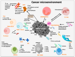
<span class="mw-page-title-main">Death receptor 4</span> Protein found in humans

Death receptor 4 (DR4), also known as TRAIL receptor 1 (TRAILR1) and tumor necrosis factor receptor superfamily member 10A (TNFRSF10A), is a cell surface receptor of the TNF-receptor superfamily that binds TRAIL and mediates apoptosis.

<span class="mw-page-title-main">Caspase 10</span> Protein-coding gene in the species Homo sapiens

Caspase-10 is an enzyme that, in humans, is encoded by the CASP10 gene.

<span class="mw-page-title-main">HU-331</span> Chemical compound

HU-331 is a quinone anticarcinogenic drug synthesized from cannabidiol, a cannabinoid in the Cannabis sativa plant. It showed a great efficacy against oncogenic human cells. HU-331 does not cause arrest in cell cycle, cell apoptosis or caspase activation. HU-331 inhibits DNA topoisomerase II even at nanomolar concentrations, but has shown a negligible effect on the action of DNA topoisomerase I. The cannabinoid quinone HU-331 is a very specific inhibitor of topoisomerase II, compared with most known anticancer quinones. One of the main objectives of these studies is the development of a new quinone derived compound that produces anti-neoplastic activity while maintaining low toxicity at therapeutic doses.

HAMLET is a complex between alpha-lactalbumin and oleic acid that has been shown in cell culture experiments to induce cell death in tumor cells, but not in healthy cells.

Cytostasis is the inhibition of cell growth and multiplication. Cytostatic refers to a cellular component or medicine that inhibits cell division.

<span class="mw-page-title-main">FL3 (flavagline)</span> Chemical compound

FL3 is a synthetic flavagline that displays potent anticancer and cardioprotectant activities. This compound induces the death of cancer cells by an original mechanism that involves the apoptosis-inducing factor and caspase 12, suggesting that it may improve the efficacy of cancer chemotherapies. It was also shown that FL3 may enhance the efficacy of one of the most widely used anticancer agents, doxorubicin, and alleviate its main adverse effect, cardiac damage.

Anticancer genes exhibit a preferential ability to kill cancer cells while leaving healthy cells unharmed. This phenomenon is achieved through various processes such as apoptosis following a mitotic catastrophe, necrosis, and autophagy. In the late 1990s, extensive research in the field of cancer cells led to the discovery of anticancer genes. Currently, 291 anticancer genes have been identified. The deregulation of these genes due to base substitutions leading to insertions, deletions, or alterations in missense amino acids can cause frameshifts, thereby altering the protein. A change in gene copy number or rearrangements is also essential for deregulating these genes. The loss or alteration of these anticancer genes due to mutations or rearrangements may lead to the development of cancer.

<span class="mw-page-title-main">Paraptosis</span> Type of programmed cell death distinct from apoptosis and necrosis

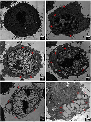
Paraptosis is a type of programmed cell death, morphologically distinct from apoptosis and necrosis. The defining features of paraptosis are cytoplasmic vacuolation, independent of caspase activation and inhibition, and lack of apoptotic morphology. Paraptosis lacks several of the hallmark characteristics of apoptosis, such as membrane blebbing, chromatin condensation, and nuclear fragmentation. Like apoptosis and other types of programmed cell death, the cell is involved in causing its own death, and gene expression is required. This is in contrast to necrosis, which is non-programmed cell death that results from injury to the cell.

References

  1. Chowdhury AR, Mandal S, Mittra B, Sharma S, Mukhopadhyay S, Majumder HK (July 2002). "Betulinic acid, a potent inhibitor of eukaryotic topoisomerase I: identification of the inhibitory step, the major functional group responsible and development of more potent derivatives". Medical Science Monitor. 8 (7): BR254–65. PMID   12118187.
  2. 1 2 Tan Y, Yu R, Pezzuto JM (July 2003). "Betulinic acid-induced programmed cell death in human melanoma cells involves mitogen-activated protein kinase activation". Clinical Cancer Research. 9 (7): 2866–75. PMID   12855667.
  3. 1 2 3 4 5 Zuco V, Supino R, Righetti SC, Cleris L, Marchesi E, Gambacorti-Passerini C, Formelli F (January 2002). "Selective cytotoxicity of betulinic acid on tumor cell lines, but not on normal cells". Cancer Letters. 175 (1): 17–25. doi:10.1016/S0304-3835(01)00718-2. PMID   11734332.
  4. Gao H, Wu L, Kuroyanagi M, Harada K, Kawahara N, Nakane T, Umehara K, Hirasawa A, Nakamura Y (November 2003). "Antitumor-promoting constituents from Chaenomeles sinensis KOEHNE and their activities in JB6 mouse epidermal cells". Chemical & Pharmaceutical Bulletin. 51 (11): 1318–21. doi: 10.1248/cpb.51.1318 . PMID   14600382. (Chaenomeles sinensis KOEHNE is now named Pseudocydonia sinensis )
  5. Abe F, Yamauchi T, Nagao T, Kinjo J, Okabe H, Higo H, Akahane H (November 2002). "Ursolic acid as a trypanocidal constituent in rosemary". Biological & Pharmaceutical Bulletin. 25 (11): 1485–7. doi: 10.1248/bpb.25.1485 . PMID   12419966.
  6. 1 2 Ji ZN, Ye WC, Liu GG, Hsiao WL (November 2002). "23-Hydroxybetulinic acid-mediated apoptosis is accompanied by decreases in bcl-2 expression and telomerase activity in HL-60 Cells". Life Sciences. 72 (1): 1–9. doi:10.1016/S0024-3205(02)02176-8. PMID   12409140.
  7. Pisha E, Chai H, Lee IS, Chagwedera TE, Farnsworth NR, Cordell GA, Beecher CW, Fong HH, Kinghorn AD, Brown DM (October 1995). "Discovery of betulinic acid as a selective inhibitor of human melanoma that functions by induction of apoptosis". Nature Medicine. 1 (10): 1046–51. doi:10.1038/nm1095-1046. PMID   7489361. S2CID   24752850.
  8. Schmidt ML, Kuzmanoff KL, Ling-Indeck L, Pezzuto JM (October 1997). "Betulinic acid induces apoptosis in human neuroblastoma cell lines". European Journal of Cancer. 33 (12): 2007–10. doi:10.1016/S0959-8049(97)00294-3. PMID   9516843.
  9. 1 2 3 4 Fulda S, Friesen C, Los M, Scaffidi C, Mier W, Benedict M, Nuñez G, Krammer PH, Peter ME, Debatin KM (November 1997). "Betulinic acid triggers CD95 (APO-1/Fas)- and p53-independent apoptosis via activation of caspases in neuroectodermal tumors". Cancer Research. 57 (21): 4956–64. PMID   9354463.
  10. Wick W, Grimmel C, Wagenknecht B, Dichgans J, Weller M (June 1999). "Betulinic acid-induced apoptosis in glioma cells: A sequential requirement for new protein synthesis, formation of reactive oxygen species, and caspase processing". The Journal of Pharmacology and Experimental Therapeutics. 289 (3): 1306–12. PMID   10336521.
  11. 1 2 3 4 Thurnher D, Turhani D, Pelzmann M, Wannemacher B, Knerer B, Formanek M, Wacheck V, Selzer E (September 2003). "Betulinic acid: a new cytotoxic compound against malignant head and neck cancer cells". Head & Neck. 25 (9): 732–40. doi:10.1002/hed.10231. PMID   12953308. S2CID   24271002.
  12. Liu X, Jutooru I, Lei P, Kim K, Lee SO, Brents LK, Prather PL, Safe S (July 2012). "Betulinic acid targets YY1 and ErbB2 through cannabinoid receptor-dependent disruption of microRNA-27a:ZBTB10 in breast cancer". Molecular Cancer Therapeutics. 11 (7): 1421–31. doi:10.1158/1535-7163.MCT-12-0026. PMC   4924623 . PMID   22553354.
  13. Raisova M, Hossini AM, Eberle J, Riebeling C, Wieder T, Sturm I, Daniel PT, Orfanos CE, Geilen CC (August 2001). "The Bax/Bcl-2 ratio determines the susceptibility of human melanoma cells to CD95/Fas-mediated apoptosis". The Journal of Investigative Dermatology. 117 (2): 333–40. doi: 10.1046/j.0022-202x.2001.01409.x . PMID   11511312.
  14. 1 2 Rieber M, Strasberg Rieber M (May 1998). "Induction of p53 without increase in p21WAF1 in betulinic acid-mediated cell death is preferential for human metastatic melanoma". DNA and Cell Biology. 17 (5): 399–406. doi:10.1089/dna.1998.17.399. PMID   9628583.
  15. 1 2 Selzer E, Pimentel E, Wacheck V, Schlegel W, Pehamberger H, Jansen B, Kodym R (May 2000). "Effects of betulinic acid alone and in combination with irradiation in human melanoma cells". The Journal of Investigative Dermatology. 114 (5): 935–40. doi: 10.1046/j.1523-1747.2000.00972.x . PMID   10771474.
  16. Novel 3,28-Disubstituted Betulinic Acid Derivatives as Potent Anti-HIV Agents Aims/Hypothesis Out-licensing. iptechex pharmalicensing, IP Technology Exchange (2013)
  17. Uldis (2022-03-10). "How to use Betulinic acid in Cosmetics". NST Chemicals. Retrieved 2023-01-07.
  18. 1 2 Li, Jing; Zhang, Yansheng (June 19, 2014). "Modulating betulinic acid production in Saccharomyces cerevisiae by managing the intracellular supplies of the co-factor NADPH and oxygen". Journal of Bioscience and Bioengineering. 119 (1): 77–81. doi:10.1016/j.jbiosc.2014.06.013. PMID   25043336.
  19. Gauthier C, Legault J, Lebrun M, Dufour P, Pichette A (October 2006). "Glycosidation of lupane-type triterpenoids as potent in vitro cytotoxic agents". Bioorganic & Medicinal Chemistry. 14 (19): 6713–25. doi:10.1016/j.bmc.2006.05.075. PMID   16787747.