Exosome complex

Last updated

"Ribbon view" of the human exosome complex. PDB 2NN6 See the legend below. The channel through which RNA passes during degradation is visible at the center of the protein complex Exosome ribbon.png
"Ribbon view" of the human exosome complex. PDB 2NN6 See the legend below. The channel through which RNA passes during degradation is visible at the center of the protein complex

The exosome complex (or PM/Scl complex, often just called the exosome) is a multi-protein intracellular complex capable of degrading various types of RNA (ribonucleic acid) molecules. Exosome complexes are found in both eukaryotic cells and archaea, while in bacteria a simpler complex called the degradosome carries out similar functions.

Contents

The core of the exosome contains a six-membered ring structure to which other proteins are attached. In eukaryotic cells, the exosome complex is present in the cytoplasm, nucleus, and especially the nucleolus, although different proteins interact with the exosome complex in these compartments regulating the RNA degradation activity of the complex to substrates specific to these cell compartments. Substrates of the exosome include messenger RNA, ribosomal RNA, and many species of small RNAs. The exosome has an exoribonucleolytic function, meaning it degrades RNA starting at one end (the 3′ end in this case), and in eukaryotes also an endoribonucleolytic function, meaning it cleaves RNA at sites within the molecule.

Several proteins in the exosome are the target of autoantibodies in patients with specific autoimmune diseases (especially the PM/Scl overlap syndrome) and some antimetabolic chemotherapies for cancer function by blocking the activity of the exosome. In addition, mutations in exosome component 3 cause pontocerebellar hypoplasia and spinal motor neuron disease.

Discovery

The exosome was first discovered as an RNase in 1997 in the budding yeast Saccharomyces cerevisiae , an often-used model organism. [1] Not long after, in 1999, it was realized that the exosome was in fact the yeast equivalent of an already described complex in human cells called the PM/Scl complex, which had been identified as an autoantigen in patients with certain autoimmune diseases years earlier (see below). [2] Purification of this "PM/Scl complex" allowed the identification of more human exosome proteins and eventually the characterization of all components in the complex. [3] [4] In 2001, the increasing amount of genome data that had become available allowed the prediction of exosome proteins in archaea, although it would take another 2 years before the first exosome complex from an archaeal organism was purified. [5] [6]

Structure

Core proteins

Top and side view of the crystal structure of the human exosome complex. See the full legend below. Exosome crystal structure.png
Top and side view of the crystal structure of the human exosome complex. See the full legend below.

The core of the complex has a ring structure consisting of six proteins that all belong to the same class of RNases, the RNase PH-like proteins. [7] In archaea there are two different PH-like proteins (called Rrp41 and Rrp42), each present three times in an alternating order. Eukaryotic exosome complexes have six different proteins that form the ring structure. [8] [9] Of these six eukaryotic proteins, three resemble the archaeal Rrp41 protein and the other three proteins are more similar to the archaeal Rrp42 protein. [10]

Subunits and organisation of the archaeal (left) and eukaryotic (right) exosome complexes. Different proteins are numbered, showing that the archaeal exosome contains 4 different proteins, but the eukaryotic exosome contains nine different proteins. See the full legend below. Exosome schematic.png
Subunits and organisation of the archaeal (left) and eukaryotic (right) exosome complexes. Different proteins are numbered, showing that the archaeal exosome contains 4 different proteins, but the eukaryotic exosome contains nine different proteins. See the full legend below.

Located on top of this ring are three proteins that have an S1 RNA binding domain (RBD). Two proteins in addition have a K-homology (KH) domain. [7] In eukaryotes, three different "S1" proteins are bound to the ring, whereas in archaea either one or two different "S1" proteins can be part of the exosome (although there are always three S1 subunits attached to the complex). [11]

This ring structure is very similar to that of the proteins RNase PH and PNPase. In bacteria, the protein RNase PH, which is involved in tRNA processing, forms a hexameric ring consisting of six identical RNase PH proteins. [12] [13] In the case of PNPase, which is a phosphorolytic RNA-degrading protein found in bacteria and the chloroplasts and mitochondria of some eukaryotic organisms, two RNase PH domains, and both an S1 and KH RNA binding domain are part of a single protein, which forms a trimeric complex that adopts a structure almost identical to that of the exosome. [14] Because of this high similarity in both protein domains and structure, these complexes are thought to be evolutionarily related and have a common ancestor. [15] The RNase PH-like exosome proteins, PNPase and RNase PH all belong to the RNase PH family of RNases and are phosphorolytic exoribonucleases, meaning that they use inorganic phosphate to remove nucleotides from the 3' end of RNA molecules. [7]

Associated proteins

Besides these nine core exosome proteins, two other proteins often associate with the complex in eukaryotic organisms. One of these is Rrp44, a hydrolytic RNase, which belongs to the RNase R family of hydrolytic exoribonucleases (nucleases that use water to cleave the nucleotide bonds). In addition to being an exoribonucleolytic enzyme, Rrp44 also has endoribonucleolytic activity, which resides in a separate domain of the protein. [16] [17] In yeast, Rrp44 is associated with all exosome complexes and has a crucial role in the activity of the yeast exosome complex. [18] While a human homologue of the protein exists, no evidence was found for a long time that its human homologue was associated with the human exosome complex. [7] In 2010, however, it was discovered that humans have three Rrp44 homologues and two of these can be associated with the exosome complex. These two proteins most likely degrade different RNA substrates due to their different cellular localization, with one being localized in the cytoplasm (DIS3L1) and the other in the nucleus (DIS3). [19] [20]

"Ribbon view" of the partial structure of the yeast exosome subunit Rrp6, 2hbj with a-helices in red and b-sheets in yellow. Partial Rrp6 structure.png
"Ribbon view" of the partial structure of the yeast exosome subunit Rrp6, 2hbj with α-helices in red and β-sheets in yellow.

The second common associated protein is called Rrp6 (in yeast) or PM/Scl-100 (in human). Like Rrp44, this protein is a hydrolytic exoribonuclease, but in this case of the RNase D protein family. [21] The protein PM/Scl-100 is most commonly part of exosome complexes in the nucleus of cells, but can form part of the cytoplasmic exosome complex as well. [22]

Regulatory proteins

Apart from these two tightly bound protein subunits, many proteins interact with the exosome complex in both the cytoplasm and nucleus of cells. These loosely associated proteins may regulate the activity and specificity of the exosome complex. In the cytoplasm, the exosome interacts with AU-rich element (ARE) binding proteins (e.g. KRSP and TTP), which can promote or prevent degradation of mRNAs. The nuclear exosome associates with RNA binding proteins (e.g. MPP6/Mpp6 and C1D/Rrp47 in humans/yeast) that are required for processing certain substrates. [7]

In addition to single proteins, other protein complexes interact with the exosome. One of those is the cytoplasmic Ski complex, which includes an RNA helicase (Ski2) and is involved in mRNA degradation. [23] In the nucleus, the processing of rRNA and snoRNA by the exosome is mediated by the TRAMP complex, which contains both RNA helicase (Mtr4) and polyadenylation (Trf4) activity. [24]

Function

Enzymatic function

Reaction diagrams for both hydrolytic (left) and phosphorolytic (right) 3' end degradation of RNA. 3' RNA degradation.png
Reaction diagrams for both hydrolytic (left) and phosphorolytic (right) 3' end degradation of RNA.

As stated above, the exosome complex contains many proteins with ribonuclease domains. The exact nature of these ribonuclease domains has changed across evolution from bacterial to archaeal to eukaryotic complexes as various activities have been gained and lost. The exosome is primarily a 3'-5' exoribonuclease, meaning that it degrades RNA molecules from their 3' end. Exoribonucleases contained in exosome complexes are either phosphorolytic (the RNase PH-like proteins) or, in eukaryotes, hydrolytic (the RNase R and RNase D domain proteins). The phosphorolytic enzymes use inorganic phosphate to cleave the phosphodiester bonds – releasing nucleotide diphosphates. The hydrolytic enzymes use water to hydrolyse these bonds – releasing nucleotide monophosphates.

In archaea, the Rrp41 subunit of the complex is a phosphorolytic exoribonuclease. Three copies of this protein are present in the ring and are responsible for the activity of the complex. [9] In eukaryotes, none of the RNase PH subunits have retained this catalytic activity, meaning the core ring structure of the human exosome has no enzymatically active protein. [25] Despite this loss of catalytic activity, the structure of the core exosome is highly conserved from archaea to humans, suggesting that the complex performs a vital cellular function. In eukaryotes, the absence of the phosphorolytic activity is compensated by the presence of the hydrolytic enzymes, which are responsible for the ribonuclease activity of the exosome in such organisms. [26] [27] [28]

As stated above, the hydrolytic proteins Rrp6 and Rrp44 are associated with the exosome in yeast and in humans, besides Rrp6, two different proteins, Dis3 and Dis3L1 can be associated at the position of the yeast Rrp44 protein. [19] [20] Although originally the S1 domain proteins were thought to have 3'-5' hydrolytic exoribonuclease activity as well, the existence of this activity has recently been questioned and these proteins might have just a role in binding substrates prior to their degradation by the complex. [26]

Schematic view of the archaeal (left) and eukaryotic (right) exosome complexes with the most common associated proteins. In color and marked with a star are the subunits of each complex that have catalytic activity. See below for a full legend. Exosome active subunits simple.png
Schematic view of the archaeal (left) and eukaryotic (right) exosome complexes with the most common associated proteins. In color and marked with a star are the subunits of each complex that have catalytic activity. See below for a full legend.

Substrates

The exosome is involved in the degradation and processing of a wide variety of RNA species. In the cytoplasm of cells, it is involved in the turn-over of messenger RNA (mRNA) molecules. The complex can degrade mRNA molecules that have been tagged for degradation because they contain errors, through interactions with proteins from the nonsense mediated decay or non-stop decay pathways. In alternative fashion, mRNAs are degraded as part of their normal turnover. Several proteins that stabilize or destabilize mRNA molecules through binding to AU-rich elements in the 3' untranslated region of mRNAs interact with the exosome complex. [29] [30] [31] In the nucleus, the exosome is required for the correct processing of several small nuclear RNA molecules. [32] Finally, the nucleolus is the compartment where the majority of the exosome complexes are found. There it plays a role in the processing of the 5.8S ribosomal RNA (the first identified function of the exosome) and of several small nucleolar RNAs. [1] [32] [33]

Although most cells have other enzymes that can degrade RNA, either from the 3' or from the 5' end of the RNA, the exosome complex is essential for cell survival. When the expression of exosome proteins is artificially reduced or stopped, for example by RNA interference, growth stops and the cells eventually die. Both the core proteins of the exosome complex, as well as the two main associated proteins, are essential proteins. [34] Bacteria do not have an exosome complex; however, similar functions are performed by a simpler complex that includes the protein PNPase, called the degradosome . [35]

Two core subunits of the archaeal exosome (Rrp41 and Rrp42), bound to a small RNA molecule (in red). Exosome core with RNA.png
Two core subunits of the archaeal exosome (Rrp41 and Rrp42), bound to a small RNA molecule (in red).

The exosome is a key complex in cellular RNA quality control. Unlike prokaryotes, eukaryotes possess highly active RNA surveillance systems that recognise unprocessed and mis-processed RNA-protein complexes (such as ribosomes) prior to their exit from the nucleus. It is presumed that this system prevents aberrant complexes from interfering with important cellular processes such as protein synthesis. [36]

In addition to RNA processing, turnover and surveillance activities, the exosome is important for the degradation of so-called cryptic unstable transcripts (CUTs) that are produced from thousands of loci within the yeast genome. [37] [38] The importance of these unstable RNAs and their degradation are still unclear, but similar RNA species have also been detected in human cells. [39]

Disease

Autoimmunity

The exosome complex is the target of autoantibodies in patients with various autoimmune diseases. These autoantibodies are mainly found in people with the PM/Scl overlap syndrome, an autoimmune disease in which patients have symptoms from both scleroderma and either polymyositis or dermatomyositis. [40] Autoantibodies can be detected in the serum of patients by a variety of assays. In the past, the most commonly used methods were double immunodiffusion using calf thymus extracts, immunofluorescence on HEp-2 cells or immunoprecipitation from human cell extracts. In immunoprecipitation assays with sera from anti-exosome positive sera, a distinctive set of proteins is precipitated. Already years before the exosome complex was identified, this pattern was termed the PM/Scl complex. [41] Immunofluorescence using sera from these patients usually shows a typical staining of the nucleolus of cells, which sparked the suggestion that the antigen recognized by autoantibodies might be important in ribosome synthesis. [42] More recently, recombinant exosome proteins have become available and these have been used to develop line immunoassays (LIAs) and enzyme linked immunosorbent assays (ELISAs) for detecting these antibodies. [7]

In these diseases, antibodies are mainly directed against two of the proteins of the complex, called PM/Scl-100 (the RNase D like protein) and PM/Scl-75 (one of the RNase PH like proteins from the ring) and antibodies recognizing these proteins are found in approximately 30% of patients with the PM/Scl overlap syndrome. [43] Although these two proteins are the main target of the autoantibodies, other exosome subunits and associated proteins (like C1D) can be targeted in these patients. [44] [45] At the current time, the most sensitive way to detect these antibodies is by using a peptide, derived from the PM/Scl-100 protein, as the antigen in an ELISA, instead of complete proteins. By this method, autoantibodies are found in up to 55% of patients with the PM/Scl overlap syndrome, but they can also be detected in patients with either scleroderma, polymyositis, or dermatomyositis alone. [46]

As the autobodies are found mainly in patients that have characteristics of several different autoimmune diseases, the clinical symptoms of these patients can vary widely. The symptoms that are seen most often are the typical symptoms of the individual autoimmune diseases and include Raynaud's phenomenon, arthritis, myositis and scleroderma. [47] Treatment of these patients is symptomatic and is similar to treatment for the individual autoimmune disease, often involving either immunosuppressive or immunomodulating drugs. [48]

Cancer treatment

The exosome has been shown to be inhibited by the antimetabolite fluorouracil, a drug used in the chemotherapy of cancer. It is one of the most successful drugs for treating solid tumors. In yeast cells treated with fluorouracil, defects were found in the processing of ribosomal RNA identical to those seen when the activity of the exosome was blocked by molecular biological strategies. Lack of correct ribosomal RNA processing is lethal to cells, explaining the antimetabolic effect of the drug. [49]

Neurological disorders

Mutations in exosome component 3 cause infantile spinal motor neuron disease, cerebellar atrophy, progressive microcephaly and profound global developmental delay, consistent with pontocerebellar hypoplasia type 1B (PCH1B; MIM 614678). [50]

List of subunits

LegendGeneral nameDomainsHumanYeast (S. cerevisiae)Archaea MW (kD) Human gene Yeast gene
1Csl4S1 RBDhCsl4Csl4p/Ski4pCsl421–32 EXOSC1 YNL232W
2Rrp4S1/KH RBDhRrp4Rrp4pRrp428–39 EXOSC2 YHR069C
3Rrp40S1/KH RBDhRrp40Rrp40p(Rrp4) A 27–32 EXOSC3 YOL142W
4Rrp41RNase PHhRrp41Rrp41p/Ski6pRrp41 C 26–28 EXOSC4 YGR195W
5Rrp46RNase PHhRrp46Rrp46p(Rrp41) A, C 25–28 EXOSC5 YGR095C
6Mtr3RNase PHhMtr3Mtr3p(Rrp41) A, C 24–37 EXOSC6 YGR158C
7Rrp42RNase PHhRrp42Rrp42pRrp4229–32 EXOSC7 YDL111C
8Rrp43RNase PHOIP2Rrp43p(Rrp42) A 30–44 EXOSC8 YCR035C
9Rrp45RNase PHPM/Scl-75Rrp45p(Rrp42) A 34–49 EXOSC9 YDR280W
10Rrp6RNase DPM/Scl-100 C Rrp6p C n/a84–100 EXOSC10 YOR001W
11Rrp44RNase RDis3 B, C

Dis3L1 B, C

Rrp44p/Dis3p C n/a105–113 DIS3

DIS3L1

YOL021C

See also

Related Research Articles

<span class="mw-page-title-main">Exonuclease</span> Class of enzymes; type of nuclease

Exonucleases are enzymes that work by cleaving nucleotides one at a time from the end (exo) of a polynucleotide chain. A hydrolyzing reaction that breaks phosphodiester bonds at either the 3′ or the 5′ end occurs. Its close relative is the endonuclease, which cleaves phosphodiester bonds in the middle (endo) of a polynucleotide chain. Eukaryotes and prokaryotes have three types of exonucleases involved in the normal turnover of mRNA: 5′ to 3′ exonuclease (Xrn1), which is a dependent decapping protein; 3′ to 5′ exonuclease, an independent protein; and poly(A)-specific 3′ to 5′ exonuclease.

<span class="mw-page-title-main">Ribonuclease P</span> Class of enzymes

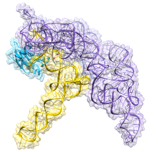
Ribonuclease P is a type of ribonuclease which cleaves RNA. RNase P is unique from other RNases in that it is a ribozyme – a ribonucleic acid that acts as a catalyst in the same way that a protein-based enzyme would. Its function is to cleave off an extra, or precursor, sequence of RNA on tRNA molecules. Further, RNase P is one of two known multiple turnover ribozymes in nature, the discovery of which earned Sidney Altman and Thomas Cech the Nobel Prize in Chemistry in 1989: in the 1970s, Altman discovered the existence of precursor tRNA with flanking sequences and was the first to characterize RNase P and its activity in processing of the 5' leader sequence of precursor tRNA. Recent findings also reveal that RNase P has a new function. It has been shown that human nuclear RNase P is required for the normal and efficient transcription of various small noncoding RNAs, such as tRNA, 5S rRNA, SRP RNA and U6 snRNA genes, which are transcribed by RNA polymerase III, one of three major nuclear RNA polymerases in human cells.

<span class="mw-page-title-main">Polynucleotide phosphorylase</span> Class of enzymes

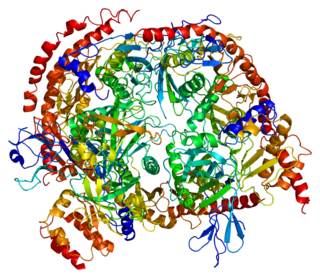
Polynucleotide Phosphorylase (PNPase) is a bifunctional enzyme with a phosphorolytic 3' to 5' exoribonuclease activity and a 3'-terminal oligonucleotide polymerase activity. That is, it dismantles the RNA chain starting at the 3' end and working toward the 5' end. It also synthesizes long, highly heteropolymeric tails in vivo. It accounts for all of the observed residual polyadenylation in strains of Escherichia coli missing the normal polyadenylation enzyme. Discovered by Marianne Grunberg-Manago working in Severo Ochoa's lab in 1955, the RNA-polymerization activity of PNPase was initially believed to be responsible for DNA-dependent synthesis of messenger RNA, a notion that was disproven by the late 1950s.

<span class="mw-page-title-main">Exoribonuclease</span> Class of enzymes that degrade RNA

An exoribonuclease is an exonuclease ribonuclease, which are enzymes that degrade RNA by removing terminal nucleotides from either the 5' end or the 3' end of the RNA molecule. Enzymes that remove nucleotides from the 5' end are called 5'-3' exoribonucleases, and enzymes that remove nucleotides from the 3' end are called 3'-5' exoribonucleases.

<span class="mw-page-title-main">Non-stop decay</span>

Non-stop decay (NSD) is a cellular mechanism of mRNA surveillance to detect mRNA molecules lacking a stop codon and prevent these mRNAs from translation. The non-stop decay pathway releases ribosomes that have reached the far 3' end of an mRNA and guides the mRNA to the exosome complex, or to RNase R in bacteria for selective degradation. In contrast to nonsense-mediated decay (NMD), polypeptides do not release from the ribosome, and thus, NSD seems to involve mRNA decay factors distinct from NMD.

<span class="mw-page-title-main">TRAMP complex</span>

TRAMP complex is a multiprotein, heterotrimeric complex having distributive polyadenylation activity and identifies wide varieties of RNAs produced by polymerases. It was originally discovered in Saccharomycescerevisiae by LaCava et al., Vanacova et al. and Wyers et al. in 2005.

<span class="mw-page-title-main">Exosome component 2</span> Protein-coding gene in the species Homo sapiens

Exosome component 2, also known as EXOSC2, is a protein which in humans is encoded by the EXOSC2 gene.

<span class="mw-page-title-main">Exosome component 10</span> Protein-coding gene in the species Homo sapiens

Exosome component 10, also known as EXOSC10, is a human gene, the protein product of which is part of the exosome complex and is an autoantigen is patients with certain auto immune diseases, most notably scleromyositis.

<span class="mw-page-title-main">DIS3</span> Protein-coding gene in the species Homo sapiens

Exosome complex exonuclease RRP44 or Dis3 is an enzyme that in humans is encoded by the DIS3 gene. Its protein product is an RNase enzyme homologous to the yeast protein Rrp44, and can be part of the exosome complex in the nucleus of eukaryotic cells.

<span class="mw-page-title-main">Exosome component 8</span> Protein-coding gene in the species Homo sapiens

Exosome component 8, also known as EXOSC8, is a human gene, the protein product of which is part of the exosome complex.

<span class="mw-page-title-main">Exosome component 7</span> Protein-coding gene in the species Homo sapiens

Exosome component 7, also known as EXOSC7, is a human gene, the protein product of which is part of the exosome complex.

<span class="mw-page-title-main">Exosome component 3</span> Protein-coding gene in the species Homo sapiens

Exosome component 3, also known as EXOSC3, is a human gene, which is part of the exosome complex.

<span class="mw-page-title-main">Exosome component 9</span> Protein-coding gene in the species Homo sapiens

Exosome component 9, also known as EXOSC9, is a human gene, the protein product of which is part of the exosome complex and is an autoantigen is patients with certain auto immune diseases, most notably scleromyositis.

<span class="mw-page-title-main">Exosome component 4</span> Protein-coding gene in the species Homo sapiens

Exosome component 4, also known as EXOSC4, is a human gene, which is part of the exosome complex.

<span class="mw-page-title-main">SKIV2L2</span> Protein-coding gene in the species Homo sapiens

Superkiller viralicidic activity 2-like 2 is a protein that in humans is encoded by the SKIV2L2 gene.

<span class="mw-page-title-main">Exosome component 1</span> Protein-coding gene in the species Homo sapiens

3'-5' exoribonuclease CSL4 homolog is an enzyme that in humans is encoded by the EXOSC1 gene.

<span class="mw-page-title-main">Exosome component 5</span> Protein-coding gene in the species Homo sapiens

Exosome component 5, also known as EXOSC5, is a human gene, which is part of the exosome complex.

<span class="mw-page-title-main">C1D</span> Protein-coding gene in the species Homo sapiens

Nuclear nucleic acid-binding protein C1D is a protein that in humans is encoded by the C1D gene. The C1D protein is encoded by a DNA binding gene traced in the nucleus. Protein C1D has a chromosomal location of 2p14. C1D has a family of proteins consisting of C1D homologues which may include Sas10 domains. C1D is thought to bind to RNA and DNA where it may be involved in mechanisms of DNA repair. Protein C1D is ubiquitously expressed in different human tissues.

<span class="mw-page-title-main">Exosome component 6</span> Protein-coding gene in the species Homo sapiens

Exosome complex exonuclease MTR3 is an enzyme that in humans is encoded by the EXOSC6 gene.

<span class="mw-page-title-main">DIS3L</span> Protein-coding gene in the species Homo sapiens

DIS3-like exonuclease 1 is an enzyme that in humans is encoded by the DIS3L gene. Its protein product is an RNase enzyme homologous to the yeast protein Rrp44, and can be part of the exosome complex in the cytoplasm of eukaryotic cells.

References

  1. 1 2 Mitchell, P; Petfalski, E; Shevchenko, A; Mann, M; Tollervey, D (1997). "The Exosome: A Conserved Eukaryotic RNA Processing Complex Containing Multiple 3′→5′ Exoribonucleases". Cell . 91 (4): 457–466. doi: 10.1016/S0092-8674(00)80432-8 . PMID   9390555. S2CID   16035676.
  2. Allmang, C; Petfalski, E; Podtelejnikov, A; Mann, M; Tollervey, D; Mitchell, P (1999). "The yeast exosome and human PM-Scl are related complexes of 3' → 5' exonucleases". Genes & Development. 13 (16): 2148–58. doi:10.1101/gad.13.16.2148. PMC   316947 . PMID   10465791.
  3. Brouwer, R; Allmang, C; Raijmakers, R; Van Aarssen, Y; Egberts, WV; Petfalski, E; Van Venrooij, WJ; Tollervey, D; Pruijn, GJ (2001). "Three novel components of the human exosome". Journal of Biological Chemistry. 276 (9): 6177–84. doi: 10.1074/jbc.M007603200 . hdl: 2066/186951 . PMID   11110791.
  4. Chen, CY; Gherzi, R; Ong, SE; Chan, EL; Raijmakers, R; Pruijn, GJ; Stoecklin, G; Moroni, C; et al. (2001). "AU binding proteins recruit the exosome to degrade ARE-containing mRNAs". Cell. 107 (4): 451–64. doi: 10.1016/S0092-8674(01)00578-5 . PMID   11719186. S2CID   14817671.
  5. Koonin, EV; Wolf, YI; Aravind, L (2001). "Prediction of the archaeal exosome and its connections with the proteasome and the translation and transcription machineries by a comparative-genomic approach". Genome Research. 11 (2): 240–52. doi:10.1101/gr.162001. PMC   311015 . PMID   11157787.
  6. Evguenieva-Hackenberg, E; Walter, P; Hochleitner, E; Lottspeich, F; Klug, G (2003). "An exosome-like complex in Sulfolobus solfataricus". EMBO Reports . 4 (9): 889–93. doi:10.1038/sj.embor.embor929. PMC   1326366 . PMID   12947419.
  7. 1 2 3 4 5 6 Schilders, G; Van Dijk, E; Raijmakers, R; Pruijn, GJ (2006). Cell and molecular biology of the exosome: how to make or break an RNA. International Review of Cytology. Vol. 251. pp. 159–208. doi:10.1016/S0074-7696(06)51005-8. ISBN   9780123646552. PMID   16939780.
  8. Lorentzen, E; Walter, P; Fribourg, S; Evguenieva-Hackenberg, E; Klug, G; Conti, E (2005). "The archaeal exosome core is a hexameric ring structure with three catalytic subunits". Nature Structural & Molecular Biology. 12 (7): 575–81. doi:10.1038/nsmb952. PMID   15951817. S2CID   2003922.
  9. 1 2 Shen, V; Kiledjian, M (2006). "A view to a kill: structure of the RNA exosome". Cell. 127 (6): 1093–5. doi:10.1016/j.cell.2006.11.035. PMC   1986773 . PMID   17174886.
  10. Raijmakers, R; Egberts, WV; Van Venrooij, WJ; Pruijn, GJ (2002). "Protein-protein interactions between human exosome components support the assembly of RNase PH-type subunits into a six-membered PNPase-like ring". Journal of Molecular Biology. 323 (4): 653–63. doi:10.1016/S0022-2836(02)00947-6. hdl: 2066/186665 . PMID   12419256.
  11. Walter, P; Klein, F; Lorentzen, E; Ilchmann, A; Klug, G; Evguenieva-Hackenberg, E (2006). "Characterization of native and reconstituted exosome complexes from the hyperthermophilic archaeon Sulfolobus solfataricus". Molecular Microbiology. 62 (4): 1076–89. doi:10.1111/j.1365-2958.2006.05393.x. PMID   17078816. S2CID   27114625.
  12. Ishii, R; Nureki, O; Yokoyama, S (2003). "Crystal structure of the tRNA processing enzyme RNase PH from Aquifex aeolicus". Journal of Biological Chemistry. 278 (34): 32397–404. doi: 10.1074/jbc.M300639200 . PMID   12746447.
  13. Harlow, LS; Kadziola, A; Jensen, KF; Larsen, S (2004). "Crystal structure of the phosphorolytic exoribonuclease RNase PH from Bacillus subtilis and implications for its quaternary structure and tRNA binding". Protein Science. 13 (3): 668–77. doi:10.1110/ps.03477004. PMC   2286726 . PMID   14767080.
  14. Symmons, MF; Jones, GH; Luisi, BF (2000). "A duplicated fold is the structural basis for polynucleotide phosphorylase catalytic activity, processivity, and regulation". Structure. 8 (11): 1215–26. doi: 10.1016/S0969-2126(00)00521-9 . PMID   11080643.
  15. Lin-Chao, S; Chiou, NT; Schuster, G (2007). "The PNPase, exosome and RNA helicases as the building components of evolutionarily-conserved RNA degradation machines". Journal of Biomedical Science. 14 (4): 523–32. doi: 10.1007/s11373-007-9178-y . PMID   17514363.
  16. Lebreton, A; Tomecki, R; Dziembowski, A; Séraphin, B (2008). "Endonucleolytic RNA cleavage by a eukaryotic exosome" (PDF). Nature. 456 (7224): 993–6. Bibcode:2008Natur.456..993L. doi:10.1038/nature07480. PMID   19060886. S2CID   1808371.
  17. Schneider, C; Leung, E; Brown, J; Tollervey, D (2009). "The N-terminal PIN domain of the exosome subunit Rrp44 harbors endonuclease activity and tethers Rrp44 to the yeast core exosome". Nucleic Acids Research. 37 (4): 1127–40. doi:10.1093/nar/gkn1020. PMC   2651783 . PMID   19129231.
  18. Schneider, C; Anderson, JT; Tollervey, D (2007). "The exosome subunit Rrp44 plays a direct role in RNA substrate recognition". Molecular Cell. 27 (2): 324–31. doi:10.1016/j.molcel.2007.06.006. PMC   7610968 . PMID   17643380.
  19. 1 2 Staals, RH; Bronkhorst, AW; Schilders, G; Slomovic, S; Schuster, G; Heck, AJ; Raijmakers, R; Pruijn, GJ (2010). "Dis3-like 1: a novel exoribonuclease associated with the human exosome". The EMBO Journal. 29 (14): 2358–67. doi:10.1038/emboj.2010.122. PMC   2910272 . PMID   20531389.
  20. 1 2 Tomecki, R; Kristiansen, MS; Lykke-Andersen, S; Chlebowski, A; Larsen, KM; Szczesny, RJ; Drazkowska, K; Pastula, A; et al. (2010). "The human core exosome interacts with differentially localized processive RNases: hDIS3 and hDIS3L". The EMBO Journal. 29 (14): 2342–57. doi:10.1038/emboj.2010.121. PMC   2910271 . PMID   20531386.
  21. Mian, IS (1997). "Comparative sequence analysis of ribonucleases HII, III, II PH and D". Nucleic Acids Research. 25 (16): 3187–3195. doi:10.1093/nar/25.16.3187. PMC   146874 . PMID   9241229.
  22. Raijmakers, R; Schilders, G; Pruijn, GJ (2004). "The exosome, a molecular machine for controlled RNA degradation in both nucleus and cytoplasm". European Journal of Cell Biology. 83 (5): 175–83. doi:10.1078/0171-9335-00385. PMID   15346807.
  23. Wang, L; Lewis, MS; Johnson, AW (2005). "Domain interactions within the Ski2/3/8 complex and between the Ski complex and Ski7p". RNA. 11 (8): 1291–302. doi:10.1261/rna.2060405. PMC   1370812 . PMID   16043509.
  24. LaCava, J; Houseley, J; Saveanu, C; Petfalski, E; Thompson, E; Jacquier, A; Tollervey, D (2005). "RNA degradation by the exosome is promoted by a nuclear polyadenylation complex". Cell. 121 (5): 713–24. doi: 10.1016/j.cell.2005.04.029 . PMID   15935758. S2CID   14898055.
  25. Liu, Q; Greimann, JC; Lima, CD (2007). "Erratum: Reconstitution, activities, and structure of the eukaryotic RNA exosome". Cell . 131 (1): 188–189. doi: 10.1016/j.cell.2007.09.019 .
  26. 1 2 Dziembowski, A; Lorentzen, E; Conti, E; Séraphin, B (2007). "A single subunit, Dis3, is in essence responsible for yeast exosome core activity". Nature Structural & Molecular Biology. 14 (1): 15–22. doi:10.1038/nsmb1184. PMID   17173052. S2CID   24691764.
  27. Liu, Q; Greimann, JC; Lima, CD (2006). "Reconstitution, activities, and structure of the eukaryotic RNA exosome". Cell . 127 (6): 1223–37. doi: 10.1016/j.cell.2006.10.037 . PMID   17174896. S2CID   62785677.
  28. Lorentzen, E; Conti, E (2005). "Structural basis of 3' end RNA recognition and exoribonucleolytic cleavage by an exosome RNase PH core". Molecular Cell. 20 (3): 473–81. doi: 10.1016/j.molcel.2005.10.020 . PMID   16285928.
  29. LeJeune, F; Li, X; Maquat, LE (2003). "Nonsense-mediated mRNA decay in mammalian cells involves decapping, deadenylating, and exonucleolytic activities". Molecular Cell. 12 (3): 675–87. doi: 10.1016/S1097-2765(03)00349-6 . PMID   14527413.
  30. Wilson, MA; Meaux, S; Van Hoof, A (2007). "A genomic screen in yeast reveals novel aspects of nonstop mRNA metabolism". Genetics. 177 (2): 773–84. doi:10.1534/genetics.107.073205. PMC   2034642 . PMID   17660569.
  31. Lin, WJ; Duffy, A; Chen, CY (2007). "Localization of AU-rich element-containing mRNA in cytoplasmic granules containing exosome subunits". Journal of Biological Chemistry. 282 (27): 19958–68. doi: 10.1074/jbc.M702281200 . PMID   17470429.
  32. 1 2 Allmang, C; Kufel, J; Chanfreau, G; Mitchell, P; Petfalski, E; Tollervey, D (1999). "Functions of the exosome in rRNA, snoRNA and snRNA synthesis". EMBO Journal. 18 (19): 5399–410. doi:10.1093/emboj/18.19.5399. PMC   1171609 . PMID   10508172.
  33. Schilders, G; Raijmakers, R; Raats, JM; Pruijn, GJ (2005). "MPP6 is an exosome-associated RNA-binding protein involved in 5.8S rRNA maturation". Nucleic Acids Research. 33 (21): 6795–804. doi:10.1093/nar/gki982. PMC   1310903 . PMID   16396833.
  34. van Dijk, EL; Schilders, G; Pruijn, GJ (2007). "Human cell growth requires a functional cytoplasmic exosome, which is involved in various mRNA decay pathways". RNA. 13 (7): 1027–35. doi:10.1261/rna.575107. PMC   1894934 . PMID   17545563.
  35. Carpousis AJ, AJ (2002). "The Escherichia coli RNA degradosome: structure, function and relationship in other ribonucleolytic multienzyme complexes". Biochem. Soc. Trans. 30 (2): 150–5. doi:10.1042/BST0300150. PMID   12035760.
  36. Houseley J, LaCava J, Tollervey D (July 2006). "RNA-quality control by the exosome". Nat. Rev. Mol. Cell Biol. 7 (7): 529–39. doi:10.1038/nrm1964. PMID   16829983. S2CID   22499032.
  37. Wyers F, F; Rougemaille, M; Badis, G; et al. (June 2005). "Cryptic pol II transcripts are degraded by a nuclear quality control pathway involving a new poly(A) polymerase". Cell. 121 (5): 725–37. doi: 10.1016/j.cell.2005.04.030 . PMID   15935759. S2CID   1390706.
  38. Neil H, Malabat C, d'Aubenton-Carafa Y, Xu Z, Steinmetz LM, Jacquier A (February 2009). "Widespread bidirectional promoters are the major source of cryptic transcripts in yeast". Nature. 457 (7232): 1038–42. Bibcode:2009Natur.457.1038N. doi:10.1038/nature07747. PMID   19169244. S2CID   4329373.
  39. Preker P, P; Nielsen, J; Kammler, S; et al. (December 2008). "RNA exosome depletion reveals transcription upstream of active human promoters". Science. 322 (5909): 1851–4. Bibcode:2008Sci...322.1851P. doi: 10.1126/science.1164096 . PMID   19056938.
  40. J.E. Pope, JE (2002). "Scleroderma overlap syndromes". Current Opinion in Rheumatology. 14 (6): 704–10. doi:10.1097/00002281-200211000-00013. PMID   12410095.
  41. Gelpi, C; Algueró, A; Angeles Martinez, M; Vidal, S; Juarez, C; Rodriguez-Sanchez, JL (1991). "Identification of protein components reactive with anti-PM/Scl autoantibodies". Clinical and Experimental Immunology. 81 (1): 59–64. doi:10.1111/j.1365-2249.1990.tb05291.x. PMC   1535032 . PMID   2199097.
  42. Targoff, IN; Reichlin, M (1985). "Nucleolar localization of the PM-Scl antigen". Arthritis & Rheumatism. 28 (2): 226–30. doi:10.1002/art.1780280221. PMID   3918546.
  43. Raijmakers, R; Renz, M; Wiemann, C; Egberts, WV; Seelig, HP; Van Venrooij, WJ; Pruijn, GJ (2004). "PM-Scl-75 is the main autoantigen in patients with the polymyositis/scleroderma overlap syndrome". Arthritis & Rheumatism. 50 (2): 565–9. doi:10.1002/art.20056. PMID   14872500.
  44. Brouwer, R; Vree Egberts, WT; Hengstman, GJ; Raijmakers, R; Van Engelen, BG; Seelig, HP; Renz, M; Mierau, R; et al. (2002). "Autoantibodies directed to novel components of the PM/Scl complex, the human exosome". Arthritis Research & Therapy . 4 (2): 134–8. doi: 10.1186/ar389 . PMC   83843 . PMID   11879549.
  45. Schilders, G; Egberts, WV; Raijmakers, R; Pruijn, GJ (2007). "C1D is a major autoantibody target in patients with the polymyositis-scleroderma overlap syndrome". Arthritis & Rheumatism. 56 (7): 2449–54. doi:10.1002/art.22710. PMID   17599775.
  46. Mahler, M; Raijmakers, R; Dähnrich, C; Blüthner, M; Fritzler, MJ (2005). "Clinical evaluation of autoantibodies to a novel PM/Scl peptide antigen". Arthritis Research & Therapy. 7 (3): R704–13. doi: 10.1186/ar1729 . PMC   1174964 . PMID   15899056.
  47. Mahler, M; Raijmakers, R (2007). "Novel aspects of autoantibodies to the PM/Scl complex: Clinical, genetic and diagnostic insights". Autoimmunity Reviews. 6 (7): 432–7. doi:10.1016/j.autrev.2007.01.013. PMID   17643929.
  48. Jablonska, S; Blaszczyk, M (1998). "Scleromyositis: a scleroderma/polymyositis overlap syndrome". Clinical Rheumatology. 17 (6): 465–7. doi:10.1007/BF01451281. PMID   9890673. S2CID   39237322.
  49. Lum, PY; Armour, CD; Stepaniants, SB; Cavet, G; Wolf, MK; Butler, JS; Hinshaw, JC; Garnier, P; et al. (2004). "Discovering modes of action for therapeutic compounds using a genome-wide screen of yeast heterozygotes". Cell. 116 (1): 121–37. doi: 10.1016/S0092-8674(03)01035-3 . PMID   14718172. S2CID   11922219.
  50. Wan, J.; Yourshaw, M.; Mamsa, H.; Rudnik-Schöneborn, S.; Menezes, M. P.; Hong, J. E.; Leong, D. W.; Senderek, J.; Salman, M. S.; Chitayat, D.; Seeman, P.; Von Moers, A.; Graul-Neumann, L.; Kornberg, A. J.; Castro-Gago, M.; Sobrido, M. A. J. S.; Sanefuji, M.; Shieh, P. B.; Salamon, N.; Kim, R. C.; Vinters, H. V.; Chen, Z.; Zerres, K.; Ryan, M. M.; Nelson, S. F.; Jen, J. C. (2012). "Mutations in the RNA exosome component gene EXOSC3 cause pontocerebellar hypoplasia and spinal motor neuron degeneration". Nature Genetics. 44 (6): 704–708. doi:10.1038/ng.2254. PMC   3366034 . PMID   22544365.

Further reading