ISCU | |||||||||||||||||||||||||||||||||||||||||||||||||||
---|---|---|---|---|---|---|---|---|---|---|---|---|---|---|---|---|---|---|---|---|---|---|---|---|---|---|---|---|---|---|---|---|---|---|---|---|---|---|---|---|---|---|---|---|---|---|---|---|---|---|---|
| |||||||||||||||||||||||||||||||||||||||||||||||||||
Identifiers | |||||||||||||||||||||||||||||||||||||||||||||||||||
Aliases | ISCU , 2310020H20Rik, HML, ISU2, NIFU, NIFUN, hnifU, iron-sulfur cluster assembly enzyme | ||||||||||||||||||||||||||||||||||||||||||||||||||
External IDs | OMIM: 611911 MGI: 1913633 HomoloGene: 6991 GeneCards: ISCU | ||||||||||||||||||||||||||||||||||||||||||||||||||
| |||||||||||||||||||||||||||||||||||||||||||||||||||
| |||||||||||||||||||||||||||||||||||||||||||||||||||
| |||||||||||||||||||||||||||||||||||||||||||||||||||
| |||||||||||||||||||||||||||||||||||||||||||||||||||
Wikidata | |||||||||||||||||||||||||||||||||||||||||||||||||||
|
Iron-sulfur cluster assembly enzyme ISCU, mitochondrial is a protein that in humans is encoded by the ISCU gene. [5] It encodes an iron-sulfur (Fe-S) cluster scaffold protein involved in [ 2Fe-2S] and [ 4Fe-4S] cluster synthesis and maturation. [6] [7] [8] [9] A deficiency of ISCU is associated with a mitochondrial myopathy with lifelong exercise intolerance where only minor exertion causes tachycardia, shortness of breath, muscle weakness and myalgia. [10]
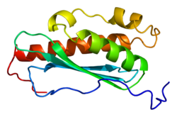
ISCU is located on the q arm of chromosome 12 in position 23.3 and has 8 exons. [7] ISCU, the protein encoded by this gene, is a member of the NifU family. It is an iron-sulfur transferase that contains binding sites for [ 2Fe-2S] and [ 4Fe-4S] clusters. ISCU contains a transit peptide, 4 beta strands, 4 alpha helixes, and 4 turns. [8] [9] Alternative splicing results in transcript variants encoding different protein isoforms that localize either to the cytosol or to the mitochondrion. A pseudogene of this gene is present on chromosome 1. [7]
ISCU encodes a component of the iron-sulfur (Fe-S) cluster scaffold responsible for the synthesis and maturation of [ 2Fe-2S] and [ 4Fe-4S] clusters. Fe-S clusters are cofactors that play a role in the function of a diverse set of enzymes, including those that regulate metabolism, iron homeostasis, and oxidative stress response. In one process, the [2Fe-2S] cluster transiently assembles on ISCU and is then transferred to GLRX5 in a cysteine desulfurase complex NFS1-LYRM4/ISD11 dependent process. [7] [6] [8] [9]
ISCU has two isoforms, isoform 1, which is found in the mitochondrion and isoform 2, which is found in the nucleus and cytoplasm. [8] [9]
ISCU mutations have been found in patients with a mitochondrial myopathy called hereditary myopathy with lactic acidosis (HML). [11] It is also known as hereditary myopathy with exercise intolerance, Swedish type; [11] myopathy with deficiency of succinate dehydrogenase and aconitase; [11] myoglobinuria due to abnormal glycolysis; [11] myopathy with deficiency of ISCU; [10] Larsson–Linderholm syndrome; [12] and Linderholm myopathy. [13]
This disease is a result of a deficiency of ISCU that corresponds to the deficiency of mitochondrial iron-sulfur proteins and impaired muscle oxidative metabolism. [7] Characteristics of mitochondrial myopathy with deficiency of ISCU may include lifelong exercise intolerance in which exertion can cause tachycardia, dyspnoea, cardiac palpitations, shortness of breath, fatigue, pain of active muscles, rhabdomyolysis, myoglobinuria, elevated lactate and pyruvate, decreased oxygen utilization, may have a pseudoathletic appearance of hypertrophic calf muscles, and possibly weakness. [14] [15] [8] [9] [10] [16] Primarily affecting the skeletal muscles, rarely there is also hypertrophic cardiomyopathy and ptosis (drooping eyelid). [11] [17]
Biopsy of skeletal muscle shows deficiency of succinate dehydrogenase and aconitase, abnormal iron deposition and lipid droplet accumulation in the mitochondria. Histochemical studies show impaired respiratory chain complexes I, II, and III, impairing oxidative phosphorylation. Electromyography normal or myopathic increased polyphasic MUAPs. EMG results may be dynamic: more likely to have increased polyphasic MUAPs after exercise. [14] There has been documented temporary restoration of succinate dehydrogenase in regenerating muscle after an episode of rhabdomyolysis; however, the effect does not last. [18]
The disease is limited to muscle, with fibroblasts from skin biopsy being unaffected. As non-muscle tissues are unaffected, this necessitates the need for muscle biopsy when DNA testing using saliva or blood is inconclusive. [19] [17]
This disorder has been associated with several different mutations and is inherited predominantly in an autosomal recessive manner. It was originally believed to affect only those of northern Swedish ancestry, however the disease has been found in those of Norwegian and Finnish decent as well. The carrier rate in northern Sweden has been estimated at 1:188. [15] ISCU deficiency has been linked to pathogenic variants including intronic variants c.418+382G>C, g.7044G>C, [19] and IVS5+382 G>C [20] as well as a c.149G>A missense mutation in exon 3. [21] The intronic mutations have been suggested to activate a cryptic splice site, resulting in the production of a splice variant that encodes a putatively non-functional protein. [10]
In 2016, an Italian male was found to have a de novo autosomal dominant mutation, c.287G>T (p.Gly96Val) in the ISCU gene, that caused hereditary mitochondrial myopathy with lactic acidosis (HML). This mutation resulted in a similar phenotype as seen in the recessive form of the disease. [17]
ISCU has been shown to have 235 binary protein-protein interactions including 79 co-complex interactions. ISCU appears to interact with ISCS, NUP62, SDHB, HPRT1, CCDC172, GOLGA2, IKZF1, KRT40, AGTRAP, NECAB2, FAM9B, BANP, LNX1, MID2, GOLGA6L9, ccdc136, KRT34, SPERT, PICK1, YWHAB, SFN, mbl, E7, dnaX, hscB, MAPk-Ak2, hale, and cv-c. [22]
The coenzyme Q : cytochrome c – oxidoreductase, sometimes called the cytochrome bc1 complex, and at other times complex III, is the third complex in the electron transport chain, playing a critical role in biochemical generation of ATP. Complex III is a multisubunit transmembrane protein encoded by both the mitochondrial and the nuclear genomes. Complex III is present in the mitochondria of all animals and all aerobic eukaryotes and the inner membranes of most eubacteria. Mutations in Complex III cause exercise intolerance as well as multisystem disorders. The bc1 complex contains 11 subunits, 3 respiratory subunits, 2 core proteins and 6 low-molecular weight proteins.
Succinate dehydrogenase (SDH) or succinate-coenzyme Q reductase (SQR) or respiratory complex II is an enzyme complex, found in many bacterial cells and in the inner mitochondrial membrane of eukaryotes. It is the only enzyme that participates in both the citric acid cycle and the electron transport chain. Histochemical analysis showing high succinate dehydrogenase in muscle demonstrates high mitochondrial content and high oxidative potential.
Succinate dehydrogenase [ubiquinone] cytochrome b small subunit, mitochondrial (CybS), also known as succinate dehydrogenase complex subunit D (SDHD), is a protein that in humans is encoded by the SDHD gene. Names previously used for SDHD were PGL and PGL1. Succinate dehydrogenase is an important enzyme in both the citric acid cycle and the electron transport chain. Hereditary PGL-PCC syndrome is caused by a parental imprint of the SDHD gene. Screening can begin by 6 years of age.
Succinate dehydrogenase complex subunit C, also known as succinate dehydrogenase cytochrome b560 subunit, mitochondrial, is a protein that in humans is encoded by the SDHC gene. This gene encodes one of four nuclear-encoded subunits that comprise succinate dehydrogenase, also known as mitochondrial complex II, a key enzyme complex of the tricarboxylic acid cycle and aerobic respiratory chains of mitochondria. The encoded protein is one of two integral membrane proteins that anchor other subunits of the complex, which form the catalytic core, to the inner mitochondrial membrane. There are several related pseudogenes for this gene on different chromosomes. Mutations in this gene have been associated with pheochromocytomas and paragangliomas. Alternatively spliced transcript variants have been described.
Mitochondrial trifunctional protein deficiency is an autosomal recessive fatty acid oxidation disorder that prevents the body from converting certain fats to energy, particularly during periods without food. People with this disorder have inadequate levels of an enzyme that breaks down a certain group of fats called long-chain fatty acids.
Mitochondrial myopathies are types of myopathies associated with mitochondrial disease. Adenosine triphosphate (ATP), the chemical used to provide energy for the cell, cannot be produced sufficiently by oxidative phosphorylation when the mitochondrion is either damaged or missing necessary enzymes or transport proteins. With ATP production deficient in mitochondria, there is an over-reliance on anaerobic glycolysis which leads to lactic acidosis either at rest or exercise-induced.
Succinate dehydrogenase [ubiquinone] iron-sulfur subunit, mitochondrial (SDHB) also known as iron-sulfur subunit of complex II (Ip) is a protein that in humans is encoded by the SDHB gene.
Succinate dehydrogenase complex, subunit A, flavoprotein variant is a protein that in humans is encoded by the SDHA gene. This gene encodes a major catalytic subunit of succinate-ubiquinone oxidoreductase, a complex of the mitochondrial respiratory chain. The complex is composed of four nuclear-encoded subunits and is localized in the mitochondrial inner membrane. SDHA contains the FAD binding site where succinate is deprotonated and converted to fumarate. Mutations in this gene have been associated with a form of mitochondrial respiratory chain deficiency known as Leigh Syndrome. A pseudogene has been identified on chromosome 3q29. Alternatively spliced transcript variants encoding different isoforms have been found for this gene.
Electron-transferring-flavoprotein dehydrogenase is an enzyme that transfers electrons from electron-transferring flavoprotein in the mitochondrial matrix, to the ubiquinone pool in the inner mitochondrial membrane. It is part of the electron transport chain. The enzyme is found in both prokaryotes and eukaryotes and contains a flavin and FE-S cluster. In humans, it is encoded by the ETFDH gene. Deficiency in ETF dehydrogenase causes the human genetic disease multiple acyl-CoA dehydrogenase deficiency.
Mitochondrially encoded tRNA leucine 1 (UUA/G) also known as MT-TL1 is a transfer RNA which in humans is encoded by the mitochondrial MT-TL1 gene.
MT-ND5 is a gene of the mitochondrial genome coding for the NADH-ubiquinone oxidoreductase chain 5 protein (ND5). The ND5 protein is a subunit of NADH dehydrogenase (ubiquinone), which is located in the mitochondrial inner membrane and is the largest of the five complexes of the electron transport chain. Variations in human MT-ND5 are associated with mitochondrial encephalomyopathy, lactic acidosis, and stroke-like episodes (MELAS) as well as some symptoms of Leigh's syndrome and Leber's hereditary optic neuropathy (LHON).
NADH dehydrogenase [ubiquinone] iron-sulfur protein 8, mitochondrial also known as NADH-ubiquinone oxidoreductase 23 kDa subunit, Complex I-23kD (CI-23kD), or TYKY subunit is an enzyme that in humans is encoded by the NDUFS8 gene. The NDUFS8 protein is a subunit of NADH dehydrogenase (ubiquinone) also known as Complex I, which is located in the mitochondrial inner membrane and is the largest of the five complexes of the electron transport chain. Mutations in this gene have been associated with Leigh syndrome.
NADH dehydrogenase [ubiquinone] iron-sulfur protein 7, mitochondrial, also knowns as NADH-ubiquinone oxidoreductase 20 kDa subunit, Complex I-20kD (CI-20kD), or PSST subunit is an enzyme that in humans is encoded by the NDUFS7 gene. The NDUFS7 protein is a subunit of NADH dehydrogenase (ubiquinone) also known as Complex I, which is located in the mitochondrial inner membrane and is the largest of the five complexes of the electron transport chain.
Cysteine desulfurase, mitochondrial is an enzyme that in humans is encoded by the NFS1 gene.
Metabolic myopathies are myopathies that result from defects in biochemical metabolism that primarily affect muscle. They are generally genetic defects that interfere with muscle's ability to create energy, causing a low ATP reservoir within the muscle cell.
Iron-sulfur protein NUBPL (IND1) also known as nucleotide-binding protein-like (NUBPL), IND1 homolog, Nucleotide-binding protein-like or huInd1 is an iron-sulfur (Fe/S) protein that, in humans, is encoded by the NUBPL gene, located on chromosome 14q12. It has an early role in the assembly of the mitochondrial complex I assembly pathway.
Aconitase 2, mitochondrial is a protein that in humans is encoded by the ACO2 gene.
Succinate dehydrogenase complex assembly factor 1 (SDHAF1), also known as LYR motif-containing protein 8 (LYRM8), is a protein that, in humans, is encoded by the SDHAF1, or LYRM8, gene. SDHAF1 is a chaperone protein involved in the assembly of the succinate dehydrogenase (SDH) complex. Mutations in this gene are associated with SDH-defective infantile leukoencephalopathy and mitochondrial complex II deficiency.
LYR motif containing 7, also known as Complex III assembly factor LYRM7 or LYR motif-containing protein 7 is a protein that in humans is encoded by the LYRM7 gene. The protein encoded by this gene is a nuclear-encoded mitochondrial matrix protein that stabilizes UQCRFS1 and chaperones it to the CIII complex. Defects in this gene are a cause of mitochondrial complex III deficiency, nuclear type 8. Three transcript variants encoding two different isoforms have been found for this gene.
Tracey Ann Rouault is an American rheumatologist and physician-scientist who researches mammalian iron-sulfur proteins. Rouault is a senior investigator at the Eunice Kennedy Shriver National Institute of Child Health and Human Development and she heads the section on human iron metabolism.
This article incorporates text from the United States National Library of Medicine, which is in the public domain.